. 1998 Sep;66(9):4331-9.
doi: 10.1128/IAI.66.9.4331-4339.1998.

Coiling phagocytosis of trypanosomatids and fungal cells

Affiliations

Coiling phagocytosis of trypanosomatids and fungal cells

M G Rittig et al. Infect Immun. 1998 Sep.

Abstract

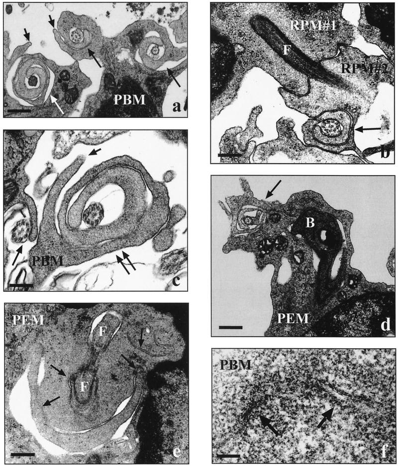
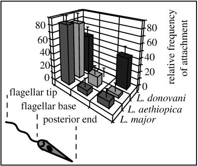
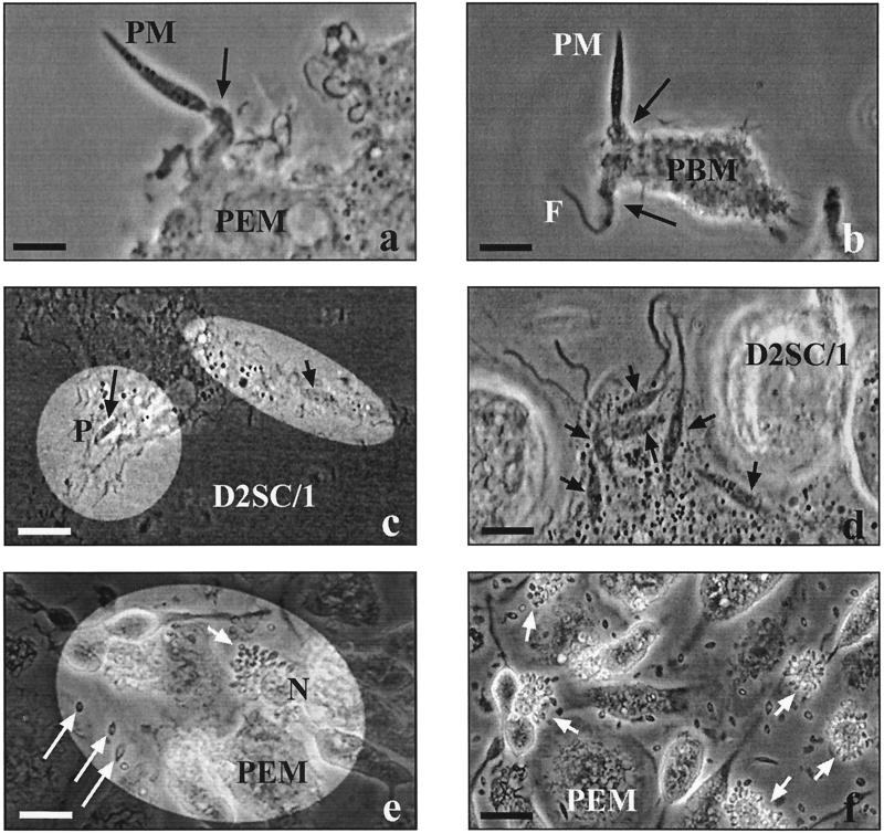
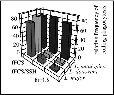
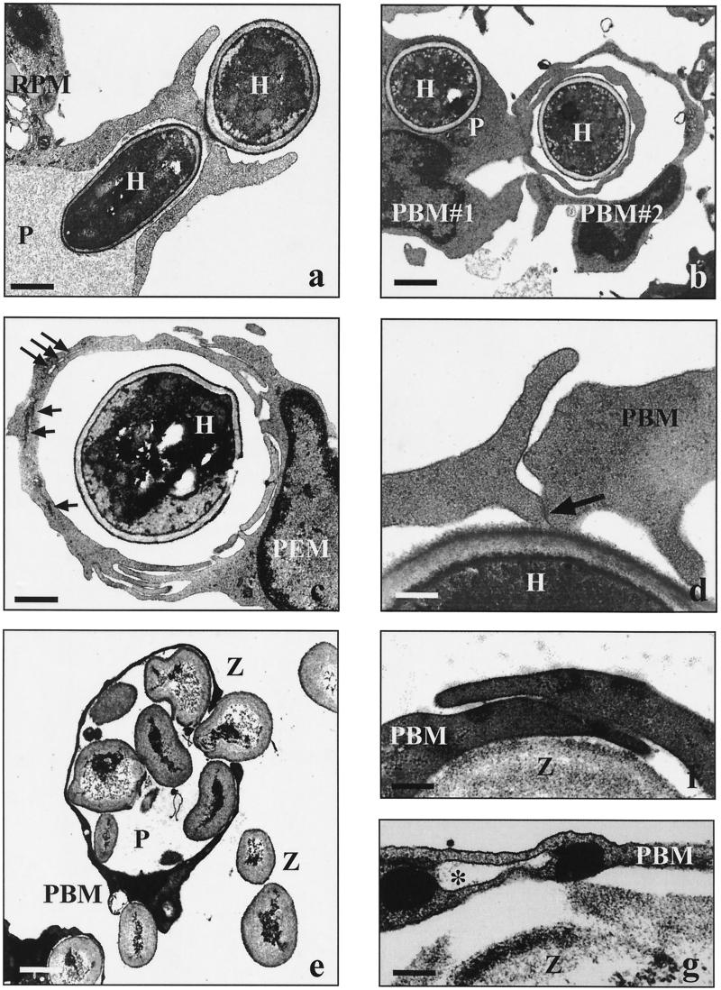
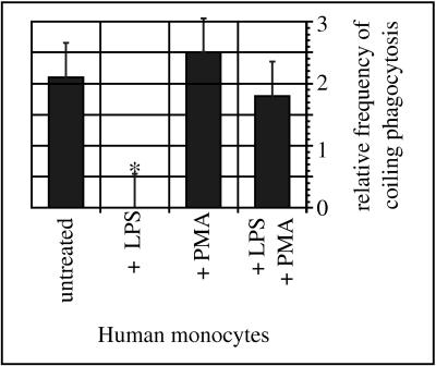
Coiling phagocytosis has previously been studied only with the bacteria Legionella pneumophila and Borrelia burgdorferi, and the results were inconsistent. To learn more about this unconventional phagocytic mechanism, the uptake of various eukaryotic microorganisms by human monocytes, murine macrophages, and murine dendritic cells was investigated in vitro by video and electron microscopy. Unconventional phagocytosis of Leishmania spp. promastigotes, Trypanosoma cruzi trypomastigotes, Candida albicans hyphae, and zymosan particles from Saccharomyces cerevisiae differed in (i) morphology (rotating unilateral pseudopods with the trypanosomatids, overlapping bilateral pseudopods with the fungi), (ii) frequency (high with Leishmania; occasional with the fungi; rare with T. cruzi), (iii) duration (rapid with zymosan; moderate with the trypanosomatids; slow with C. albicans), (iv) localization along the promastigotes (flagellum of Leishmania major and L. aethiopica; flagellum or posterior pole of L. donovani), and (v) dependence on complement (strong with L. major and L. donovani; moderate with the fungi; none with L. aethiopica). All of these various types of unconventional phagocytosis gave rise to similar pseudopod stacks which eventually transformed to a regular phagosome. Further video microscopic studies with L. major provided evidence for a cytosolic localization, synchronized replication, and exocytic release of the parasites, extending traditional concepts about leishmanial infection of host cells. It is concluded that coiling phagocytosis comprises phenotypically similar consequences of various disturbances in conventional phagocytosis rather than representing a single separate mechanism.


Figures

FIG. 1
FIG. 1
Electron micrographs showing the uptake of Leishmania spp. by human and murine phagocytes. Human peripheral blood monocytes (PBM) and murine resident (RPM) or thioglycolate-elicited (PEM) peritoneal macrophages (2 × 10⁶ each) were incubated with live promastigotes (2 × 10⁷ each) from three Leishmania spp. in the presence of fFCS for 30 min unless otherwise indicated. (a) Unilateral pseudopods (long arrows) rotating around flagella of heat-killed parasites (L. donovani; short arrows, additional contrarotating pseudopods; bar = 0.4 μm). (b) Two phagocytes (RPM#1 and RPM#2) simultaneously internalizing a parasite via conventional phagocytosis (L. major; F, flagellum; arrow, pseudopod coil; bar = 0.2 μm). (c) Beginning (single long arrow) and advanced (double long arrows) coiling phagocytosis of parasite flagella in presence of hiFCS (L. aethiopica; short arrow, additional contrarotating pseudopod; bar = 0.2 μm). (d) Parasite with its flagellum engulfed by a pseudopod coil (arrow) and its body (B) located in a regular phagosome (L. aethiopica; bar = 0.6 μm). (e) Contorted coils (arrows) of a pseudopod whorl starting to transform into a confluent phagosome wall (L. major; F, flagellum; bar = 0.2 μm). (f) Fused self-apposed membranes (arrows) in the transition zone of a transforming pseudopod coil (L. major; bar = 0.01 μm).
FIG. 2
FIG. 2
Regional differences in the attachment of promastigotes from various Leishmania spp. to human monocytes. Human peripheral blood monocytes (106) were incubated with either L. major, L. donovani, or L. aethiopica promastigotes (107) in the presence of fFCS for 15 min. The bars represent the relative frequencies of the attachment sites observed with randomly chosen promastigotes, given as mean ± SEM of duplicate determinations. Essentially the same results were observed in two additional experiments using phagocytes from different donors. Promastigotes from L. major and L. aethiopica attach predominantly with the flagellar tip and only occasionally with the flagellar base or posterior pole, whereas L. donovani promastigotes attach with either the flagellar tip or the posterior pole in approximately equal proportions.
FIG. 3
FIG. 3
Video microscopic observations on the infection of various host cells by L. major. Human peripheral blood monocytes (PBM), murine peritoneal macrophages (PEM), or D2SC/1 cells (106 each) were pulsed with L. major promastigotes (PM; 107) in presence of fFCS for 30 min and subsequently chased by video-enhanced phase-contrast microscopy for several days. (a) Funnel-like extension (arrow) of a phagocyte moving along a radiating PM which has been attached with its flagellar tip (bar = 10 μm). (b) Funnel-like extensions (arrows) of a phagocyte moving along in either direction of a PM which has been attached with its flagellar base (F, flagellum; bar = 10 μm). (c) Two PM inside a host cell, one (long arrow) clearly located within a phagosome (P) and the other (short arrow) obviously not bounded by a host cell membrane (bar = 15 μm). (d) Multiple intracellular PM (arrows) obviously not bounded by host cell membranes (bar = 10 μm). (e) Asymmetrical accumulation of small vacuoles (arrow) at the periphery of the host cell (5 days postinfection; N, host cell nucleus; long arrows, released amastigotes; bar = 20 μm). (f) Numerous host cells (arrows) simultaneously releasing replicated parasites; the pericellular fluid is full of amastigotes (5 days postinfection; bar = 20 μm). Reprinted from “Macrophages: Infection with Leishmania major” (28a) with permission of the publisher.
FIG. 4
FIG. 4
Influence of complement on the frequency of coiling phagocytosis of promastigotes from various Leishmania spp. Human peripheral blood monocytes (2 × 10⁶) were incubated with promastigotes from either L. major, L. donovani, or L. aethiopica (2 × 10⁷ each) for 30 min in the presence of 10% (vol/vol) hiFCS or fFCS, the latter with or without 1 mM SSH. Phagocytosis was evaluated in a blinded fashion by electron microscopy of randomly chosen ultrathin sections. The bars represent the relative frequencies of coiling phagocytosis, given as mean ± SEM of duplicate determinations in a typical experiment. Essentially the same results were observed in additional two experiments using phagocytes from different donors. Coiling and conventional phagocytosis of L. major and L. donovani, but not of L. aethiopica, was strongly reduced in presence of heat-inactivated or SSH-neutralized serum.
FIG. 5
FIG. 5
Frequency of coiling phagocytosis observed with L. major, T. cruzi, and C. albicans. Human peripheral blood monocytes (PBM) and resident (RPM) or thioglycolate-elicited (PEM) macrophages from the peritoneal cavities of mice (2 × 10⁶ each) were incubated with either L. major promastigotes, T. cruzi trypomastigotes, or C. albicans hyphae (2 × 10⁷ each) in the presence of fFCS for 30 min. Phagocytosis was evaluated by electron microscopy of randomly chosen ultrathin sections. The bars represent the relative frequencies of coiling phagocytosis, given as mean ± SEM of duplicate determinations in a typical experiment. Essentially the same results were observed in additional two experiments using phagocytes from different donors and animals. The majority of the L. major promastigotes are engulfed via pseudopod coils by all phagocyte populations, whereas this is a very rare event with T. cruzi trypomastigotes. Coiling phagocytosis is only occasionally observed with C. albicans hyphae by PBM and RPM but not by PEM.
FIG. 6
FIG. 6
Electron micrographs showing the uptake of C. albicans and zymosan particles by human and murine phagocytes. Human peripheral blood monocytes (PBM) and murine resident (RPM) or thioglycolate-elicited (PEM) peritoneal macrophages (2 × 10⁶ each) were incubated with live C. albicans hyphae (H) or zymosan particles (Z) (2 × 10⁷) in the presence of fFCS for incubation periods of 20 min to 8 h unless otherwise indicated. (a) Phagocytosis of C. albicans via a symmetrical phagocytic cup (30 min; P, phagosome; bar = 1.7 μm). (b) Two phagocytes (PBM#1 and PBM#2) simultaneously engulfing C. albicans via overlapping pseudopods (60 min; P, conventional phagosome; bar = 3.0 μm). (c) Various stages of fusion events along the overlapping pseudopods enclosing a heat-killed C. albicans cell (240 min; long arrows, remaining membrane fissures; short arrows, closed membrane fissures; bar = 4.0 μm). (d) Slightly overlapping pseudopods of a PBM in the presence of 1 μg of LPS per ml (30 min; arrow, starting membrane fusion; bar = 0.2 μm). (e) Giant phagosome (P) filled with several zymosan particles (20 min; bar = 2.5 μm). (f) Overlapping pseudopods along a zymosan particle in the absence of serum (20 min; bar = 0.5 μm). (g) Remaining membrane-bounded fissure (asterisk) in the phagosome wall enclosing a zymosan particle, indicative of incomplete fusion of overlapping pseudopods (20 min; bar = 0.1 μm).
FIG. 7
FIG. 7
Influence of LPS and PMA on the frequency of coiling phagocytosis of C. albicans. Untreated human peripheral blood monocytes (PBM; 2 × 10⁶) and PBM pretreated with LPS (1 μg/ml) and/or PMA (10 ng/ml) for 10 min were incubated with C. albicans hyphae (2 × 10⁷) in the presence of fFCS for another 30 min. Phagocytosis was evaluated in a blinded fashion by electron microscopy of randomly chosen ultrathin sections. The bars represent the normalized frequencies of coiling phagocytosis, expressed as mean ± SEM of three separate experiments with monocytes from different donors. Pretreatment of PBM with LPS abolished coiling phagocytosis of C. albicans hyphae. The effect of LPS could be neutralized by additional treatment with PMA, whereas PMA alone did not affect the frequency of coiling phagocytosis to a significant extent (∗, P < 0.05 in the χ2 test).

Similar articles

Cited by

References

    1. Akiyama H J, Haight R D. Interaction of Leishmania donovani and hamster peritoneal macrophages. A phase-contrast microscopical study. Am J Trop Med Hyg. 1971;20:539–545. - PubMed
    1. Akuffo H, Maasho K, Blostedt M, Höjeberg B, Britton S, Bakhiet M. Leishmania aethiopica derived from diffuse leishmaniosis patients preferentially induce mRNA for interleukin-12 while those from localized leishmaniosis patients induce interferon-γ. J Infect Dis. 1997;175:737–741. - PubMed
    1. Barker K, Fan H, Carroll C, Kaplan G, Barker J, Hellmann W, Cohn Z A. Nonadherent cultures of human monocytes kill Mycobacterium smegmatis, but adherent cultures do not. Infect Immun. 1996;64:428–433. - PMC - PubMed
    1. Behrendt H, Seemayer N H, Braumann A, Nissen M. Electron microscopy investigations of the effect of quartz dust DQ 12 on human monocytes/macrophages in vitro. Silikoserep Nordrhein-Westphalen. 1987;16:171–183.
    1. Beverley S M, Turco S J. Lipophosphoglycan (LPG) and the identification of virulence genes in the protozoan parasite Leishmania. Trends Microbiol. 1998;6:35–40. - PubMed

Publication types

LinkOut - more resources