

Adhesion of lens capsule to intraocular lenses of polymethylmethacrylate, silicone, and acrylic foldable materials: an experimental study
- PMID: 9713064
- PMCID: PMC1722579
- DOI: 10.1136/bjo.82.5.549
Adhesion of lens capsule to intraocular lenses of polymethylmethacrylate, silicone, and acrylic foldable materials: an experimental study
Abstract
Aims: To investigate the adhesion characteristics of several intraocular lenses (IOLs) to the simulated and rabbit lens capsule.
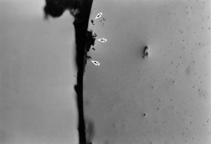
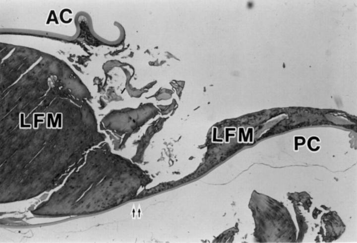
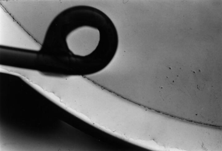
Methods: Adhesive force to bovine collagen sheets was measured in water with polymethylmethacrylate (PMMA), three piece silicone, and acrylic foldable IOLs. In rabbit eyes, phacoemulsification and IOL implantation were performed. Three weeks later, adhesion between the anterior/posterior capsules and IOL optic was tested, and the capsule was examined histologically.
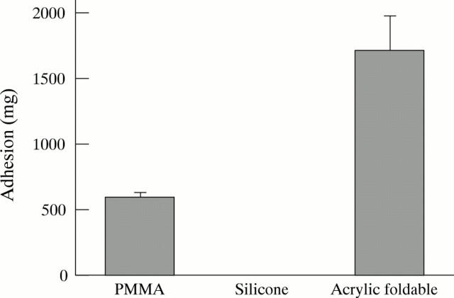
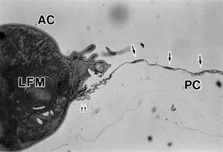
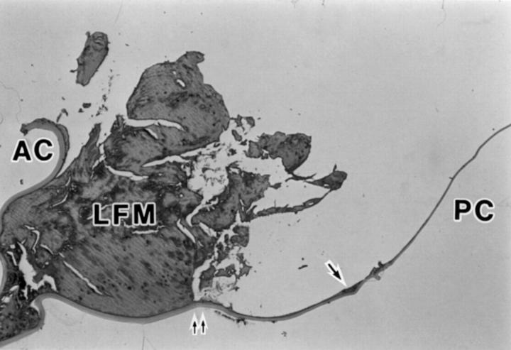
Results: The mean adhesive force to the collagen sheet was 1697 (SD 286) mg for acrylic foldable, 583 (49) mg for PMMA, and 0 mg for silicone IOLs (p = 0.0003, Kruskal-Wallis test). Scores (0-5) of adhesion between rabbit anterior capsule and IOL optic were 4.50 (0.55) for acrylic foldable, 3.20 (0.84) for PMMA, and 0.40 (0.55) for silicone IOLs (p = 0.004). Scores between rabbit posterior capsule and IOL optic displayed a similar tendency; 4.50 (0.84) for acrylic foldable, 3.00 (1.00) for PMMA, and 0.40 (0.55) for silicone IOLs (p = 0.021). Histological observation indicated that the edge of IOL optic suppressed the migration of lens epithelial cells towards the centre of the posterior capsule. This inhibitory effect was most pronounced with acrylic foldable IOL and least with silicone IOL.
Conclusions: The acrylic foldable IOL adhered to the lens capsule more than the PMMA IOL, and the silicone IOL showed no adhesiveness. These differences seem to play a role in preventing lens epithelial cells from migrating and forming posterior capsule opacification.
Figures
Comment in
-
Adhesion of IOLs to the posterior capsule.Br J Ophthalmol. 1998 May;82(5):468. doi: 10.1136/bjo.82.5.468. Br J Ophthalmol. 1998. PMID: 9713048 Free PMC article. No abstract available.
References
MeSH terms
Substances
LinkOut - more resources
Full Text Sources
Other Literature Sources