In vivo comparison of three different porous materials intended for use in a keratoprosthesis
- PMID: 9713067
- PMCID: PMC1722587
- DOI: 10.1136/bjo.82.5.569
In vivo comparison of three different porous materials intended for use in a keratoprosthesis
Abstract
Aim: The goal was to compare the biological response of the corneal stroma with three porous materials: a melt blown microfibre web of polybutylene:polypropylene (80:20); a polyester spun laced fabric (polyethylene terephthalate), and an expanded polytetrafluoroethylene. Fifty per cent of each of the materials were modified using argon radio frequency.
Methods: Discs (6 mm in diameter) were inserted into interlamellar stromal pockets and followed for a period of 12 weeks. Clinical evaluations were performed weekly. At 6 and 12 weeks, fibroplasia and the distribution of matrix proteins and growth factors (bFGF and TGF-beta) were evaluated immunohistochemically. The characterisation of glycosaminoglycans was determined after selective extraction and digestion.
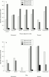
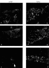
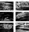
Results: The response to the disc resembled that of a wound with a decrease in keratan sulphate and an increase in dermatan sulphate. Pretreatment of the disc reduced corneal oedema and neovascularisation. Heparan sulphate, not normally detected in the corneal stroma, was detected in the region immediately surrounding the disc and in the discs of some materials. The presence of glycosaminoglycans and collagens in the disc indicated that cells had migrated into the disc and deposited a complex matrix in all three materials. The collagen response was not surface specific. bFGF and TGF-beta were detected in the region between the disc and the stroma in the polybutylene material and became diffuse with time.
Conclusion: Fibroplasia occurred most rapidly into the polyester discs but was accompanied by a large number of inflammatory cells. While the distribution of collagens was not altered by the material, the expression and distribution of growth factors was material dependent. bFGF was expressed transiently and occurred before that of TGF-beta. It is predicted that the transient expression of growth factors mediates the regulation of matrix proteins.
Figures


Similar articles
-
Comparative experiments for in vivo fibroplasia and biological stability of four porous polymers intended for use in the Seoul-type keratoprosthesis.Br J Ophthalmol. 2002 Jul;86(7):809-14. doi: 10.1136/bjo.86.7.809. Br J Ophthalmol. 2002. PMID: 12084755 Free PMC article.
-
Effect of pretreating porous webs on stromal fibroplasia in vivo.J Biomed Mater Res. 1994 Feb;28(2):195-202. doi: 10.1002/jbm.820280209. J Biomed Mater Res. 1994. PMID: 8207031
-
In vivo fibroplasia of a porous polymer in the cornea.Invest Ophthalmol Vis Sci. 1991 Dec;32(13):3245-51. Invest Ophthalmol Vis Sci. 1991. PMID: 1748554
-
[Development of better tolerated prosthetic materials: applications in gynecological surgery].J Gynecol Obstet Biol Reprod (Paris). 2002 Oct;31(6):527-40. J Gynecol Obstet Biol Reprod (Paris). 2002. PMID: 12407323 Review. French.
-
Corneal collagens.Pathol Biol (Paris). 2001 May;49(4):353-63. doi: 10.1016/s0369-8114(01)00144-4. Pathol Biol (Paris). 2001. PMID: 11428172 Review.
Cited by
-
Comparative experiments for in vivo fibroplasia and biological stability of four porous polymers intended for use in the Seoul-type keratoprosthesis.Br J Ophthalmol. 2002 Jul;86(7):809-14. doi: 10.1136/bjo.86.7.809. Br J Ophthalmol. 2002. PMID: 12084755 Free PMC article.
-
Histopathologic Evaluation of Polymer Supports for Pintucci-type Keratoprostheses: An Animal Study.J Ophthalmic Vis Res. 2019 Jul 18;14(3):243-250. doi: 10.18502/jovr.v14i3.4779. eCollection 2019 Jul-Sep. J Ophthalmic Vis Res. 2019. PMID: 31660102 Free PMC article. Review.
References
Publication types
MeSH terms
Substances
Grants and funding
LinkOut - more resources
Full Text Sources
Other Literature Sources
