

Review
doi: 10.1136/bjo.82.5.577.
Regeneration and transplantation of the optic nerve: developing a clinical strategy
Affiliations
- PMID: 9713068
- PMCID: PMC1722609
- DOI: 10.1136/bjo.82.5.577
Item in Clipboard
Review
Regeneration and transplantation of the optic nerve: developing a clinical strategy
Br J Ophthalmol.
1998 May.
Abstract
Three separate experimental models of optic nerve regeneration have been presented--along the existing pathway in the presence of antibodies to neutralise inhibitory molecules, along peripheral nerve grafts and from retinal transplants. Each offers a theoretical clinical strategy for restoration of vision, if the mechanism of re-establishment of maps and reconnection to appropriate targets during regeneration can be determined. This is the process of axon guidance, and underlines the importance of our research into the molecular determinants that guide normal development of the visual system.
Figures
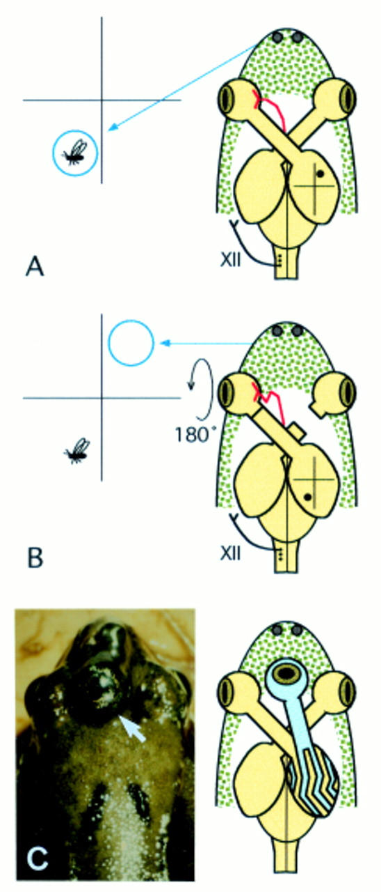
Optic nerve regeneration in the frog and the recreation of visual maps. (A) In normal circumstances, the left visual field maps precisely to the right optic tectum. Movement of a fly lure detected in the visual field will elicit a response at a corresponding location in the contralateral tectum. The tongue (blue arrow) will thus be directed appropriately towards the target. (B) If the left optic nerve is rotated about 180 degrees and cut (preserving the blood supply), following regeneration, an inverted map of visual space is reformed on the contralateral tectum. With the normal right optic nerve disconnected, the frog will perceive the lure to be in the opposite quadrant of visual space and will repeatedly miss the target with its tongue (blue arrow). This experiment therefore shows that regenerating ganglion cells in the frog retain an intrinsic ability to navigate back to appropriate central targets, despite complete misalignment of cut optic nerve fibres. Similar processes of axon pathfinding would most likely be necessary for successful optic nerve regeneration in primates. (C) A third eye transplanted to the top of the head at embryonic stages can become fully functional (arrow). The transplanted eye (shown in blue) connects with correct retinal topography to the optic tectum, although tectal sites are shared with the host eye in an arrangement similar to ocular dominance columns (blue stripes). This observation suggests that guidance cues within a host tectum are also able to direct ganglion cells from a transplanted eye to appropriate targets.
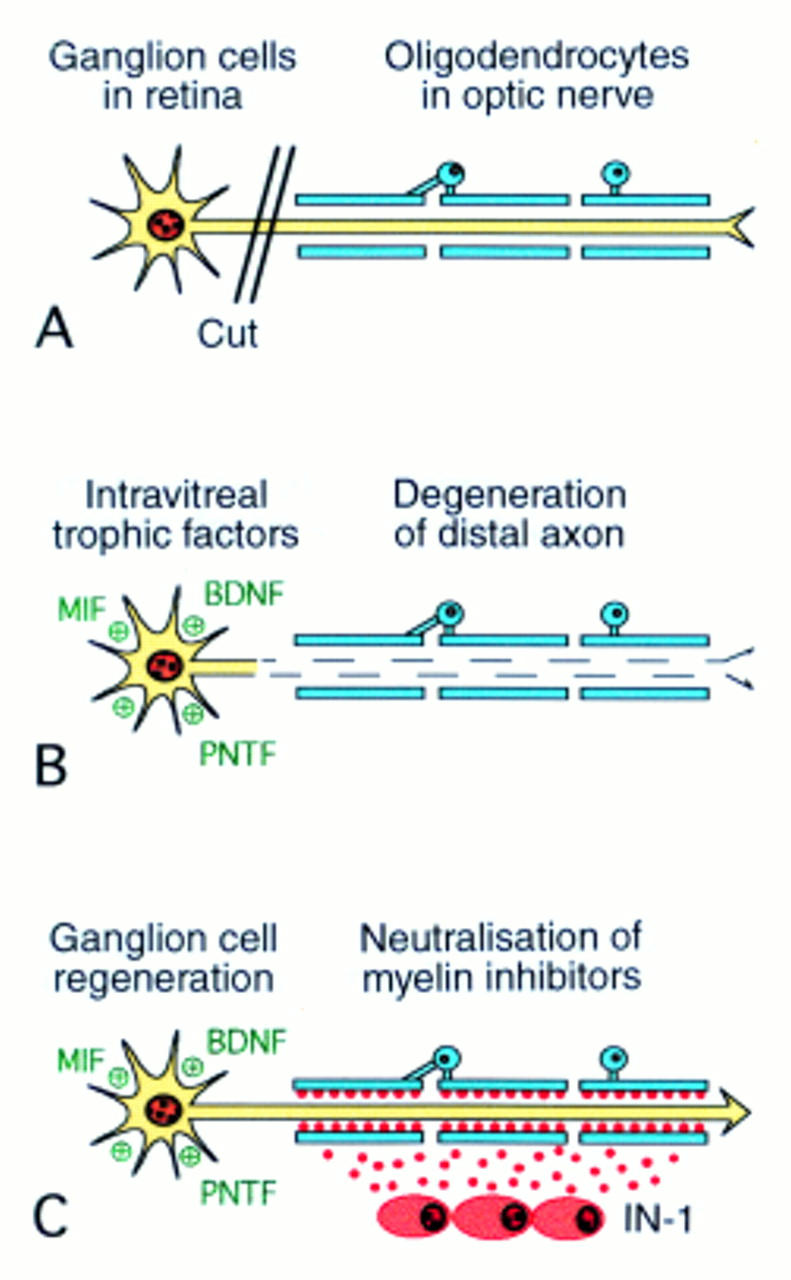
Optic nerve regeneration in mammals by the neutralisation of myelin associated inhibitory proteins. (A) Ganglion cell axons in the optic nerve are surrounded by oligodendrocytes which normally inhibit axon growth. (B) After axotomy, the distal axon will degenerate but the soma can be prevented from undergoing cell death by the intravitreal injection of certain growth factors and immunosuppressants. (C) In order to re-extend an axon along the optic nerve, the inhibitory myelin proteins in oligodendrocytes need to be neutralised. This can be done experimentally by the surgical implantation of hybridoma cells continually secreting monoclonal antibodies (IN-1) directed against and blocking the inhibitory proteins. The axon can then regenerate a significant distance along the optic nerve, although reconnection to central targets has not yet been achieved (MIF = macrophage inhibitory factor; BDNF = brain derived neurotrophic factor; PNTF = peripheral nerve trophic factors).
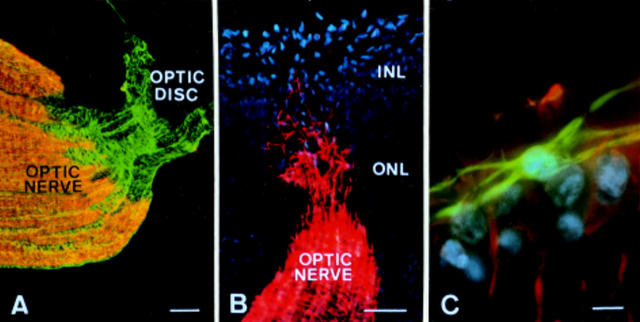
Glial cells within the visual system. (A) The optic nerve is densely myelinated (orange) and is thus inhibitory to axonal growth, although the optic nerve head and adjacent retina contain only astrocytes (green) and are therefore preferred regions for the insertion of peripheral nerve grafts. (B) Astrocytes (red) migrate into the retina through the optic nerve head at early stages of development (INL = inner neuroblastic layer; ONL = outer neuroblastic layer) to surround retinal blood vessels. (C) A mature astrocyte (green) shown close to the ganglion cell layer (blue nuclei) and perpendicular to the radial processes of Müller cells (red). After CNS injury, astrocytes normally express inhibitory proteins to block regeneration, but this has not been observed in the retina. Glial cell differences may therefore explain why the retina is probably a more favourable site for regeneration than elsewhere in the CNS. Scale bars: A = 50 µm; B = 25 µm; C = 10 µm (opossum)
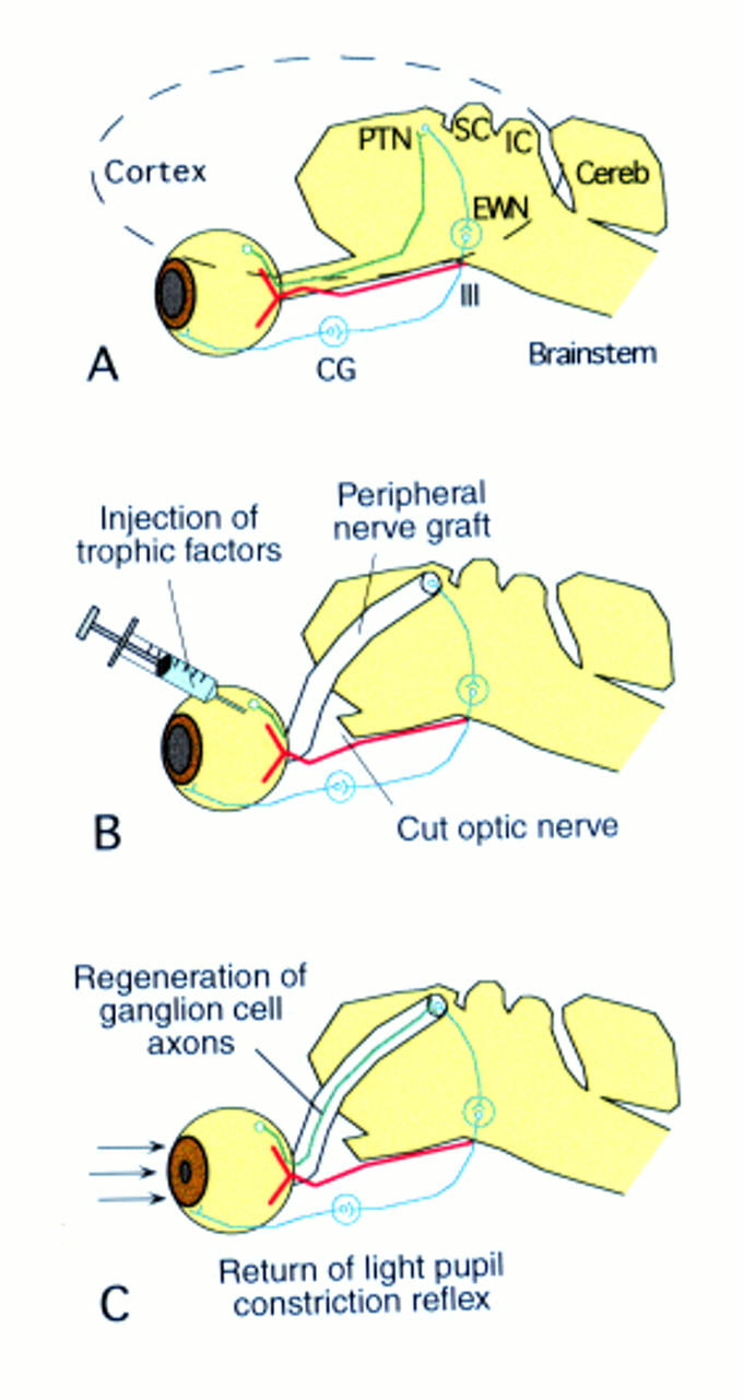
Utilisation of a peripheral nerve graft to connect regenerating ganglion cells directly to central targets. (A) The normal light reflex is mediated by ganglion cells through synapses in the pretectal nucleus (PTN), Edinger-Westphal nucleus (EWN), and ciliary ganglion (CG) of the oculomotor nerve (III). The positions of the superior and inferior colliculi (SC and IC) and cerebellum (Cereb) are also shown. (B) After optic nerve transection, intravitreal growth factors can sustain the lesioned ganglion cells in the retina, provided that the retinal blood supply remains viable (shown in red). A peripheral nerve transplant can provide a conduit between the optic nerve head and pretectal nucleus. (C) Ganglion cell axons are able to regenerate within the favourable environment of the peripheral nerve graft to reform functional connections in the pretectal nucleus and restore the light reflex. The graft thus facilitates regeneration by bypassing the inhibitory CNS environment altogether.
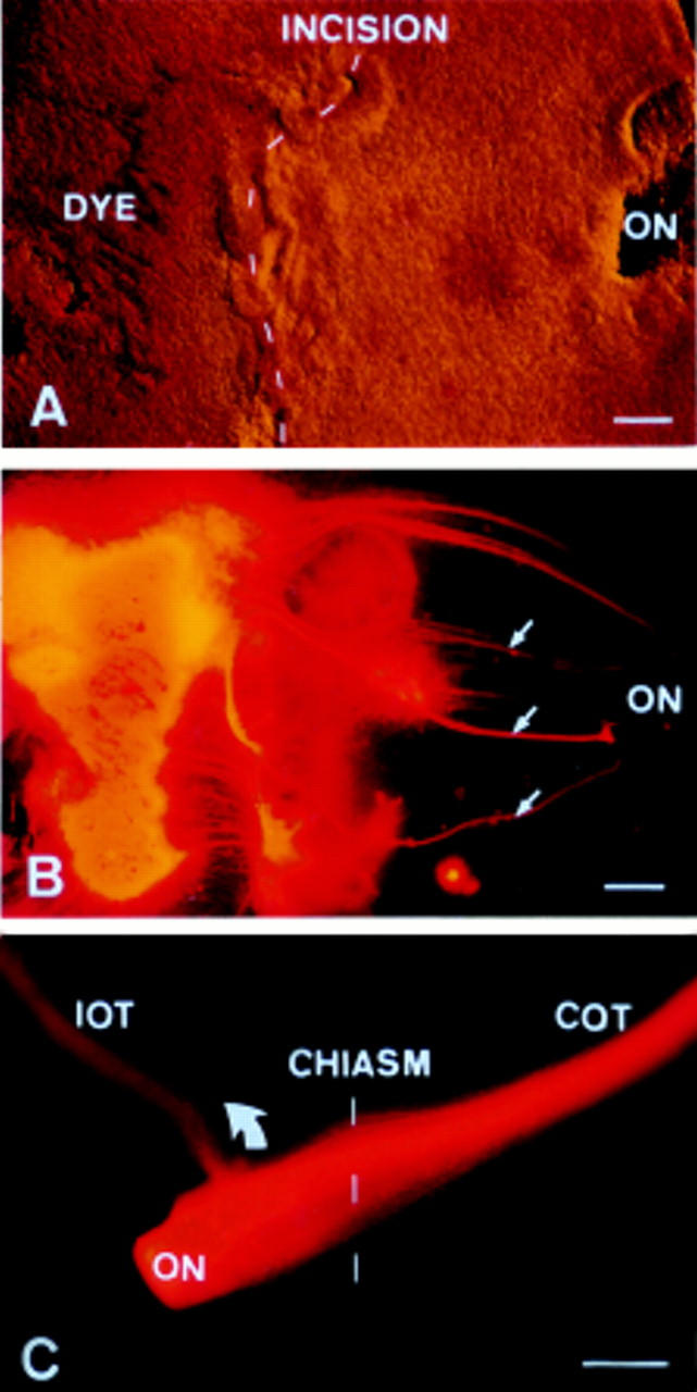
Navigation of regenerating ganglion cells in the developing mammalian CNS. (A) At early stages of development, ganglion cells can regenerate across an incision made in the retina. The optic nerve (ON) is shown to the right and an axon labelling dye is shown embedded in the temporal retina to the left. (B) Under fluorescent light, the precise path of regenerating axons can be seen. Some axons have regrown directly across the incision (arrows) and all are appropriately guided into the optic nerve. (C) At the chiasm, axons originating from the temporal retina turn appropriately (arrow) to enter the ipsilateral optic tract (IOT), while the remainder project to the contralateral optic tract (COT). This study not only demonstrates that mammalian ganglion cells can regenerate at early stages of development, but more importantly, also have a capacity for navigation back to appropriate targets. Scale bars: A = 100 µm; B = 100 µm; C = 250 µm (opossum).
Similar articles
-
Regeneration of rat optic axons into peripheral nerve grafts.Exp Neurol. 1986 Jan;91(1):52-9. doi: 10.1016/0014-4886(86)90025-7. Exp Neurol. 1986. PMID: 3940879
-
Regeneration in the optic nerve of adult rats: influences of cultured astrocytes and optic nerve grafts of different ontogenetic stages.J Neurocytol. 1995 Oct;24(10):783-93. doi: 10.1007/BF01191214. J Neurocytol. 1995. PMID: 8586998
-
Advances in experimental optic nerve regeneration.Curr Opin Ophthalmol. 2017 Nov;28(6):558-563. doi: 10.1097/ICU.0000000000000417. Curr Opin Ophthalmol. 2017. PMID: 28795960 Review.
-
[Optic nerve regeneration by nerve transplantation].Nippon Ganka Gakkai Zasshi. 1996 Dec;100(12):956-71. Nippon Ganka Gakkai Zasshi. 1996. PMID: 9022308 Review. Japanese.
-
[Optic nerve regeneration and functional recovery of vision following peripheral nerve transplant].No To Shinkei. 1998 Mar;50(3):227-35. No To Shinkei. 1998. PMID: 9565997 Review. Japanese. No abstract available.
Cited by
-
Emerging options for the management of age-related macular degeneration with stem cells.Stem Cells Cloning. 2010 Dec 22;4:1-10. doi: 10.2147/SCCAA.S7674. Stem Cells Cloning. 2010. PMID: 24198525 Free PMC article. Review.
-
Impediments to eye transplantation: ocular viability following optic-nerve transection or enucleation.Br J Ophthalmol. 2009 Sep;93(9):1134-40. doi: 10.1136/bjo.2008.155267. Epub 2009 Mar 13. Br J Ophthalmol. 2009. PMID: 19286686 Free PMC article. Review.
-
Synergetic effects of ciliary neurotrophic factor and olfactory ensheathing cells on optic nerve reparation (complete translation).Neural Regen Res. 2016 Jun;11(6):1006-12. doi: 10.4103/1673-5374.184505. Neural Regen Res. 2016. PMID: 27482233 Free PMC article.
-
Early decompression of the injured optic nerve reduces axonal degeneration and improves functional outcome in the adult rat.Exp Brain Res. 2007 May;179(1):121-30. doi: 10.1007/s00221-006-0775-1. Epub 2006 Nov 14. Exp Brain Res. 2007. PMID: 17103208
References
Publication types
MeSH terms
LinkOut - more resources
Full Text Sources