. 1998 Sep 15;511 ( Pt 3)(Pt 3):947-56.
doi: 10.1111/j.1469-7793.1998.947bg.x.

Inhibitory action of forearm flexor muscle afferents on corticospinal outputs to antagonist muscles in humans

Affiliations

Inhibitory action of forearm flexor muscle afferents on corticospinal outputs to antagonist muscles in humans

L Bertolasi et al. J Physiol. .

Abstract

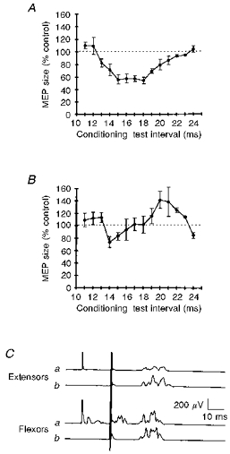
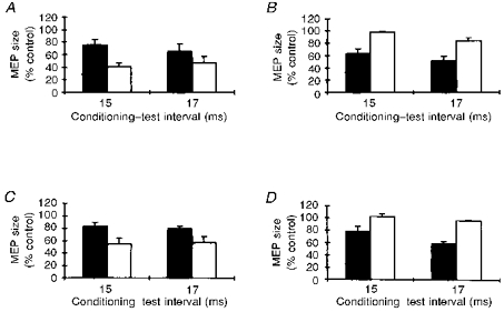
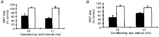
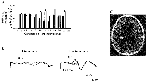
1. To find out whether muscle afferents influence the excitability of corticospinal projections to antagonist muscles, we studied sixteen healthy subjects and one patient with a focal brain lesion. 2. Using transcranial magnetic and electrical brain stimulation we tested the excitability of corticomotoneuronal connections to right forearm muscles at rest after conditioning stimulation of the median nerve at the elbow. Somatosensory potentials evoked by median nerve stimulation were also recorded in each subject. 3. Test stimuli delivered at 13-19 ms after median nerve stimulation significantly inhibited EMG responses elicited in forearm extensor muscles by transcranial magnetic stimulation, but did not inhibit responses to electrical stimulation. In contrast, magnetically and electrically elicited responses in forearm flexor muscles were suppressed to the same extent. 4. The higher the intensity of the test shocks, the smaller was the amount of median nerve-elicited inhibition. Inhibition in extensor muscles was also smaller during tonic wrist extension, or if the induced electrical stimulating current in the brain flowed from posterior to anterior over the motor strip rather than vice versa. Test responses evoked by magnetic transcranial stimulation in the first dorsal interosseous and in brachioradialis muscles were not inhibited after median nerve stimulation at the elbow. Stimulation of digital nerves failed to inhibit motor potentials in extensor muscles. 5. Test stimuli delivered at 15 and 17 ms after radial nerve stimulation significantly inhibited EMG responses elicited in forearm flexor muscles by magnetic transcranial stimulation. 6. In the patient with a focal thalamic lesion, who had dystonic postures and an absent N20 component of the somatosensory-evoked potentials but normal strength, median nerve stimulation failed to inhibit magnetically evoked responses in forearm extensor muscles. 7. We propose that activation of median nerve muscle afferents can suppress the excitability of cortical areas controlling the antagonist forearm extensor muscles acting on the hand. The inhibitory effect occurs at short latency and might assist spinal pathways mediating reciprocal inhibition by contrasting the co-activation of antagonistic pools of corticospinal cells.


Figures

Figure 1
Figure 1. Magnetic MEPs in forearm extensor and flexor muscles conditioned by median nerve stimulation
The average time course of the effect of a conditioning stimulus delivered at motor threshold intensity over the median nerve at the elbow on the size of magnetically evoked MEPs in forearm extensor (A) and flexor muscles (B). In A and B, the x-axis shows the conditioning-test intervals (ms), and the y-axis the MEP size as a percentage of the area of the unconditioned control response. The graphs show that median nerve stimulation at conditioning-test intervals of 15–18 ms produced a marked decrease in the MEP size in extensors, whereas it induced far less pronounced inhibition in flexors. Each point is the average of 10 subjects, except the 22 and 23 ms intervals, which were studied in 6 subjects. Error bars show 1 s.e.m.C, magnetic MEPs in forearm extensors and flexors conditioned (a) and unconditioned (b) by a stimulus delivered over the median nerve at the elbow at the conditioning-test interval of 18 ms in a representative subject. Note that the electrodes over forearm extensor muscles do not pick up the small M wave and the H reflex evoked in flexors by conditioning stimulation of the median nerve. Each trace is the average of 8 sweeps.
Figure 2
Figure 2. Characteristics of MEP inhibition in extensors after median nerve stimulation
The effects of a slight voluntary contraction (A), of test stimulus intensity (B), and of the direction of the current flow in the coil (C) on the MEP inhibition in forearm extensors after median nerve stimulation at the most effective conditioning-test intervals, 15 and 17 ms. A: contraction, ▪; at rest, □. B: 60 % of stimulator output (clockwise current), ▪; 85 % of stimulator output (clockwise currents), □. C: anticlockwise currents, ▪; clockwise currents, □. The graphs show that conditioning stimulation of the median nerve produced a larger inhibition at rest than during contraction (A), with lower test stimulus intensities (B), and with current flowing clockwise in the coil. D, the effect of conditioning stimulation delivered to the median nerve at the elbow on magnetic MEPs in forearm extensor muscles (▪) and in the first dorsal interosseous muscle (□) at conditioning-test intervals of 15 and 17 ms. The graph shows that a conditioning stimulus over the median nerve selectively inhibits magnetic MEPs in the forearm extensors but leaves responses in a small hand muscle unchanged. Each bar is the mean of 5 subjects, and error bars show 1 s.e.m.
Figure 3
Figure 3. Magnetic versus electrical test TCS, and mixed nerve versus cutaneous conditioning input
A, comparison of magnetic MEPs (▪) and electrical MEPs (□) in forearm extensor muscles after a stimulus delivered over the median nerve at the elbow at the most significant conditioning-test intervals of 15 and 17 ms. The graph shows that a conditioning stimulus over the median nerve selectively reduces the size of magnetic MEPs, whereas it does not inhibit electrical MEPs. B, comparison between stimulation of the median nerve at the elbow at motor threshold intensities (▪) and cutaneous stimulation of the fingers (□). Conditioning-test intervals for finger stimulation were corrected by adding the conduction time from the fingers to the elbow (see Methods). The graph shows that conditioning median nerve stimulation decreases the MEP size whereas finger stimulation does not. Each bar is the mean of 5 subjects, and error bars show 1 s.e.m.
Figure 4
Figure 4. Time course of magnetic MEP inhibition in forearm extensors after a conditioning stimulus delivered to the median nerve in a patient with a thalamic lesion
A, test responses in the affected upper limb contralateral to the lesion (□) and in the sound upper limb ipsilateral to the lesion (▪). Each bar is the mean size of 8 conditioned MEPs, and error bars show 1 s.e.m.B, SEPs evoked by median nerve stimulation contralateral (on the left) and ipsilateral (on the right) to the lesion. In A, note that when conditioning stimulation and test responses are in the (affected) forearm contralateral to the lesion and the N20 component is absent there is no magnetic MEP inhibition. Conversely, when conditioning stimulation and test responses are in the (sound) forearm ipsilateral to the thalamic lesion and the N20 is regularly present, test responses are regularly inhibited. The P14 component (generated in the brainstem) is bilaterally present. C, the brain CT scan showing a left thalamic haemorrhage.

Similar articles

Cited by

References

    1. Baldissera F, Leocani L. Afferent excitation of human motor cortex as revealed by enhancement of direct cortico-spinal actions on motoneurones. Electroencephalography and Clinical Neurophysiology. 1995;97:394–401. - PubMed
    1. Berardelli A, Day BL, Marsden CD, Rothwell JC. Evidence favouring presynaptic inhibition between antagonist muscle afferents in the human forearm. The Journal of Physiology. 1987;391:71–83. - PMC - PubMed
    1. Bertrand L, Capaday C, Devanne H, Lavoie BA. Intracortical connections between motor cortical zones controlling antagonist muscles. Society for Neuroscience Abstracts. 1996;22:1083.
    1. Caccia MR, McComas AJ, Upton ARM, Blog T. Cutaneous reflexes in small muscles of the hand. Journal of Neurology Neurosurgery and Psychiatry. 1970;36:960–977. - PMC - PubMed
    1. Clouston PD, Kiers L, Menkes D, Sander H, Chiappa K, Cros D. Inhibition of motor evoked potential by digital electrical stimulation. Electroencephalography and Clinical Neurophysiology. 1995;97:114–125. - PubMed

Publication types

MeSH terms

LinkOut - more resources