. 1998 Aug 15;12(16):2469-74.
doi: 10.1101/gad.12.16.2469.

Reduced skin tumor development in cyclin D1-deficient mice highlights the oncogenic ras pathway in vivo

Affiliations

Reduced skin tumor development in cyclin D1-deficient mice highlights the oncogenic ras pathway in vivo

A I Robles et al. Genes Dev. .

Abstract

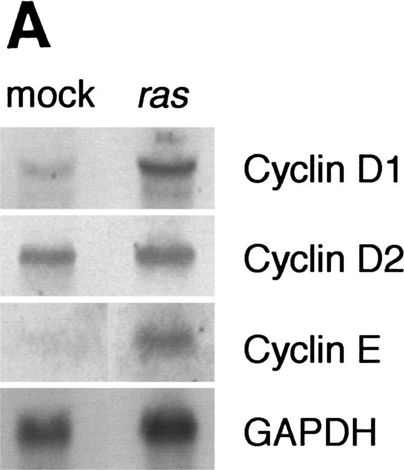
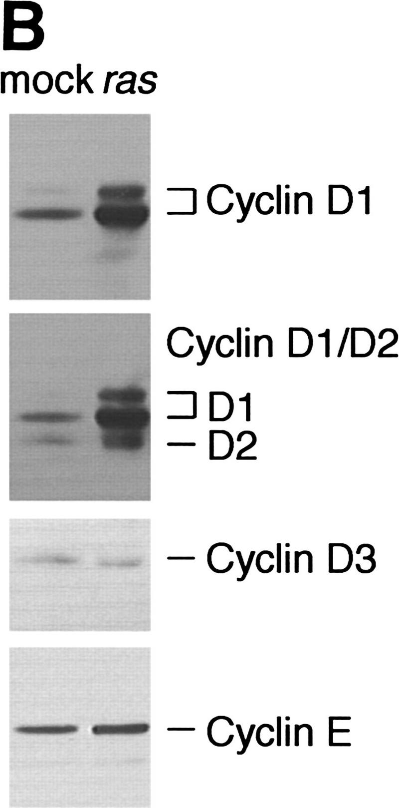
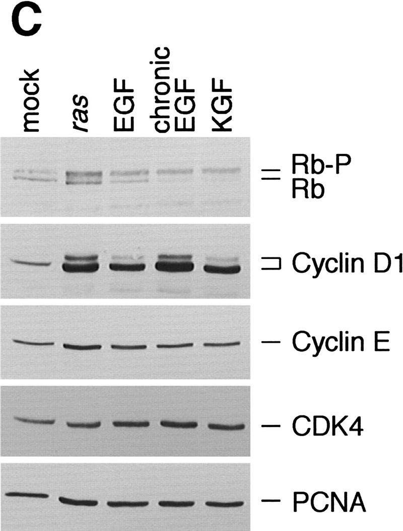
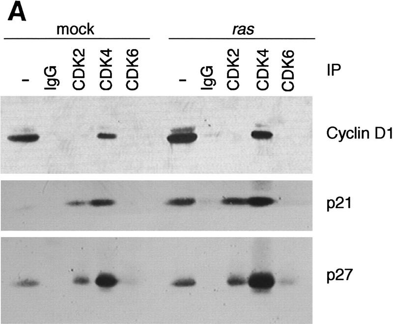
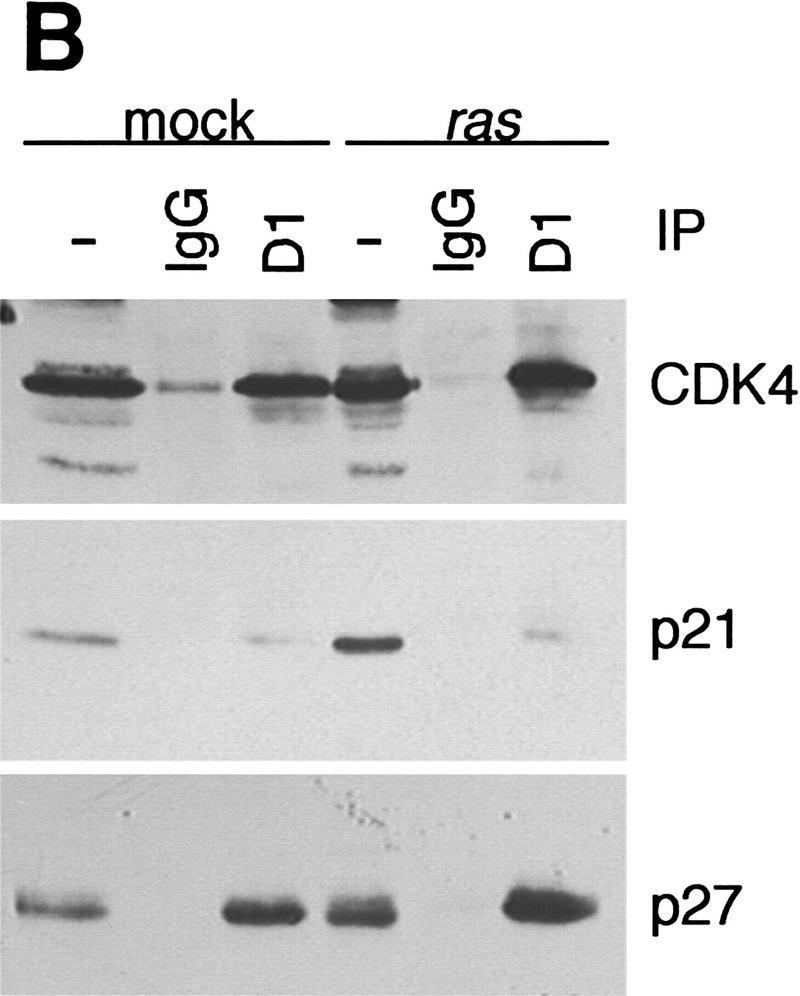
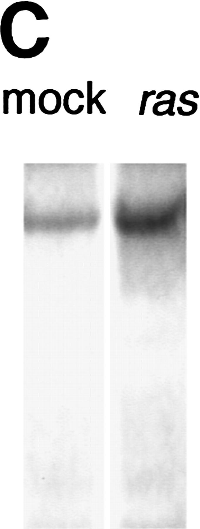
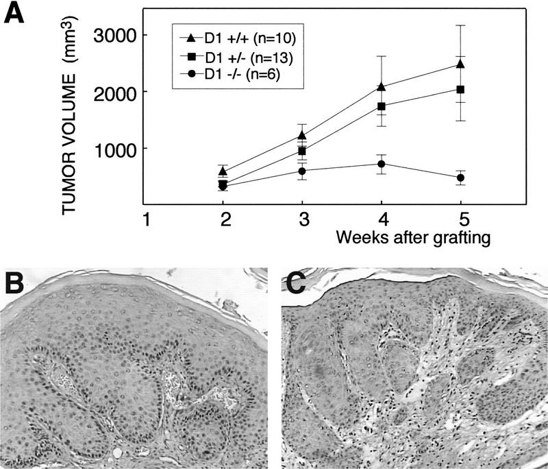
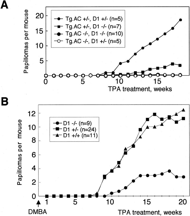
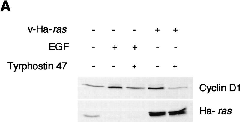
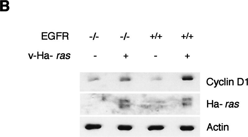
Cyclin D1 is part of a cell cycle control node consistently deregulated in most human cancers. However, studies with cyclin D1-null mice indicate that it is dispensable for normal mouse development as well as cell growth in culture. Here, we provide evidence that ras-mediated tumorigenesis depends on signaling pathways that act preferentially through cyclin D1. Cyclin D1 expression and the activity of its associated kinase are up-regulated in keratinocytes in response to oncogenic ras. Furthermore, cyclin D1 deficiency results in up to an 80% decrease in the development of squamous tumors generated through either grafting of retroviral ras-transduced keratinocytes, phorbol ester treatment of ras transgenic mice, or two-stage carcinogenesis.


Figures

Figure 1
Figure 1
Expression of G1 cyclins mRNA (A) and proteins (B) in mock and v-Ha-ras-infected murine primary keratinocytes. (C) Comparative induction of cyclin D1 protein expression and pRb hyperphosphorylation in mock-infected (mock), v-Ha-ras-infected (ras), and after single-dose EGF (EGF), continuous presence of EGF in the media (chronic EGF), and single-dose KGF (KGF) treatments of primary murine keratinocytes.
Figure 1
Figure 1
Expression of G1 cyclins mRNA (A) and proteins (B) in mock and v-Ha-ras-infected murine primary keratinocytes. (C) Comparative induction of cyclin D1 protein expression and pRb hyperphosphorylation in mock-infected (mock), v-Ha-ras-infected (ras), and after single-dose EGF (EGF), continuous presence of EGF in the media (chronic EGF), and single-dose KGF (KGF) treatments of primary murine keratinocytes.
Figure 2
Figure 2
Analysis of G1 cyclin–CDK complex formation in mock and v-Ha-ras-infected murine primary keratinocytes. Whole cell extracts and CDK (A) or cyclin D1 (B) immunoprecipitates were subjected to immunoblot analysis with cyclin D1, p21, and p27 (A), or CDK4, p21, and p27 (B). (C) Phosphorylation of pRb by CDK4 immunocomplex.
Figure 2
Figure 2
Analysis of G1 cyclin–CDK complex formation in mock and v-Ha-ras-infected murine primary keratinocytes. Whole cell extracts and CDK (A) or cyclin D1 (B) immunoprecipitates were subjected to immunoblot analysis with cyclin D1, p21, and p27 (A), or CDK4, p21, and p27 (B). (C) Phosphorylation of pRb by CDK4 immunocomplex.
Figure 3
Figure 3
(A) Primary keratinocytes from each cyclin D1 genotype were infected with v-Ha-ras retrovirus and grafted onto nude mice. Approximate tumor volume was calculated as tumor height × length × width in mm. Error bars, s.e.m. Immunohistochemical staining of cyclin D1 in a cyclin D1 heterozygous (B) and a cyclin D1 knockout (C) graft.
Figure 4
Figure 4
(A) Papilloma development in isogenic female mice generated through backcrossing of the Tg.AC transgene onto cyclin D1 knockout background and treated with TPA. (B) Papilloma development in isogenic cyclin D1 knockout (−/−), heterozygous (+/−), and wild-type (+/+) mice subjected to a two-stage carcinogenesis protocol using DMBA as initiator and TPA as promoter.
Figure 5
Figure 5
(A) v-Ha-ras-infected primary murine keratinocytes were treated with 50 μm of tyrphostin 47 or DMSO for 18 hr. As a control, primary murine keratinocytes were treated with 10 ng/ml EGF for 18 hr in addition to tyrphostin. (B) Expression of cyclin D1 and v-Ha-ras in mock- and v-Ha-ras-infected primary murine keratinocytes isolated from EGFR knockout mice and wild type. Expression of actin was used for equal loading correction.
Figure 5
Figure 5
(A) v-Ha-ras-infected primary murine keratinocytes were treated with 50 μm of tyrphostin 47 or DMSO for 18 hr. As a control, primary murine keratinocytes were treated with 10 ng/ml EGF for 18 hr in addition to tyrphostin. (B) Expression of cyclin D1 and v-Ha-ras in mock- and v-Ha-ras-infected primary murine keratinocytes isolated from EGFR knockout mice and wild type. Expression of actin was used for equal loading correction.

References

    1. Aagaard L, Lukas J, Bartkova J, Kjerulff AA, Strauss M, Bartek J. Aberrations of p16Ink4 and retinoblastoma tumour-suppressor genes occur in distinct sub-sets of human cancer cell lines. Int J Cancer. 1995;61:115–120. - PubMed
    1. Albanese C, Johnson J, Watanabe G, Eklund N, Vu D, Arnold A, Pestell RG. Transforming p21ras mutants and c-Ets-2 activate the cyclin D1 promoter through distinguishable regions. J Biol Chem. 1995;270:23589–23597. - PubMed
    1. Bartkova J, Lukas J, Strauss M, Bartek J. The PRAD-1/cyclin D1 oncogene product accumulates aberrantly in a subset of colorectal carcinomas. Int J Cancer. 1994;58:568–573. - PubMed
    1. Bianchi AB, Fischer SM, Robles AI, Rinchik EM, Conti CJ. Overexpression of cyclin D1 in mouse skin carcinogenesis. Oncogene. 1993;8:1127–1133. - PubMed
    1. Dlugosz AA, Cheng C, Denning MF, Dempsey PJ, Coffey RJ, Jr, Yuspa SH. Keratinocyte growth factor-receptor ligands induce TGFα expression and activate the epidermal growth factor-receptor signaling pathway in cultured epidermal keratinocytes. Cell Growth & Differ. 1994;5:1283–1292. - PubMed

Publication types