. 1998 Aug 15;12(16):2610-22.
doi: 10.1101/gad.12.16.2610.

Differential recruitment of Dishevelled provides signaling specificity in the planar cell polarity and Wingless signaling pathways

Affiliations

Differential recruitment of Dishevelled provides signaling specificity in the planar cell polarity and Wingless signaling pathways

J D Axelrod et al. Genes Dev. .

Abstract

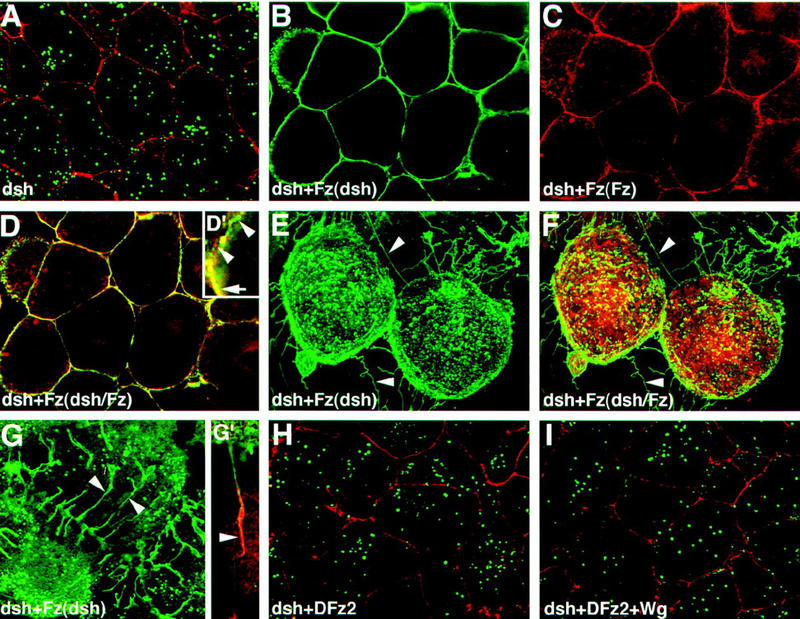
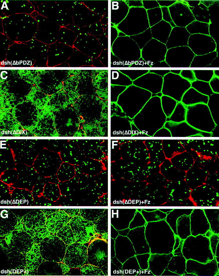
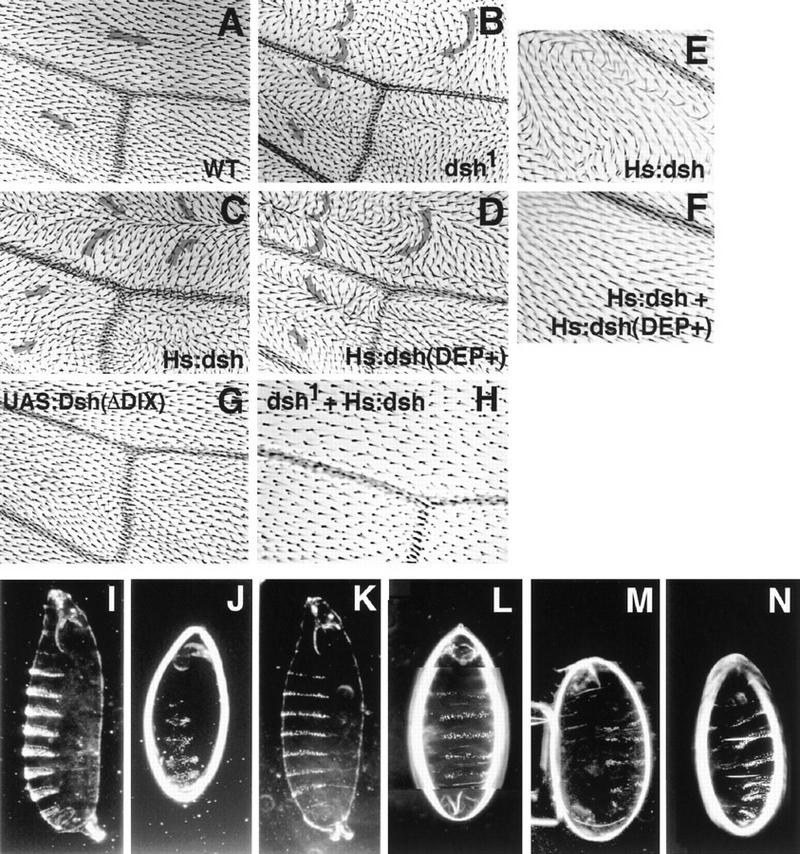
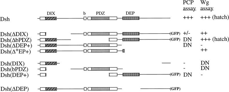
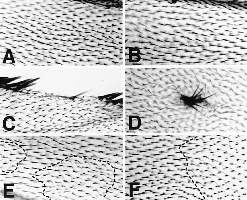
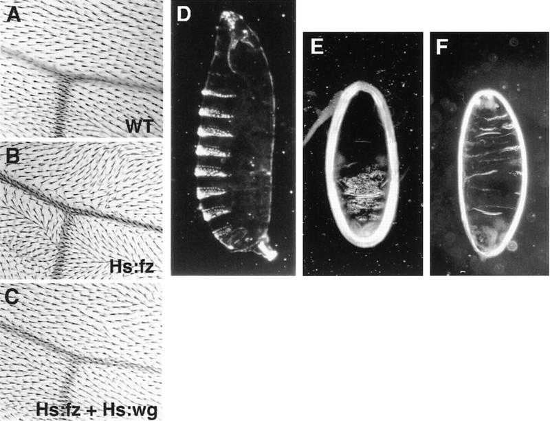
In Drosophila, planar cell polarity (PCP) signaling is mediated by the receptor Frizzled (Fz) and transduced by Dishevelled (Dsh). Wingless (Wg) signaling also requires Dsh and may utilize DFz2 as a receptor. Using a heterologous system, we show that Dsh is recruited selectively to the membrane by Fz but not DFz2, and this recruitment depends on the DEP domain but not the PDZ domain in Dsh. A mutation in the DEP domain impairs both membrane localization and the function of Dsh in PCP signaling, indicating that translocation is important for function. Further genetic and molecular analyses suggest that conserved domains in Dsh function differently during PCP and Wg signaling, and that divergent intracellular pathways are activated. We propose that Dsh has distinct roles in PCP and Wg signaling. The PCP signal may selectively result in focal Fz activation and asymmetric relocalization of Dsh to the membrane, where Dsh effects cytoskeletal reorganization to orient prehair initiation.


Figures

Figure 1
Figure 1
Dsh–GFP localizes to the membrane and filopodia in response to Fz in Xenopus animal cap cells. (A) Dsh–GFP (green) localizes in the cytoplasm in an apparent association with intracellular vesicles (Texas Red phalloidin labels the cell cortex). (B) In response to Fz, Dsh–GFP redistributes to the plasma membrane. (C) Fz localizes predominantly to the plasma membrane in the presence (shown) or absence (not shown) of Dsh–GFP. (D, D′) Dsh–GFP colocalizes with Fz at the plasma membrane. Yellow staining represents colocalization of Dsh–GFP (green from B) and Fz (red from C) and is marked by an arrow in D′. However, regions of the plasma membrane are also stained only by Dsh–GFP or Fz, demonstrating that the colocalization of Dsh–GFP and Fz is not absolute (arrowheads in D′). (E–G) In response to Fz, Dsh–GFP also accumulates in filopodia that extend from the free surface of the animal cap cells (arrowheads). These filopodia contain actin (stained with phalloidin; arrowhead in G′) but lack Fz (note lack of yellow staining in filopodia in F). (H,I) In contrast to the effects of Fz on the localization of Dsh–GFP, neither DFz2 (H) nor the combination of DFz2 and Wg (I) results in a change in the localization of Dsh–GFP (Dsh–GFP = green). Texas Red phalloidin was used to visualize cell outlines in A, H, and I, and the filopodium in G′.
Figure 2
Figure 2
Analysis of Dsh domains required for Fz-dependent relocalization of Dsh in Xenopus animal cap cells. (A) Dsh(ΔbPDZ)–GFP localizes to the cytoplasm in a punctate pattern. (B) In response to Fz, Dsh(ΔbPDZ)–GFP relocalizes to the plasma membrane (shown) and is also present in filopodia (not shown). (C) Dsh(ΔDIX)–GFP does not display a punctate pattern and instead is distributed diffusely throughout the cytoplasm (but is excluded from yolk granules). (D) In response to Fz, Dsh(ΔDIX)–GFP relocalizes to the plasma membrane (shown) and is also present in filopodia (not shown). (E) Dsh(ΔDEP)–GFP localizes to the cytoplasm in a punctate pattern. (F) Dsh(ΔDEP)–GFP, however, does not relocalize to the plasma membrane in response to Fz and displays a punctate pattern similar to that seen in the absence of Fz. (G) Dsh(DEP+) is diffusely cytoplasmic in the absence of Fz, and is sufficient to promote membrane localization in the presence of Fz (H). Texas Red phalloidin was used to visualize cell outlines in A, C, E, and G.
Figure 3
Figure 3
Sequence of the dsh1 mutant allele. The dsh1 allele carries a Lys → Met mutation in the DEP domain, at position 417. The conserved Dsh domains are boxed, and the recognized motifs are shaded.
Figure 4
Figure 4
The dsh1 mutation affects Fz-dependent localization. (A,B) In response to Fz, Dsh–GFP redistributes to the plasma membrane in a diffuse pattern. (D,E) In contrast, Dsh1–GFP redistributes to the membrane but remains associated with vesicles at the cell periphery. This effect was seen at all ratios of dsh to fz RNAs; the experiment shown used a ratio of 2:1. (C) In the absence of Fz, Dsh1–GFP is vesicular, and is indistinguishable from wild-type Dsh–GFP.
Figure 5
Figure 5
Dsh structure function analyses. Analysis of function in PCP signaling. Regions of wings from (A) wild type, (B) dsh1, (C) T8Hs:dsh/+ heat-shocked for 1 hr at 24 hr AP, (D) Hs:dsh(DEP+) heat-shocked for 2.5 hr at 24 hr AP. Note that the Hs:dsh(DEP+) wing resembles the dsh1wing, rather than the Hs:dsh wing. A different region of a T15Hs:dsh/+ wing shows a pattern (E) that is suppressed by simultaneous expression of Hs:dsh(DEP+) (F). (The flies in E and F were heat-shocked for 1 hr at 24 hr AP. A slightly higher magnification is shown.) Expression of Dsh(DEP+) in a dsh1 mutant failed to alter the dsh1 phenotype (not shown). By these criteria, Dsh(DEP+) behaves as a dominant negative. Expression of wild-type Dsh suppresses the dsh1 phenotype (H; the wing is marked with yellow; thus the hairs are finer and have less contrast. A slightly higher magnification is shown). Dsh(ΔDIX) produced only a weak phenotype when expressed by UAS and a range of GAL4 drivers (Hs:GAL4 is shown, G). Analysis of function in Wg signaling. (I) A wild-type cuticle; (J) a svb, dsh cuticle. The dsh mutant cuticle shows shortening, fusion of denticle bands, and absence of the head skeleton and filzkorper characteristic of mutants in the Wg pathway. Injection of wild type mRNA into the mutant embryos results in complete rescue of segmentation, the head skeleton and the filzkörper (K). Cuticles injected with Dsh(ΔbPDZ) are identical. Similarly, injection of partially functional constructs partially rescues segmentation, the head skeleton and the filzkörper to varying extents; e.g., (L) Dsh(Δ*EP+) and (M) Dsh(ΔDIX). Conversely, injection of dominant-negative constructs causes wild-type embryos to show a segmentation phenotype and loss of the head skeleton and filzkörper similar to the mutant embryos; (N) Dsh(DIX) and Dsh(bPDZ) (not shown).
Figure 6
Figure 6
Structures of the Dsh constructs. The conserved domains are as in Fig. 3, and the nonconserved basic domain is indicated (b). Results of the structure–function analyses are shown in semiquantitative form. Embryos that hatched are indicated; (DN) dominant negative. A detailed summary is found in Materials and Methods. (GFP) Some of the same constructs were fused to GFP at their carboxyl termini and used in the animal cap assays.
Figure 7
Figure 7
Arm and Zw3 have no role in PCP signaling. Wings from flies in which ArmS10 (A) or Zw3 (B) were overexpressed show a wild-type polarity phenotype. Clones of armH8.6 (C) or zw3 (D) mutant cells marked with f36a produce wing margin nicks and tufts of bristles, respectively. The zw3 mutant cells appear to cause polarity distortions in the surrounding wild-type tissue, probably by grossly distorting the architecture of the epithelium. However, cells in arm f36a mutant clones in the interior of the wing produce hairs of wild-type polarity (E), as do cells in scB57 zw3 f36a-mutant clones (F).
Figure 8
Figure 8
Reciprocal titration of the PCP and Wg pathways. Wings from wild-type (A), Hs:Fz/+ (B), and Hs:Fz/+; Hs:wg/+ (C), each heat-shocked for 1.5 hr at 24 hr AP. Overexpression of Hs:wg alone at this time has no phenotype (not shown). However, it suppresses the Hs:fz overexpression phenotype. Shown are embryos that are wild type (D), U32A/U32A; UASfz/+ (E), and U32A/U32A; UASdsh43-1-B-1/+ (F). Whereas overexpression of Dsh produces naked cuticle (F), overexpression of Fz leads to lawns of denticles (E).

Similar articles

Cited by

References

    1. Adler PN. The genetic control of tissue polarity in Drosophila. BioEssays. 1992;14:735–741. - PubMed
    1. Adler PN, Vinson C, Park WJ, Conover S, Klein L. Molecular structure of frizzled, a Drosophila tissue polarity gene. Genetics. 1990;126:401–416. - PMC - PubMed
    1. Adler PN, Krasnow RE, Liu J. Tissue polarity points from cells that have higher Frizzled levels towards cells that have lower Frizzled levels. Curr Biol. 1997;7:940–949. - PubMed
    1. Axelrod JD, Matsuno K, Artavanis-Tsakonas S, Perrimon N. Dishevelled mediates interaction between Wingless and Notch signaling pathways. Science. 1996;271:1826–1832. - PubMed
    1. Bejsovec A, Wieschaus E. Signaling activities of the Drosophila gene wingless are separably mutable and appear to be transduced at the cell surface. Genetics. 1995;139:309–320. - PMC - PubMed

Publication types

MeSH terms