Compartmentalized transgene expression of granulocyte-macrophage colony-stimulating factor (GM-CSF) in mouse lung enhances allergic airways inflammation
- PMID: 9717963
- PMCID: PMC1905049
- DOI: 10.1046/j.1365-2249.1998.00652.x
Compartmentalized transgene expression of granulocyte-macrophage colony-stimulating factor (GM-CSF) in mouse lung enhances allergic airways inflammation
Abstract
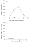
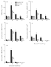
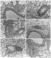
To investigate the role of GM-CSF in asthmatic airways inflammation, we have targeted GM-CSF transgene to the airway cells in a mouse model of ovalbumin (OVA)-induced allergic airways inflammation, a model in which there is marked induction of endogenous IL-5 and IL-4 but not GM-CSF. Following intranasal delivery of a replication-deficient adenoviral gene transfer vector (Ad), transgene expression was found localized primarily to the respiratory epithelial cells. Intranasal delivery of 0.03 x 10(9) plaque-forming units (PFU) of AdGM-CSF into naive BALB/c mice resulted in prolonged and compartmentalized release of GM-CSF transgene protein with a peak concentration of approximately 80 pg/ml detected in bronchoalveolar lavage fluid (BALF) at day 7, but little in serum. These levels of local GM-CSF expression per se resulted in no eosinophilia and only a minimum of tissue inflammatory responses in the lung of naive mice, similar to those induced by the control vector. However, such GM-CSF expression in the airways of OVA-sensitized mice resulted in a much greater and sustained accumulation of various inflammatory cell types, most noticeably eosinophils, both in BALF and airway tissues for 15-21 days post-OVA aerosol challenge, at which times airways inflammation had largely resolved in control mice. While the levels of IL-5 and IL-4 in BALF and the rate of eosinophil apoptosis were found similar between different treatments, there was an increased number of proliferative leucocytes in the lung receiving GM-CSF gene transfer. Our results thus provide direct experimental evidence that GM-CSF can significantly contribute to the development of allergic airways inflammation through potentiating and prolonging inflammatory infiltration induced by cytokines such as IL-5 and IL-4.
Figures


Similar articles
-
GM-CSF transgene expression in the airway allows aerosolized ovalbumin to induce allergic sensitization in mice.J Clin Invest. 1998 Nov 1;102(9):1704-14. doi: 10.1172/JCI4160. J Clin Invest. 1998. PMID: 9802884 Free PMC article.
-
Interleukin-10 gene transfer to the airway regulates allergic mucosal sensitization in mice.Am J Respir Cell Mol Biol. 1999 Nov;21(5):586-96. doi: 10.1165/ajrcmb.21.5.3755. Am J Respir Cell Mol Biol. 1999. PMID: 10536118
-
Cytokine and eosinophil responses in the lung, peripheral blood, and bone marrow compartments in a murine model of allergen-induced airways inflammation.Am J Respir Cell Mol Biol. 1997 May;16(5):510-20. doi: 10.1165/ajrcmb.16.5.9160833. Am J Respir Cell Mol Biol. 1997. PMID: 9160833
-
Gene transfer for cytokine functional studies in the lung: the multifunctional role of GM-CSF in pulmonary inflammation.J Leukoc Biol. 1996 Apr;59(4):481-8. doi: 10.1002/jlb.59.4.481. J Leukoc Biol. 1996. PMID: 8613693 Review.
-
GM-CSF, IL-3, and IL-5 Family of Cytokines: Regulators of Inflammation.Immunity. 2019 Apr 16;50(4):796-811. doi: 10.1016/j.immuni.2019.03.022. Immunity. 2019. PMID: 30995500 Review.
Cited by
-
Intranasal mucosal boosting with an adenovirus-vectored vaccine markedly enhances the protection of BCG-primed guinea pigs against pulmonary tuberculosis.PLoS One. 2009 Jun 10;4(6):e5856. doi: 10.1371/journal.pone.0005856. PLoS One. 2009. PMID: 19516906 Free PMC article.
-
Replication-defective adenovirus infection reduces Helicobacter felis colonization in the mouse in a gamma interferon- and interleukin-12-dependent manner.Infect Immun. 1999 Sep;67(9):4539-44. doi: 10.1128/IAI.67.9.4539-4544.1999. Infect Immun. 1999. PMID: 10456897 Free PMC article.
-
Type 1 interferon gene transfer enhances host defense against pulmonary Streptococcus pneumoniae infection via activating innate leukocytes.Mol Ther Methods Clin Dev. 2014 Mar 19;1:5. doi: 10.1038/mtm.2014.5. eCollection 2014. Mol Ther Methods Clin Dev. 2014. PMID: 26015944 Free PMC article.
-
Glutathione peroxidase-1 primes pro-inflammatory cytokine production after LPS challenge in vivo.PLoS One. 2012;7(3):e33172. doi: 10.1371/journal.pone.0033172. Epub 2012 Mar 6. PLoS One. 2012. PMID: 22412999 Free PMC article.
-
Transient overexpression of gamma interferon promotes Aspergillus clearance in invasive pulmonary aspergillosis.Clin Exp Immunol. 2005 Nov;142(2):233-41. doi: 10.1111/j.1365-2249.2005.02828.x. Clin Exp Immunol. 2005. PMID: 16232209 Free PMC article.
References
-
- Bochner BS, Undem BJ, Lichtenstein LM. Immunological aspects of allergic asthma. Ann Rev Immunol. 1994;12:295–335. - PubMed
-
- Bousquet J, Chanez P, Vignola AM, Lacoste J-V, Michel FB. Eosinophil inflammation in asthma. Am J Respir Crit Care Med. 1994;150:S33–S38. - PubMed
-
- Walker C, Kaegi MK, Braun P, Blaser K. Activated T cells and eosinophilia in bronchoalveolar lavages from subjects with asthma correlated with disease severity. J Allergy Clin Immunol. 1991;88:935–42. - PubMed
-
- Weller PF. The immunobiology of eosinophils. N Engl J Med. 1991;324:1110–8. - PubMed
Publication types
MeSH terms
Substances
LinkOut - more resources
Full Text Sources
Other Literature Sources
Medical

