. 1998 Sep;180(17):4497-507.
doi: 10.1128/JB.180.17.4497-4507.1998.

Molecular characterization and sequence of a methionine biosynthetic locus from Pseudomonas syringae

Affiliations

Molecular characterization and sequence of a methionine biosynthetic locus from Pseudomonas syringae

G L Andersen et al. J Bacteriol. 1998 Sep.

Abstract

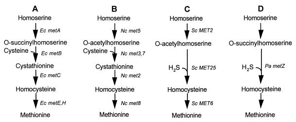
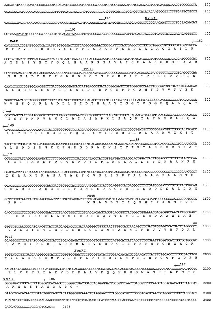
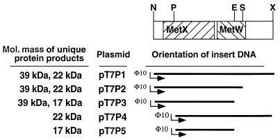
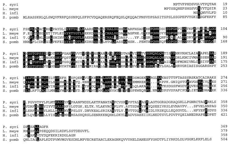
Two methionine biosynthetic genes in Pseudomonas syringae pv. syringae, metX and metW, were isolated, sequenced, and evaluated for their roles in methionine biosynthesis and bacterial fitness on leaf surfaces. The metXW locus was isolated on a 1.8-kb DNA fragment that was required for both methionine prototrophy and wild-type epiphytic fitness. Sequence analysis identified two consecutive open reading frames (ORFs), and in vitro transcription-translation experiments provided strong evidence that the ORFs encode proteins with the predicted molecular masses of 39 and 22.5 kDa. The predicted amino acid sequence of MetX (39 kDa) showed homology to several known and putative homoserine O-acetyltransferases. This enzyme is the first enzyme in the methionine biosynthetic pathway of fungi, gram-negative bacteria of the genus Leptospira, and several gram-positive bacterial genera. Both metX and metW were required for methionine biosynthesis, and transcription from both genes was not repressed by methionine. MetW (22.5 kDa) did not show significant homology to any known protein, including prokaryotic and eukaryotic methionine biosynthetic enzymes. Several classes of methionine auxotrophs, including metX and metW mutants, exhibit reduced fitness on leaf surfaces, indicating a requirement for methionine prototrophy in wild-type epiphytic fitness. This requirement is enhanced under environmentally stressful conditions, suggesting a role for methionine prototrophy in bacterial stress tolerance.


Figures

FIG. 1
FIG. 1
Methionine biosynthetic pathways in different species. (A) E. coli (Ec) (41), (B) N. crassa (Nc) (41), (C) S. cerevisiae (Sc) (41), and Brevibacterium flavum (38), and (D) P. aeruginosa (Pa) (15) pathways are shown. Note that N. crassa can also convert O-acetylhomoserine to homocysteine via direct sulfhydrylation (27) and S. cerevisiae can perform the conversion through transsulfuration (45), but these conversions are not quantitatively important (41).
FIG. 2
FIG. 2
Population sizes of P. syringae pv. syringae B728a (■) and Tn5 mutant derivatives mutant 42 (▴) and MX7 (○) on bean plants exposed to wet and dry conditions. Each point represents the mean ± standard error of the mean of 20 leaf samples.
FIG. 3
FIG. 3
Influence of methionine on the growth and survival of P. syringae pv. syringae B728a and MX7 on bean plants exposed to wet and dry conditions. Within 4 h of inoculation, 30 mM l-methionine or sterile H2O was sprayed onto plants. Symbols: B728a with H2O (○), B728a with methionine (•), MX7 with H2O (□), and MX7 with methionine (■).
FIG. 4
FIG. 4
Location and orientation of Tn3-Spice insertions in pCM2 (A) and pVGA7 (B). Closed circles represent insertions that prevented restoration of methionine prototrophy after the plasmids were mobilized into MX7, while open circles represent those plasmid-borne insertions that had no effect on prototrophy. Closed diamonds represent insertions that prevented restoration of methionine prototrophy after introduction into the B728a chromosome by marker-exchange mutagenesis, while open diamonds represent those insertions that had no effect on prototrophy after chromosomal introduction. The location of the metXW locus is relative to selected Tn3-Spice insertion points in pVGA7 and comparison with the nucleotide sequence of this locus (shaded bar in panel B). Insertions in which the ice nucleation reporter gene inaZ is oriented from left to right are placed above the line and oppositely oriented insertions are placed below. The restriction sites of BamHI (B), EcoRI (E), HindIII (H), and XhoI (X) are indicated.
FIG. 5
FIG. 5
Hybridization of a 1.2-kb PvuII-PstI restriction fragment internal to the P. syringae methionine biosynthesis locus to EcoRI-digested genomic DNA of the following. Lanes: A, P. syringae pv. phaseolicola (NPS3121); B, Pseudomonas syringae pv. glycinia; C, P. syringae pv. tabaci; D, Pseudomonas syringae subsp. savastanoi; E, R. solanacearum; F, P. syringae pv. syringae (B728a control); G, Pseudomonas syringae pv. tomato; H, Pseudomonas syringae pv. maculicola; I, P. syringae pv. lachrymans; J, P. syringae pv. pisi; K, P. syringae pv. aptata; L, P. viridiflava; M, P. cichorii; N, Xanthomonas campestris pv. vesicatoria; O, X. campestris pv. campestris; P, Xanthomonas campestris pv. phaseoli; Q, Pantoea agglomerans; R, E. coli; S, P. syringae pv. phaseolicola (4324); T, P. syringae pv. syringae (990).
FIG. 6
FIG. 6
Nucleotide and deduced amino acid sequences of a 2,426-bp region of pVGA7 containing a 1.8-kb region required for methionine prototrophy. The positions of two ORFs are labeled, and a potential ς70 promoter sequence is underlined upstream from metX at nucleotide positions 307 to 312 and 332 to 337. The insertion points and orientations of various Tn3-Spice insertions, as well as selected restriction sites, are indicated.
FIG. 7
FIG. 7
Protein expression from metX and metW. An E. coli S30 transcription-translation system was used to express proteins from the T7 promoter fusions constructed with the fragments indicated by the restriction map shown at the top. The direction of transcription is from left to right, and radiolabeled protein products were visualized on a denaturing polyacrylamide gel. Restriction enzyme sites are as follows: N, NruI; P, PvuII; E, EcoRI; S, SmaI; X, XhoI.
FIG. 8
FIG. 8
Optimal alignment (PILEUP) of the deduced amino acid sequence of MetX from P. syringae (P. syri) to the homoserine O-acetyltransferases of L. meyeri (L. meye) (accession no. Y10744), H. influenzae (H. infl) (accession no. L42023), and Schizosaccharomyces pombe (S. pomb) (accession no. Z69909). Residues identical in three or all of the sequences are shown in blackened areas.

References

    1. Anderson D M, Mills D. The use of transposon mutagenesis in the isolation of nutritional and virulent mutants in two pathovars of Pseudomonas syringae. Phytopathology. 1985;75:104–108.
    1. Baroni M, Livian S, Martegani E, Alberghina L. Molecular cloning and regulation of the expression of the MET2 gene of Saccharomyces cerevisiae. Gene. 1986;46:71–78. - PubMed
    1. Beattie G A, Lindow S E. Survival, growth, and localization of epiphytic fitness mutants of Pseudomonas syringae on leaves. Appl Environ Microbiol. 1994;60:3790–3798. - PMC - PubMed
    1. Beattie G A, Lindow S E. Comparison of the behavior of epiphytic fitness mutants of Pseudomonas syringae under controlled and field conditions. Appl Environ Microbiol. 1994;60:3799–3808. - PMC - PubMed
    1. Bender C L, Cooksey D A. Indigenous plasmids in Pseudomonas syringae pv. tomato: conjugative transfer and role in copper resistance. J Bacteriology. 1986;165:534–541. - PMC - PubMed

LinkOut - more resources