. 1998 Sep;180(17):4628-37.
doi: 10.1128/JB.180.17.4628-4637.1998.

SdeK is required for early fruiting body development in Myxococcus xanthus

Affiliations

SdeK is required for early fruiting body development in Myxococcus xanthus

A G Garza et al. J Bacteriol. 1998 Sep.

Abstract

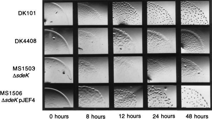
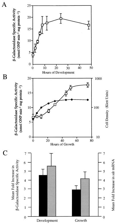
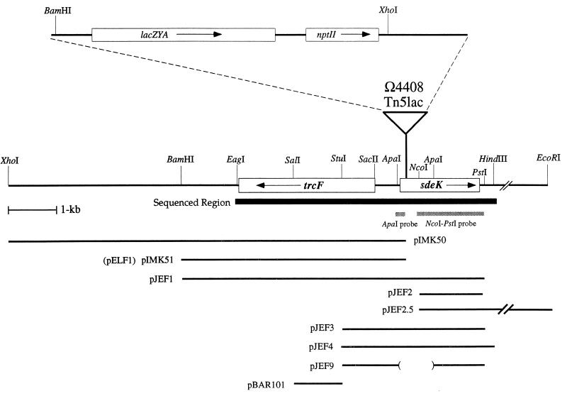
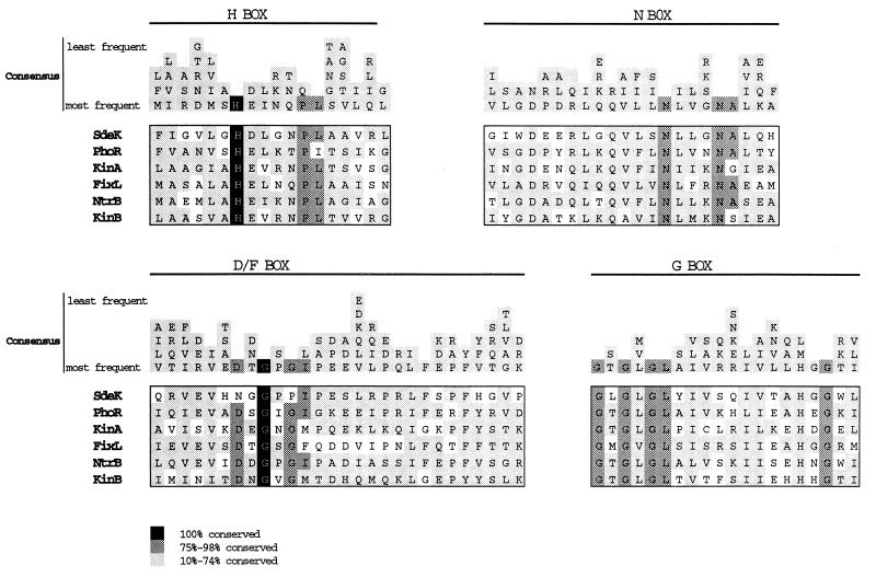
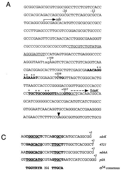
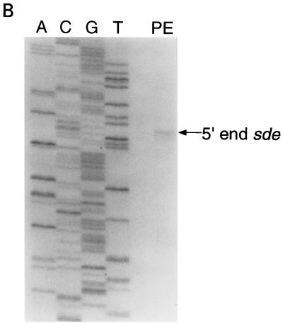
Myxococcus xanthus cells carrying the Omega4408 Tn5lac insertion at the sde locus show defects in fruiting body development and sporulation. Our analysis of sde expression patterns showed that this locus is induced early in the developmental program (0 to 2 h) and that expression increases approximately fivefold after 12 h of development. Further studies showed that expression of sde is induced as growing cells enter stationary phase, suggesting that activation of the sde locus is not limited to the developmental process. Because the peak levels of sde expression in both an sde+ and an sde mutant background were similar, we conclude that the sde locus is not autoregulated. Characterization of the sde locus by DNA sequence analysis indicated that the Omega4408 insertion occurred within the sdeK gene. Primer extension analyses localized the 5' end of sde transcript to a guanine nucleotide 307 bp upstream of the proposed start for the SdeK coding sequence. The DNA sequence in the -12 and -24 regions upstream of the sde transcriptional start site shows similarity to the sigma54 family of promoters. The results of complementation studies suggest that the defects in development and sporulation caused by the Omega4408 insertion are due to an inactivation of sdeK. The predicted amino acid sequence of SdeK was found to have similarity to the sequences of the histidine protein kinases of two-component regulatory systems. Based on our results, we propose that SdeK may be part of a signal transduction pathway required for the activation and propagation of the early developmental program.


Figures

FIG. 1
FIG. 1
Behavior of M. xanthus cells during development on TPM agar plates. The developmental progress of wild-type DK101 cells, DK4408 cells, MS1503 cells, and MS1506 cells is shown. Cells were spotted on TPM starvation agar and monitored visually as described in Materials and Methods. Photographs were taken at the times indicated.
FIG. 2
FIG. 2
Patterns of sde expression. The mean β-galactosidase specific activities found in DK4408 cells and the mean sde mRNA levels found in DK101 cells were determined from three independent experiments. Error bars are the standard deviations of the mean. The open squares represent β-galactosidase specific activity, and solid diamonds represent cell density. The mean β-galactosidase specific activities found at the indicated times during development on TPM agar (A) and during vegetative growth in CTT broth (B) are shown. The mean fold increase in β-galactosidase specific activity (■) and the mean fold increase in sde mRNA (▨) are compared for 0 to 12 h of development on TPM agar plates and 0 to 53 h of growth in CTT liquid (C).
FIG. 3
FIG. 3
Physical map of the Ω4408 insertion region. The broadened black line indicates the region that has been characterized by DNA sequence analysis. Boxes show the locations of the indicated ORF. The arrow inside each box shows the predicted orientation of transcription of the ORF. The location of the Ω4408 insertion (triangle) was determined by DNA sequencing and Southern blot analysis as described in Materials and Methods. An enlargement of Tn5lac is shown above the triangle. Plasmids containing the indicated DNA fragments of the sde locus are shown below the map of the Ω4408 insertion region. The parentheses indicate the extent of the deleted region in plasmid pJEF9, and the parallel slashes indicate contiguous DNA sequence that extends to the EcoRI site, approximately 24 kb downstream from the end of the sdeK ORF. The 300-bp ApaI fragment and the 1.1-kb NcoI-PstI used for slot blot hybridization analysis of sde mRNA are represented by stippled boxes.
FIG. 4
FIG. 4
Alignment of the SdeK C terminus with conserved regions in the transmitter domain of known histidine kinase proteins. Conserved sequences of FixL from Rhizobium meliloti (1, 5), KinA from Bacillus subtilis (2, 39), KinB from B. subtilis (54), NtrB from E. coli (35), PhoR from E. coli (30), and SdeK from M. xanthus are included. The four highly conserved regions (H-Box, N-Box, D/F-Box, and G-Box) in histidine kinase proteins are shown (38, 51). Amino acid residues found in 10 to 100% of the histidine kinase proteins are presented above the sequence alignment as presented by Stock et al. (51). Black boxes indicate that a residue is absolutely conserved among all histidine kinases. The dark gray boxes indicate that a residue is found in 75 to 98% of histidine kinases. The light gray boxes indicate that a residue is found in 10 to 74% of histidine kinases.
FIG. 5
FIG. 5
(A) DNA sequence of the region upstream of the Ω4408 Tn5lac insertion. The black triangle shows the location of the Ω4408 insertion. The bent arrow above the +1 shows the 5′ end of the predicted sde transcript, and the −12 and −24 regions are indicated. The stop codon (TAG) that defines the 5′ end of the sdeK ORF (+169) is shown in lowercase. Potential ribosome binding sites and translational start codons for the SdeK protein are in boldface. Nucleotides marked with an asterisk are complementary to the 3′ end of M. xanthus 16S rRNA (37). The dashed arrow above the GTG at position +307 is the proposed start of the SdeK coding sequence. The underlined regions show the sequences that are complementary to the two primers (ORF12 and ORF12-2) used to identify the 5′ end of the sde transcript. (B) Mapping the 5′ end of the sde developmental transcript by primer extension analysis. A, C, G, and T show the DNA sequencing ladders. PE, primer extension with total RNA prepared from DK101 cells developing on TPM agar for 12 h and primer ORF12 (Materials and Methods). (C) Comparison of the sde promoter region to the ς54 consensus sequence and three M. xanthus ς54 promoters: mbhA (21), 4521 (21), and pilA (55). The proposed start of transcription is designated by the +1.
FIG. 5
FIG. 5
(A) DNA sequence of the region upstream of the Ω4408 Tn5lac insertion. The black triangle shows the location of the Ω4408 insertion. The bent arrow above the +1 shows the 5′ end of the predicted sde transcript, and the −12 and −24 regions are indicated. The stop codon (TAG) that defines the 5′ end of the sdeK ORF (+169) is shown in lowercase. Potential ribosome binding sites and translational start codons for the SdeK protein are in boldface. Nucleotides marked with an asterisk are complementary to the 3′ end of M. xanthus 16S rRNA (37). The dashed arrow above the GTG at position +307 is the proposed start of the SdeK coding sequence. The underlined regions show the sequences that are complementary to the two primers (ORF12 and ORF12-2) used to identify the 5′ end of the sde transcript. (B) Mapping the 5′ end of the sde developmental transcript by primer extension analysis. A, C, G, and T show the DNA sequencing ladders. PE, primer extension with total RNA prepared from DK101 cells developing on TPM agar for 12 h and primer ORF12 (Materials and Methods). (C) Comparison of the sde promoter region to the ς54 consensus sequence and three M. xanthus ς54 promoters: mbhA (21), 4521 (21), and pilA (55). The proposed start of transcription is designated by the +1.

References

    1. Anthamatten D, Hennecke H. The regulatory status of the fixL- and fixJ-like genes in Bradyrhizobium japonicum may be different from Rhizobium meliloti. Mol Gen Genet. 1991;225:38–48. - PubMed
    1. Antoniewski C, Savelli B, Stragier P. The spoIIJ gene, which regulates early developmental steps in Bacillus subtilis, belongs to a class of environmentally responsive genes. J Bacteriol. 1990;172:86–93. - PMC - PubMed
    1. Bibb M J, Findlay P R, Johnson M W. The relationship between base composition and codon usage in bacterial genes and its use for the simple and reliable identification of protein coding sequences. Gene. 1984;30:157–166. - PubMed
    1. Blanco G, Drummond M, Woodley P, Kennedy C. Sequence and molecular analysis of the nifL gene of Azotobacter vinelandii. Mol Microbiol. 1993;9:869–879. - PubMed
    1. David M, Daveran M L, Batut J, Dedieu A, Domergue O, Ghai J, Hertig C. Cascade regulation of nif gene expression in Rhizobium meliloti. Cell. 1988;54:671–683. - PubMed

Publication types

LinkOut - more resources