. 1998 Aug 24;142(4):887-98.
doi: 10.1083/jcb.142.4.887.

The human polycomb group complex associates with pericentromeric heterochromatin to form a novel nuclear domain

Affiliations

The human polycomb group complex associates with pericentromeric heterochromatin to form a novel nuclear domain

A J Saurin et al. J Cell Biol. .

Abstract

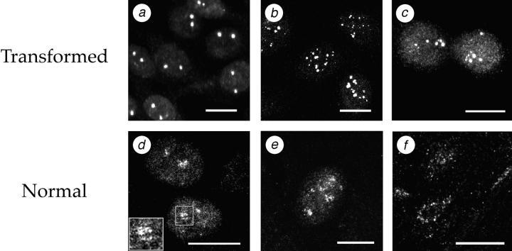
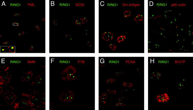
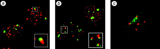
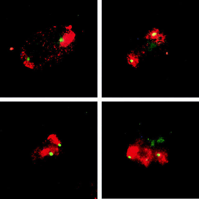
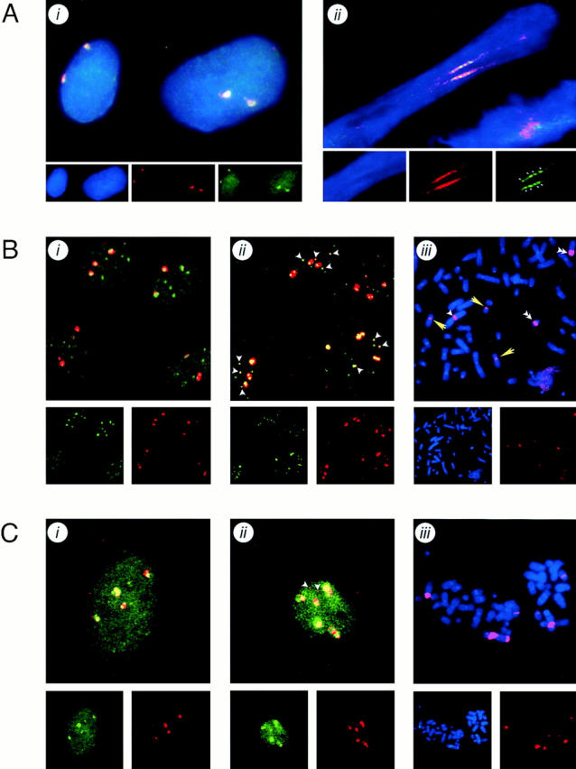
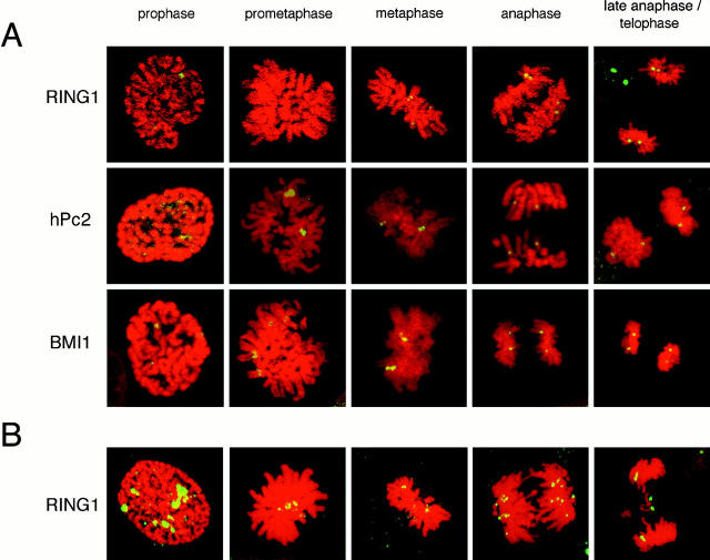
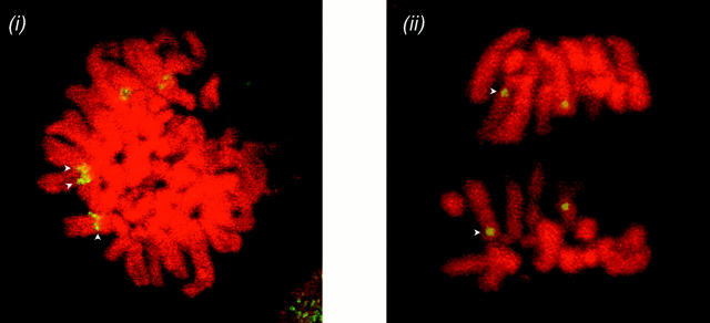
The Polycomb group (PcG) complex is a chromatin-associated multiprotein complex, involved in the stable repression of homeotic gene activity in Drosophila. Recently, a mammalian PcG complex has been identified with several PcG proteins implicated in the regulation of Hox gene expression. Although the mammalian PcG complex appears analogous to the complex in Drosophila, the molecular mechanisms and functions for the mammalian PcG complex remain unknown. Here we describe a detailed characterization of the human PcG complex in terms of cellular localization and chromosomal association. By using antibodies that specifically recognize three human PcG proteins- RING1, BMI1, and hPc2-we demonstrate in a number of human cell lines that the PcG complex forms a unique discrete nuclear structure that we term PcG bodies. PcG bodies are prominent novel nuclear structures with the larger PcG foci generally localized near the centromeres, as visualized with a kinetochore antibody marker. In both normal fetal and adult fibroblasts, PcG bodies are not randomly dispersed, but appear clustered into defined areas within the nucleus. We show in three different human cell lines that the PcG complex can tightly associate with large pericentromeric heterochromatin regions (1q12) on chromosome 1, and with related pericentromeric sequences on different chromosomes, providing evidence for a mammalian PcG-heterochromatin association. Furthermore, these heterochromatin-bound PcG complexes remain stably associated throughout mitosis, thereby allowing the potential inheritance of the PcG complex through successive cell divisions. We discuss these results in terms of the known function of the PcG complex as a transcriptional repression complex.


Figures

Figure 1
Figure 1
The human RING1 protein localizes to discrete nuclear concentrated foci in a number of human cell lines. Polyclonal antisera against the human RING1 protein was used to immunolocalize endogenous RING1 in: (a) 2C4 human fibrosarcoma cells, a derivative of the HT1080 cell line; (b) U-2 OS human osteosarcoma cells; (c) SAOS-2 human osteosarcoma cells; (d) MRC-5 normal human fetal lung fibroblast cells; (e) CS22F normal human adult fibroblast cells; and (f) human neonatal foreskin tissue section. Images represent a projection of multiple optical sections through the cell. Bar, 10 μm in all panels.
Figure 2
Figure 2
The mammalian PcG complex forms a novel nuclear structure. The mammalian PcG complex was immunolocalized in human cells using the RING1-specific polyclonal antibody ASA3, and was stained using an FITC-conjugated secondary antibody. By double immunofluorescence microscopy, the staining pattern obtained for RING1 in 2C4 human fibrosarcoma cells was compared with that of known nuclear structures. The green signal shows labeled RING1, and the red signal given by Texas red–conjugated secondary antibodies shows immunolocalization of the following: (A) PML nuclear bodies from the anti-PML antibody 5E10 (Stuurman et al., 1992); (B) speckled domains recognized by the anti-SC35 antibody (Fu and Maniatis, 1990); (C) the snRNP family of proteins, recognized by the anti-Sm antigen antibody Y12 (Lerner et al., 1981); (D) coiled bodies recognized by the anti-p80 coilin antibody p-δ; (E) gems recognized by the anti-SMN antibody 2B1 (Liu and Dreyfuss, 1996); (F) PTB protein (Huang et al., 1997); (G) DNA replication foci detected using antibodies to PCNA; (H) sites of RNA synthesis localized with antibodies to in vivo–incorporated bromo-UTP nucleotides (note this panel is shown for U-2 OS cells). All images are a digital overlay of the two optical channels obtained from a single optical confocal section.
Figure 3
Figure 3
PcG bodies are associated with kinetochores in three different cell lines. Human centromeres were localized using an autoimmune sera against kinetochores, and are shown in red. Their spatial relationship with PcG bodies, labeled with the anti-RING1 antibody (green), are shown in (a) 2C4 cells, (b) U-2 OS cells, and (c) CS22F cells. All images are a digital overlay of the two optical channels obtained from a single 1-μm optical confocal section. Any colocalization observed is shown by a yellow signal.
Figure 4
Figure 4
PcG bodies preferentially associate to chromosome 1 territories in 2C4 cells. Digitally merged images of numerous interphase 2C4 cells localizing the chromosome 1 in situ–hybridized probe by immunofluorescence showing the entire chromosome 1 territories (red) and immunofluorescent localization of PcG bodies using the anti-RING1 antibody (green) are shown. The green channel–labeled PcG bodies can always be seen associated with the red channel–labeled chromosome 1 territories in nuclei of 2C4 cells (yellow).
Figure 5
Figure 5
PcG bodies localize specifically to the pericentromeric region of chromosome 1 in a number of cell lines. Digitally merged fluorescent-channel images are shown of interphase cell nuclei showing immunofluorescent localization of the in situ–hybridized pUC1.77 probe, specific for the q12 pericentromeric region of chromosome 1 (red) and immunofluorescent localization of PcG bodies using anti-RING1 antibodies (green). Nuclei were counterstained with DAPI (blue channel) showing the nuclear volume. The separate color channels are shown under the main merged images. (A) Interphase 2C4 cell nuclei showing (i) normal interphase nuclei and (ii) interphase chromatin from a released nucleus by a cytospin where the nuclear architecture and territories are compromised, thereby releasing the chromosome domains throughout the length of the nucleus. (B) U-2 OS cells labeled after (i) the normal high-stringency immuno-FISH protocol, and (ii) lower stringency immuno-FISH protocol showing additional regions of pUC1.77 hybridization that colocalize with smaller PcG bodies (arrowheads); (iii) low-stringency hybridizations of the pUC1.77 probe were localized on metaphase chromosome spreads counterstained with DAPI (blue channel) showing the three large 1q12-specific hybridizing regions (white arrowheads) that are due to breakage of the q12 region in one chromosome 1 (double white arrowheads), together with the additional sites of hybridization during low-stringency conditions (yellow arrowheads). (C) SAOS-2 cells labeled after hybrdization of the pUC1.77 probe (i) and hybridization at lower stringency showing an additional two sites of hybridization (ii; arrowheads); (iii) low-stringency hybridizations of the pUC1.77 probe were localized on metaphase chromosome spreads counterstained with DAPI (blue channel), showing the multiple rearrangements of the 1q12 region.
Figure 6
Figure 6
PcG bodies remain chromosomally associated throughout mitosis. Mitotic chromosomes were visualized by propidium iodide staining of formaldehyde-fixed cells, and are shown in red. The various stages of mitosis were distinguished according to criteria used by Chaly et al. (1984). PcG bodies were visualized by immunofluorescent labeling of (A) 2C4 cells with antibodies against either RING1 (top; green channel), hPc2 (middle; green channel) or BMI1 (bottom; green channel) and (B) U-2 OS cells with antibodies against RING1 (green channel). A digital overlay of the two-color channels shows the relation of PcG bodies with the condensed chromosomes during the given stages of mitosis. (i) Enlargement of BMI1 during prometaphase in 2C4 cells in the main figure shows direct chromosome association with concentration of the protein on the sister chromatids (arrowheads). (ii) Enlargement of hPc2 during anaphase in 2C4 cells in the main figure shows direct chromosome association of hPc2 to a pericentromeric region on chromosome 1 (arrowheads).
Figure 6
Figure 6
PcG bodies remain chromosomally associated throughout mitosis. Mitotic chromosomes were visualized by propidium iodide staining of formaldehyde-fixed cells, and are shown in red. The various stages of mitosis were distinguished according to criteria used by Chaly et al. (1984). PcG bodies were visualized by immunofluorescent labeling of (A) 2C4 cells with antibodies against either RING1 (top; green channel), hPc2 (middle; green channel) or BMI1 (bottom; green channel) and (B) U-2 OS cells with antibodies against RING1 (green channel). A digital overlay of the two-color channels shows the relation of PcG bodies with the condensed chromosomes during the given stages of mitosis. (i) Enlargement of BMI1 during prometaphase in 2C4 cells in the main figure shows direct chromosome association with concentration of the protein on the sister chromatids (arrowheads). (ii) Enlargement of hPc2 during anaphase in 2C4 cells in the main figure shows direct chromosome association of hPc2 to a pericentromeric region on chromosome 1 (arrowheads).

References

    1. Alkema MJ, Bronk M, Verhoeven E, Otte A, van't Veer LJ, Berns A, van Lohuizen M. Identification of Bmi1-interacting proteins as constituents of a multimeric mammalian Polycomb complex. Genes Dev. 1997;11:226–240. - PubMed
    1. Bienz M, Müller J. Transcriptional silencing of homeotic genes in Drosophila. . Bioessays. 1995;17:775–784. - PubMed
    1. Brown KE, Guest SS, Smale ST, Hahm K, Merkenschlager M, Fisher AG. Association of transcriptionally silent genes with Ikaros complexes at centromeric heterochromatin. Cell. 1997;91:845–854. - PubMed
    1. Buchenau P, Hodgson J, Strutt H, Arndt-Jovin DJ. The distribution of polycomb-group proteins during cell division and development in Drosophilaembryos: impact on models for silencing. J Cell Biol. 1998;141:469–481. - PMC - PubMed
    1. Chaly N, Bladon T, Setterfield G, Little JE, Kaplan JG, Brown DL. Changes in distribution of nuclear matrix antigens during the mitotic cell cycle. J Cell Biol. 1984;99:661–671. - PMC - PubMed

Publication types