. 1998 Aug 24;142(4):913-22.
doi: 10.1083/jcb.142.4.913.

Acidic di-leucine motif essential for AP-3-dependent sorting and restriction of the functional specificity of the Vam3p vacuolar t-SNARE

Affiliations

Acidic di-leucine motif essential for AP-3-dependent sorting and restriction of the functional specificity of the Vam3p vacuolar t-SNARE

T Darsow et al. J Cell Biol. .

Abstract

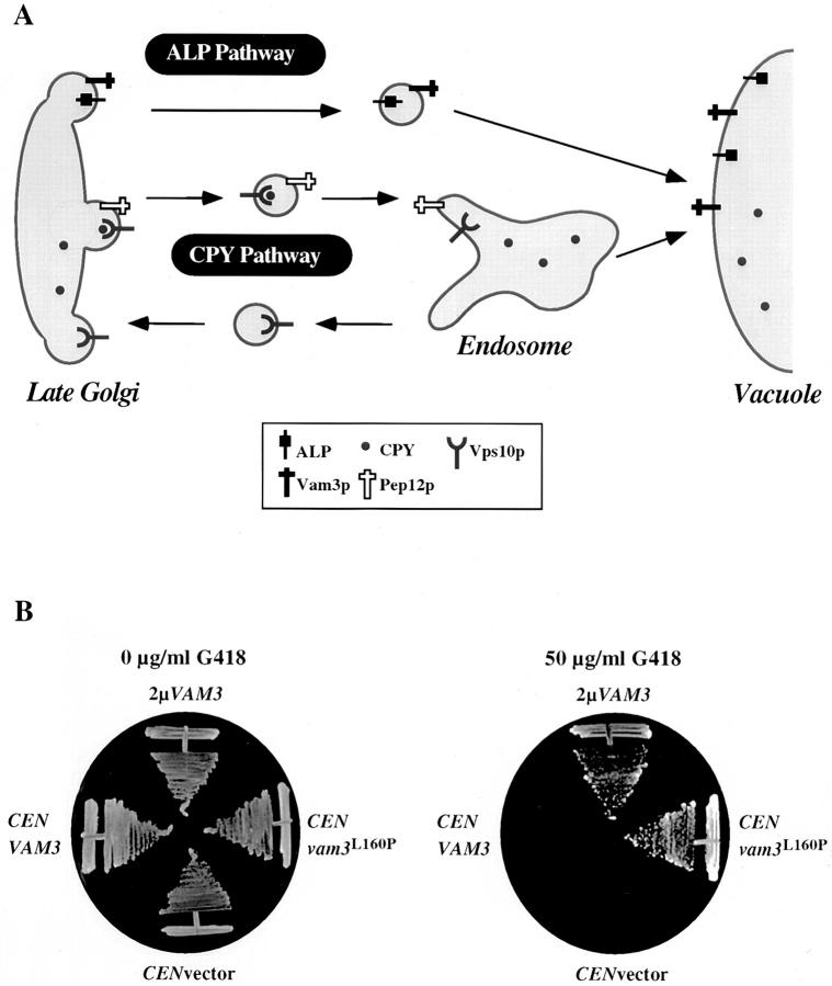
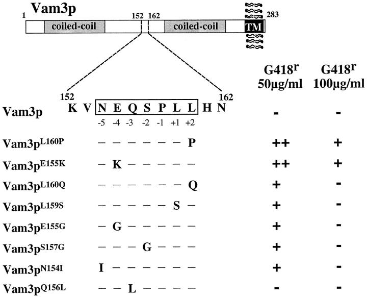
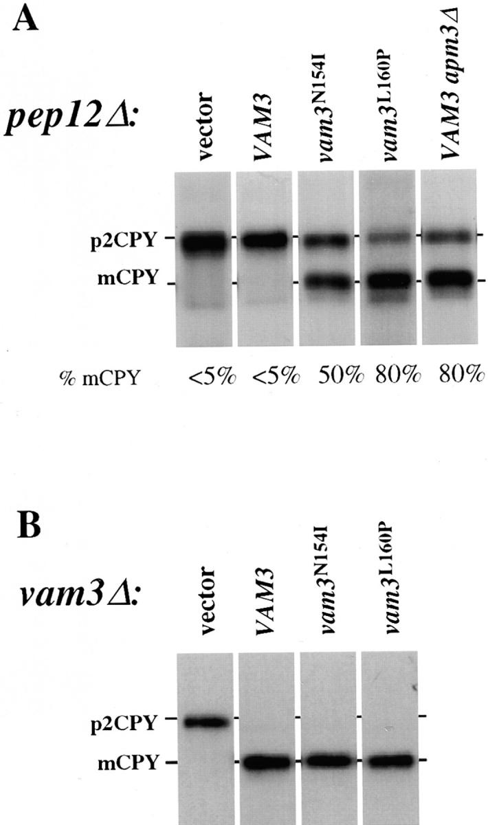
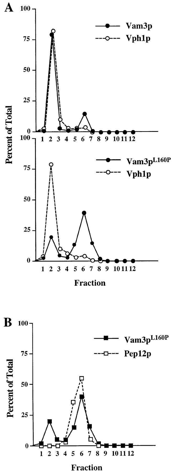
The transport of newly synthesized proteins through the vacuolar protein sorting pathway in the budding yeast Saccharomyces cerevisiae requires two distinct target SNAP receptor (t-SNARE) proteins, Pep12p and Vam3p. Pep12p is localized to the pre-vacuolar endosome and its activity is required for transport of proteins from the Golgi to the vacuole through a well defined route, the carboxypeptidase Y (CPY) pathway. Vam3p is localized to the vacuole where it mediates delivery of cargoes from both the CPY and the recently described alkaline phosphatase (ALP) pathways. Surprisingly, despite their organelle-specific functions in sorting of vacuolar proteins, overexpression of VAM3 can suppress the protein sorting defects of pep12Delta cells. Based on this observation, we developed a genetic screen to identify domains in Vam3p (e.g., localization and/or specific protein-protein interaction domains) that allow it to efficiently substitute for Pep12p. Using this screen, we identified mutations in a 7-amino acid sequence in Vam3p that lead to missorting of Vam3p from the ALP pathway into the CPY pathway where it can substitute for Pep12p at the pre-vacuolar endosome. This region contains an acidic di-leucine sequence that is closely related to sorting signals required for AP-3 adaptor-dependent transport in both yeast and mammalian systems. Furthermore, disruption of AP-3 function also results in the ability of wild-type Vam3p to compensate for pep12 mutants, suggesting that AP-3 mediates the sorting of Vam3p via the di-leucine signal. Together, these data provide the first identification of an adaptor protein-specific sorting signal in a t-SNARE protein, and suggest that AP-3-dependent sorting of Vam3p acts to restrict its interaction with compartment-specific accessory proteins, thereby regulating its function. Regulated transport of cargoes such as Vam3p through the AP-3-dependent pathway may play an important role in maintaining the unique composition, function, and morphology of the vacuole.


Figures

Figure 1
Figure 1
Novel genetic screen to identify regions of Vam3p required for function and localization at the vacuole. (A) In wild-type cells, Pep12p, the endosomal t-SNARE, directs traffic from the Golgi complex to the endosome through the CPY pathway. Vam3p, the vacuolar t-SNARE, is transported through the AP-3–dependent ALP pathway to the vacuole where it functions to receive incoming vesicular traffic from both the ALP and CPY pathways. We designed a G418-based selection scheme to identify VAM3 mutants that were able to compensate for the loss of PEP12 and thus identify regions of Vam3p sequence responsible for restricting Vam3p activity to the AP-3–dependent pathway. (B) Growth of both pep12Δ (CBY31) cells alone or pep12Δ (CBY31) cells expressing VAM3 from a single-copy plasmid (pVAM3.414) is compromised at concentrations of 50 μg/ml G418, while growth on media containing no G418 is normal. However, pep12Δ (CBY31) cells either overexpressing VAM3 from a 2μ plasmid (pVAM3.424) or expressing a mutant derived from the screen, vam3 L160P, on a single-copy plasmid (pVAM3L160P.414) results in growth at 50 μg/ml G418, as well as on media containing no G418.
Figure 2
Figure 2
Vam3p mutants define a di-leucine sorting signal. Vam3 protein domain structure is shown including the extreme COOH-terminal transmembrane domain and the two coiled-coil domains. The location of the region containing the mutations recovered from the G418 resistance screen is denoted by the dashed box. The wild-type sequence at the mutated region is shown in detail. Single mutations that were recovered from the G418 resistance screen are indicated in capital letters designating the amino acid changes. The glutamine 156 to leucine mutation was not recovered in the screen but was made by site-directed mutagenesis. The level of G418 resistance conferred by each of the mutants is as follows: ++, wild-type growth; +, slower growth to single colonies; and −, no growth.
Figure 3
Figure 3
CPY sorting of vam3 mutants in both pep12Δ and vam3Δ mutant cells. (A) pep12Δ (CBY31) cells and pep12Δaps3Δ (GOY8) double-mutant cells transformed with a single-copy plasmid containing wild-type VAM3 (pVAM3.414), and pep12Δ (CBY31) cells transformed with vector (pRS414), vam3 L160P or vam3 N154I mutant isolates (pVAM3L160P. 414 and pVAM3N154I.414, respectively) were spheroplasted, and then metabolically labeled and chased for 45 min. (B) vam3Δ (TDY2) cells were transformed with the identical plasmids from A and were labeled and chased as whole cells for 30 min. CPY was immunoprecipitated and examined by autoradiography. For both A and B, mature and Golgi-modified precursor CPY are indicated as mCPY and p2CPY, respectively. In A, the percent of mature CPY is denoted beneath each individual lane.
Figure 4
Figure 4
Localization of Vam3 mutant proteins. Cleared cell lysates generated from vam3Δ (TDY2) cells containing either wild-type VAM3 (pVAM3.414) or the vam3 L160P mutant (pVAM3L160P.414) were loaded onto the top of an Accudenz step gradient and centrifuged to equilibrium. Fractions 1–12 were collected from the top of the gradient. Proteins were precipitated from the fractions and then separated by SDS-PAGE and transferred to nitrocellulose. Vam3p, Vph1p, and Pep12p were detected by immunoblotting and visualized by ECL fluorography. The distribution of both wild-type and mutant (Vam3pL160P) Vam3p and Vph1p are shown graphically in A and the colocalization of Vam3pL160P and Pep12p are shown in B.
Figure 5
Figure 5
Localization of Vam3 mutant proteins in vps24Δ cells. vam3Δ (TDY2) and vps24Δ (BW102) cells were transformed with plasmids containing both wild-type (pGFPVAM3.426) and mutant (pGFPVAM3L160P.426) GFP-Vam3 fusion proteins. These strains were grown in selective media to exponential phase, harvested, and then resuspended in YNB for examination by microscopy. vps24Δ cultures were further labeled with 16 μM FM4-64 for a period of 1 h at 26°C in YPD. Labeled cells were then harvested, resuspended in YPD, and then chased for 1.5 h. After chase, cells were examined by Nomarski and fluorescence/ confocal microscopy for both GFP and FM4-64 fluorescence. Arrows in A indicate vacuoles and in B indicate class E compartments.
Figure 6
Figure 6
Vam3p di-leucine sequence alignment with putative AP-3 cargoes. The Vam3p di-leucine sequence shares significant sequence similarity to di-leucine sequences in both lysosomal and melanosomal proteins. The consensus motif (*Ex*xLL) is derived from mutagenesis data. An asterisk denotes a bias toward charged, polar amino acids, while x can be any amino acid. Residues that were mutated in the Vam3p sequence and are also conserved in other cargo proteins are indicated by shaded regions. ALP shares sequence similarity with Vam3p at every base that was recovered in our Vam3p mutagenesis. The acidic amino acid at −4, the polar amino acids at −5 and −3, and the proline at −1 as well as the di-leucine sequence, are conserved in the majority of the proteins that have been defined as potential AP-3 cargoes.

References

    1. Babst M, Sato TK, Banta LM, Emr SE. Endosomal transport function in yeast requires a novel AAA-type ATPase, Vps4p. EMBO (Eur Mol Biol Organ) J. 1997;16:1820–1831. - PMC - PubMed
    1. Babst M, Wendland B, Estepa EJ, Emr SD. The Vps4p AAA ATPase regulates membrane association of a Vps protein complex required for normal endosomal function. EMBO (Eur Mol Biol Organ) J. 1998;17:2982–2993. - PMC - PubMed
    1. Banfield DK, Lewis MJ, Rabouille C, Warren G, Pelham HR. Localization of Sed5, a putative vesicle targeting molecule, to the cis-Golgi network involves both its transmembrane and cytoplasmic domains. J Cell Biol. 1994;127:357–371. - PMC - PubMed
    1. Becherer KA, Reider S, Emr SD, Jones EW. Novel syntaxin homologue, Pep12p, required for the sorting of lumenal hydrolases to the lysosome-like vacuole in yeast. Mol Biol Cell. 1996;7:579–594. - PMC - PubMed
    1. Bennett MK, Scheller RH. The molecular machinery for secretion is conserved from yeast to neurons. Proc Natl Acad Sci USA. 1993;90:2559–2563. - PMC - PubMed

Publication types

MeSH terms