

Crystal structures of eukaryotic translation initiation factor 5A from Methanococcus jannaschii at 1.8 A resolution
- PMID: 9724718
- PMCID: PMC27909
- DOI: 10.1073/pnas.95.18.10419
Crystal structures of eukaryotic translation initiation factor 5A from Methanococcus jannaschii at 1.8 A resolution
Abstract
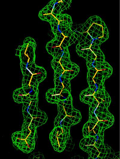
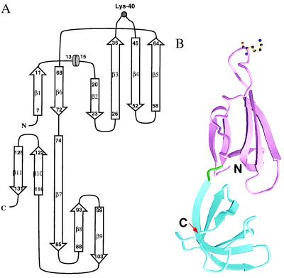
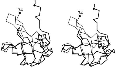
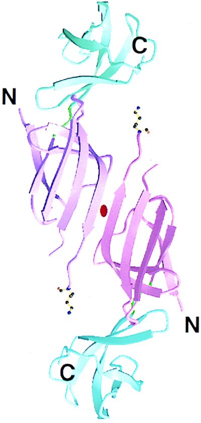
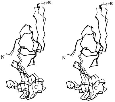
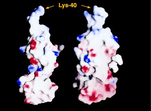
Eukaryotic translation initiation factor 5A (eIF-5A) is a ubiquitous protein found in all eukaryotic cells. The protein is closely associated with cell proliferation in the G1-S stage of the cell cycle. Recent findings show that the eIF-5A proteins are highly expressed in tumor cells and act as a cofactor of the Rev protein in HIV-1-infected cells. The mature eIF is the only protein known to have the unusual amino acid hypusine, a post-translationally modified lysine. The crystal structure of eIF-5A from Methanococcus jannaschii (MJ eIF-5A) has been determined at 1.9 A and 1.8 A resolution in two crystal forms by using the multiple isomorphous replacement method and the multiwavelength anomalous diffraction method for the first crystal form and the molecular replacement method for the second crystal form. The structure consists of two folding domains, one of which is similar to the oligonucleotide-binding domain found in the prokaryotic cold shock protein and the translation initiation factor IF1 despite the absence of any significant sequence similarities. The 12 highly conserved amino acid residues found among eIF-5As include the hypusine site and form a long protruding loop at one end of the elongated molecule.
Figures
References
-
- Park M H, Wolff E C, Folk J E. Biofactors. 1993;4:95–104. - PubMed
-
- Schumann H, Klink F. System Appl Microbiol. 1989;11:103–107.
-
- Bartig D, Lemkemeier K, Frank J, Lottspeich F, Klink F. Eur J Biochem. 1992;204:751–758. - PubMed
-
- Kemper W M, Berry K W, Merrick W C. J Biol Chem. 1976;251:5551–5557. - PubMed
-
- Benne R, Brown-Luedi M L, Hershey J W. J Biol Chem. 1978;253:3070–3077. - PubMed
Publication types
MeSH terms
Substances
Associated data
- Actions
LinkOut - more resources
Full Text Sources
Other Literature Sources
Molecular Biology Databases
