. 1998 Sep 1;95(18):10425-30.
doi: 10.1073/pnas.95.18.10425.

Protein kinase A-catalyzed phosphorylation of heat shock protein 60 chaperone regulates its attachment to histone 2B in the T lymphocyte plasma membrane

Affiliations

Protein kinase A-catalyzed phosphorylation of heat shock protein 60 chaperone regulates its attachment to histone 2B in the T lymphocyte plasma membrane

I U Khan et al. Proc Natl Acad Sci U S A. .

Abstract

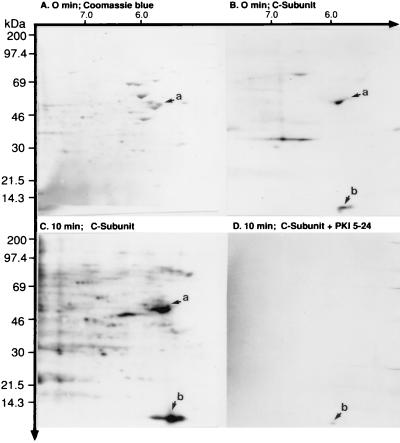
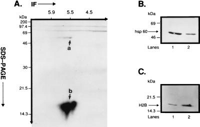
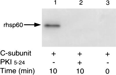
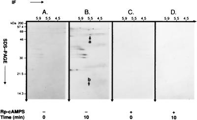
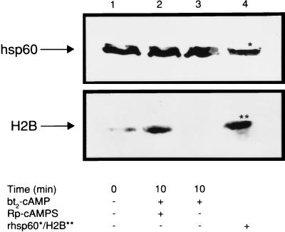
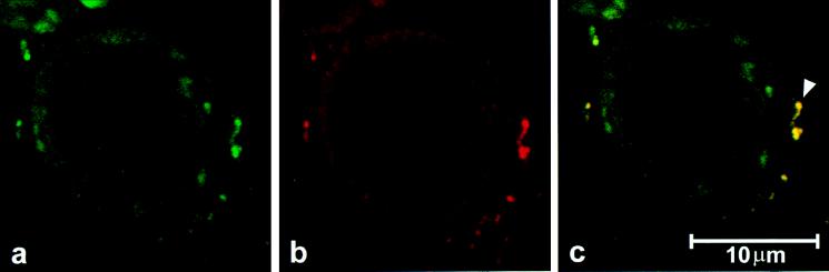
Accumulating evidence suggests that the mitochondrial molecular chaperone heat shock protein 60 (hsp60) also can localize in extramitochondrial sites. However, direct evidence that hsp60 functions as a chaperone outside of mitochondria is presently lacking. A 60-kDa protein that is present in the plasma membrane of a human leukemic CD4(+) CEM-SS T cell line and is phosphorylated by protein kinase A (PKA) was identified as hsp60. An 18-kDa plasma membrane-associated protein coimmunoprecipitated with hsp60 and was identified as histone 2B (H2B). Hsp60 physically associated with H2B when both molecules were in their dephospho forms. By contrast, PKA-catalyzed phosphorylation of both hsp60 and H2B caused dissociation of H2B from hsp60 and loss of H2B from the plasma membrane of intact T cells. These results suggest that (i) hsp60 and H2B can localize in the T cell plasma membrane; (ii) hsp60 functions as a molecular chaperone for H2B; and (iii) PKA-catalyzed phosphorylation of both hsp60 and H2B appears to regulate the attachment of H2B to hsp60. We propose a model in which phosphorylation/dephosphorylation regulates chaperoning of H2B by hsp60 in the plasma membrane.


Figures

Figure 1
Figure 1
In vitro PKA C-subunit-catalyzed phosphorylation of CEM-SS T cell plasma membrane-associated proteins. Phosphorylation was performed as described in the Methods section. The phosphorylated plasma membrane fragments were analyzed by 10% 2-D SDS/PAGE, the proteins were transblotted to Immobilon-PSQ membrane, the membranes were stained with CBB, and the blot was exposed to x-ray film. (A) CBB stain showing the distribution of plasma membrane-associated proteins. The arrow marked a points to a 60-kDa protein with a pI of 5.5. (B) Autoradiogram of the same blot at 0 min revealing constitutively phosphorylated substrates. The arrow marked a corresponds to the CBB-stained protein shown in A and reveals that the 60-kDa protein has a low level of constitutive phosphorylation. Arrow b points to a protein with an apparent Mr of 14 kDa and a pI of 5.5, is not stained by CBB in A, and is constitutively phosphorylated. (C) In vitro phosphorylation of plasma membrane-associated proteins by 50 nM purified PKA C-subunit over 10 min at 30°C. Arrows a and b reveal enhanced phosphorylation of the 60- and 14-kDa proteins compared with B. (D) Specific inhibition of C-subunit-catalyzed phosphorylation by PKI-(5–24).
Figure 2
Figure 2
Immunoprecipitation of plasma membrane-associated phosphoproteins by mAb II-13 and immunoblotting of p60 and p18. (A) Isolated CEM-SS T cell plasma membrane fragments (60 μg protein) were phosphorylated in vitro by 50 nM PKA C-subunit in the presence of γ-[32P]ATP. In vitro phosphorylated fragments were immunoprecipitated with mAb II-13, and the immunoprecipitate was resolved on a 15% 2-D SDS/PAGE. Coimmunoprecipitation of phsp60 and pp18 was observed on the autoradiogram. Also notable was that both phsp60 and pp18 were comprised of isoforms whose pI values varied between ≈5.5 and 5.8. Differences in the relative intensity of phosphorylation of phsp60 and pp18 are addressed in Discussion. (B) Isolated CEM-SS T cell plasma membrane fragments (lane 1, 60 μg protein) and 1 μg of rhsp60 (lane 2) were run on a 15% 1-D SDS/PAGE and were immunoblotted with mAb II-13. (C) Isolated CEM-SS T cell plasma membrane fragments (lane 1, 60 μg protein) and 2 μg of purified human H2B (lane 2) were run on a 15% 1-D SDS/PAGE and were immunoblotted with affinity-purified polyclonal anti-H2B Ab.
Figure 3
Figure 3
In vitro phosphorylation of rhsp60. rhsp60 (1 μg) was phosphorylated by 50 nM of purified PKA C-subunit, and phosphorylated rhsp60 was resolved by 10% 1-D SDS/PAGE. Resolved prhsp60 then was transferred to poly(vinylidene difluoride) membrane, and autoradiography was performed. Lanes: 1, 10 min without PKI-(5–24); lane 2, 10 min with 10 μM PKI-(5–24); lane 3, 0 min without PKI-(5–24).
Figure 4
Figure 4
cAMP-dependent, PKA-catalyzed phosphorylation of hsp60 in intact CEM-SS T cell isolated plasma membrane fragments. After loading with [32Pi]orthophosphate for 3 hr, cells were exposed to 2.5 mM bt2-cAMP in the presence of 200 μM isobutylmethylxanthine and 10 μM okadaic acid for 0 min and 10 min. Plasma membrane fragments then were isolated, and phsp60 was immunoprecipitated with mAb II-13. Immunoprecipitated pp60 was analyzed by 15% 2-D SDS/PAGE. (A) At 0 min, shows the absence of basal phosphorylation of hsp60. Note that other, unidentified phosphoproteins are coimmunoprecipitated with hsp60, but pH2B is not observed. (B) At 10 min, shows immunoprecipitated phsp60 (arrow a). Although other, unidentified phosphoproteins are coimmunoprecipitated with phsp60, pH2B is not observed (arrow b). (C) At 0 min with 500 μM Rp-cAMPS. (D) At 10 min with 500 μM Rp-cAMPS blocks cAMP-dependent, PKA-catalyzed phosphorylation of hsp60.
Figure 5
Figure 5
Effect of cAMP-dependent, PKA-catalyzed phosphorylation on the presence of H2B and hsp60 in the T cell plasma membrane. T cells were either pretreated or not pretreated with 500 μM Rp-cAMPS and subsequently were exposed to 2.5 mM bt2-cAMP for 0 or 10 min at 30°C in the absence or presence of Rp-cAMPS, and plasma membrane fragments were isolated. The membrane fragments were run on a 10% 1-D SDS/PAGE, were transferred, and were immunoblotted with polyclonal anti-H2B Ab or mAb II-13. Lanes: 1, constitutive H2B and hsp60; 2, immunoblots of pH2B and phsp60 at 10 min after initiation of phosphorylation in the presence of Rp-cAMPS; 3, immunoblots of pH2B and phsp60 at 10 min after initiation of phosphorylation in the absence of Rp-cAMPS; 4, rhsp60 and H2B controls.
Figure 6
Figure 6
Confocal laser photomicrograph shows the distribution of (a) hsp60 (green) and (b) H2B (red) in the CEM-SS T cell. (c) An image generated by computer-assisted superimposing a and b. Colocalization of hsp60 and H2B is recognized as yellow (▹) in the representative cell.

Similar articles

Cited by

References

    1. Rothman J E. Cell. 1989;59:591–601. - PubMed
    1. Gething M-J, Sambrook J. Nature (London) 1992;355:33–45. - PubMed
    1. Ellis R J, van der Vies S M. Annu Rev Biochem. 1991;60:321–347. - PubMed
    1. Hartl F-U. Nature (London) 1996;381:571–580. - PubMed
    1. Neupert W. Annu Rev Biochem. 1997;66:863–917. - PubMed

Publication types