

The location of the carboxy-terminal region of gamma chains in fibrinogen and fibrin D domains
- PMID: 9724734
- PMCID: PMC27925
- DOI: 10.1073/pnas.95.18.10511
The location of the carboxy-terminal region of gamma chains in fibrinogen and fibrin D domains
Abstract
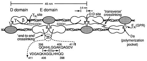
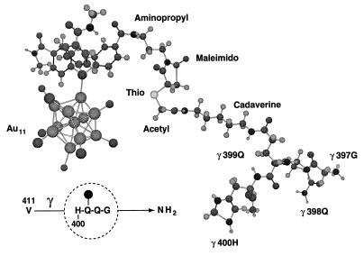
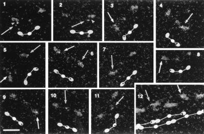
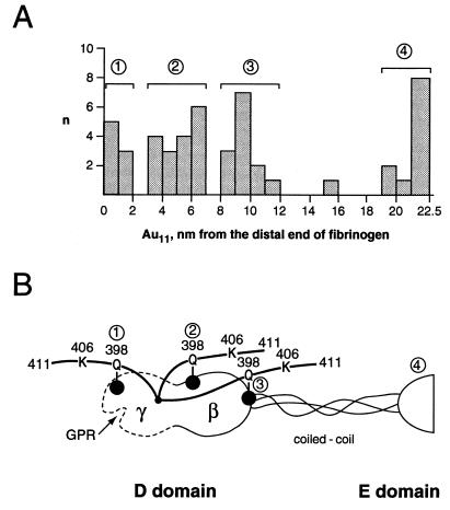
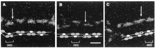
Elongated fibrinogen molecules are comprised of two outer "D" domains, each connected through a "coiled-coil" region to the central "E" domain. Fibrin forms following thrombin cleavage in the E domain and then undergoes intermolecular end-to-middle D:E domain associations that result in double-stranded fibrils. Factor XIIIa mediates crosslinking of the C-terminal regions of gamma chains in each D domain (the gammaXL site) by incorporating intermolecular epsilon-(gamma-glutamyl)lysine bonds between amine donor gamma406 lysine of one gamma chain and a glutamine acceptor at gamma398 or gamma399 of another. Several lines of evidence show that crosslinked gamma chains extend "transversely" between the strands of each fibril, but other data suggest instead that crosslinked gamma chains can only traverse end-to-end-aligned D domains within each strand. To examine this issue and determine the location of the gammaXL site in fibrinogen and assembled fibrin fibrils, we incorporated an amine donor, thioacetyl cadaverine, into glutamine acceptor sites in fibrinogen in the presence of XIIIa, and then labeled the thiol with a relatively small (0.8 nm diameter) electron dense gold cluster compound, undecagold monoaminopropyl maleimide (Au11). Fibrinogen was examined by scanning transmission electron microscopy to locate Au11-cadaverine-labeled gamma398/399 D domain sites. Seventy-nine percent of D domain Au11 clusters were situated in middle to proximal positions relative to the end of the molecule, with the remaining Au11 clusters in a distal position. In fibrin fibrils, D domain Au11 clusters were located in middle to proximal positions. These findings show that most C-terminal gamma chains in fibrinogen or fibrin are oriented toward the central domain and indicate that gammaXL sites in fibrils are situated predominantly between strands, suitably aligned for transverse crosslinking.
Figures
References
-
- Mosesson M W, Siebenlist K R, Hainfeld J F, Wall J S. J Struct Biol. 1995;115:88–101. - PubMed
-
- Chen R, Doolittle R F. Biochemistry. 1971;10:4486–4491. - PubMed
-
- Doolittle R F, Chen R, Lau F. Biochem Biophys Res Comm. 1971;44:94–100. - PubMed
-
- Purves L R, Purves M, Brandt W. Biochemistry. 1987;26:4640–4646. - PubMed
-
- Kloczewiak M, Timmons S, Hawiger J. Thromb Res. 1983;29:249–255. - PubMed
Publication types
MeSH terms
Substances
Grants and funding
LinkOut - more resources
Full Text Sources
