. 1998 Sep 1;95(18):10535-40.
doi: 10.1073/pnas.95.18.10535.

Footprints on the viral DNA ends in moloney murine leukemia virus preintegration complexes reflect a specific association with integrase

Affiliations

Footprints on the viral DNA ends in moloney murine leukemia virus preintegration complexes reflect a specific association with integrase

S Q Wei et al. Proc Natl Acad Sci U S A. .

Abstract

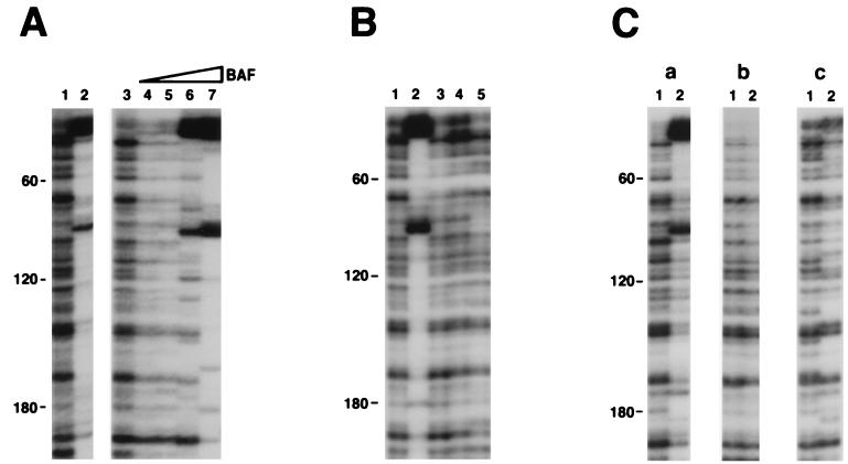
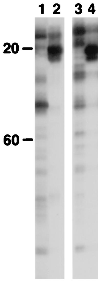
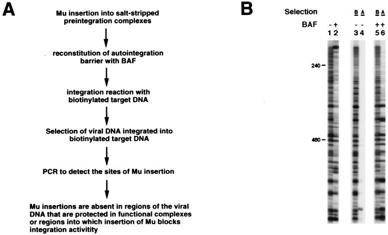
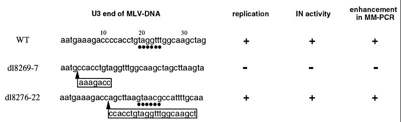
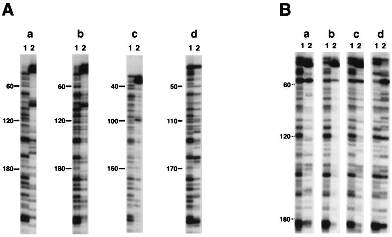
Retroviral DNA integration is mediated by the preintegration complex, a large nucleoprotein complex derived from the core of the infecting virion. We previously have used Mu-mediated PCR to probe the nucleoprotein organization of Moloney murine leukemia virus preintegration complexes. A region of protection spans several hundred base pairs at each end of the viral DNA, and strong enhancements are present near the termini. Here, we show that these footprints reflect a specific association between integrase and the viral DNA ends in functional preintegration complexes. Barrier-to-autointegration factor, a cellular protein that blocks autointegration of Moloney murine leukemia virus DNA, also plays an indirect role in generating the footprints at the ends of the viral DNA. We have exploited Mu-mediated PCR to examine the effect of mutations at the viral DNA termini on complex formation. We find that a replication competent mutant with a deletion at one end of the viral DNA still exhibits a strong enhancement about 20 bp from the terminus of the mutant DNA end. The site of the enhancement therefore appears to be at a fixed distance from the ends of the viral DNA. We also find that a mutation at one end of the viral DNA, which renders the virus incompetent for replication, abolishes the enhancements and protection at both the U3 and U5 ends. A pair of functional viral DNA ends therefore are required to interact before the chemical step of 3' end processing.


Figures

Figure 1
Figure 1
Reconstitution of the footprints on the viral DNA U3 end. All preintegration complexes were treated with 750 mM KCl before separation from free proteins on a 10–50% Nycodenz gradient. Reconstitution reactions then were carried out on the gradient purified complexes, followed by MM-PCR footprinting (see Materials and Methods for details). End-labeled primer SW2 (13) was used for PCR. Numbers on the left side of each panel indicate the distance from the very end of the viral DNA. Distinctive enhancements are located at ≈20 bp and ≈100 bp from the U3 end. (A) Reconstitution with BAF protein. Samples are naked viral DNA (lane 1) and MLV preintegration complexes (lane 2). Reconstitution reactions included: lane 3, buffer only control, and lanes 4–7, BAF (0.2, 0.6, 2, and 6 ng, respectively). (B) Reconstitution with various protein factors. Reconstitution reactions included: lane 1, buffer only control; lane 2, BAF (2 ng); lane 3, murine HMG Y; lane 4, human HMG I(Y); and lane 5, human HMG1. Purified protein factors shown in lanes 3–5 have been tested in a range of 0.5 to 5 μg, and no differences were observed in this range (data not shown); therefore only reactions with 1 μg of protein are shown. (C) Enhancements are observed only with complexes containing integrase. Reconstitution reactions with BAF were carried out with (a) salt-stripped MLV preintegration complexes, (b) naked viral DNA, and (c) salt-stripped integrase-minus MLV preintegration complexes. Lanes 1 and 2 included 0 and 2 ng BAF, respectively.
Figure 2
Figure 2
Footprinting of MLV integrase on terminal viral DNA. Footprinting was carried out as described in Materials and Methods. End-labeled primer SW6 (13) was used for PCR to detect the U5 viral DNA end. Strong enhancements are located at ≈20 bp from the tip of the U5 end. Controls are with naked viral DNA (lane 1) and MLV preintegration complexes (lane 2). Footprinting in the absence (lane 3) or presence (lane 4) of purified integrase is shown.
Figure 3
Figure 3
Extensive regions at the ends of the viral DNA are required for integration activity. (A) Schematic depiction of the functional footprinting assay. (B) Functional footprints on the viral DNA U3 end. Lanes 1 and 2 are controls of standard MM-PCR footprinting with salt-stripped MLV preintegration complexes reconstituted with 0 or 2 ng BAF, respectively. Reconstitution reactions in the functional footprinting experiments contained 0 (lanes 3 and 4) or 20 ng BAF (lanes 5 and 6). DNA samples before and after biotin selection are marked with B and A, respectively. In the absence of reconstitution of the autointegration barrier with BAF, intermolecular integration was very inefficient, and therefore little signal is seen in lane 4.
Figure 4
Figure 4
Summary of MLV U3 end mutants used in this study. Deletions shown in the boxes are at the U3 ends of both LTRs. ▴ indicates the position of the deletion, and • indicates the strong enhancement observed at ≈20 bp from the U3 end. Viral replication was determined by measuring the reverse transcriptase activity in culture supernatants. Integration activity was assayed with preintegration complexes in the corresponding cytoplasmic extracts. MM-PCR was used to assay for the presence of the characteristic enhancements (see Fig. 5).
Figure 5
Figure 5
Footprints on the viral DNA ends of various MLV mutants. All complexes were salt-stripped and reconstituted with BAF followed by MM-PCR. Characteristic enhancements are located at ≈20 bp and ≈100 bp from the U3 end, and ≈20 bp from the U5 end. Shown are footprints on the U3 (A) and U5 (B) viral DNA ends. (a) Control MLV preintegration complexes made by coculturing clone 4 with NIH 3T3 cells. (b-d) Preintegration complexes obtained by transient transfection of 293T cells with plasmid (b) pNCA (wild-type MLV); (c) pSQ70 (active dl8276–22 MLV); and (d) pJM1X1 (inactive dl8269–7 MLV). Reconstitution was done in the absence of BAF in lanes 1 and with 2 ng BAF in lanes 2.

Similar articles

Cited by

References

    1. Varmus H, Brown P O. In: Mobile DNA. Berg D E, Howe M M, editors. Washington, DC: Am. Soc. Microbiol.; 1989. pp. 53–108.
    1. Goff S P. Annu Rev Genet. 1992;26:527–544. - PubMed
    1. Brown P O, Bowerman B, Varmus H E, Bishop J M. Cell. 1987;49:347–356. - PubMed
    1. Bowerman B, Brown P O, Bishop J M, Varmus H E. Genes Dev. 1989;3:469–478. - PubMed
    1. Ellison V, Abrams H, Roe T, Lifson J, Brown P. J Virol. 1990;64:2711–2715. - PMC - PubMed

Publication types