

The domain structure of protoporphyrinogen oxidase, the molecular target of diphenyl ether-type herbicides
- PMID: 9724741
- PMCID: PMC27932
- DOI: 10.1073/pnas.95.18.10553
The domain structure of protoporphyrinogen oxidase, the molecular target of diphenyl ether-type herbicides
Abstract
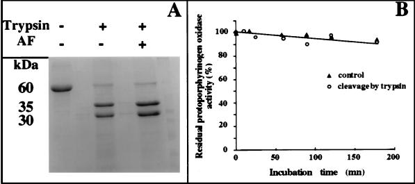
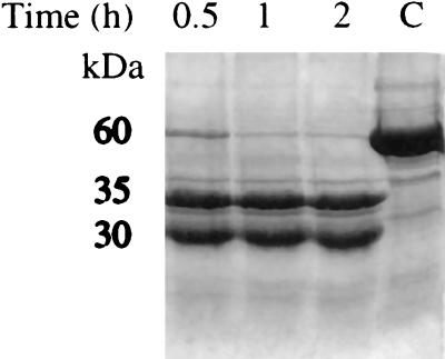
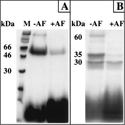
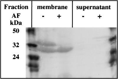
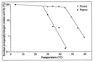
Protoporphyrinogen oxidase (EC 1-3-3-4), the 60-kDa membrane-bound flavoenzyme that catalyzes the final reaction of the common branch of the heme and chlorophyll biosynthesis pathways in plants, is the molecular target of diphenyl ether-type herbicides. It is highly resistant to proteases (trypsin, endoproteinase Glu-C, or carboxypeptidases A, B, and Y), because the protein is folded into an extremely compact form. Trypsin maps of the native purified and membrane-bound yeast protoporphyrinogen oxidase show that this basic enzyme (pI > 8.5) was cleaved at a single site under nondenaturing conditions, generating two peptides with relative molecular masses of 30,000 and 35,000. The endoproteinase Glu-C also cleaved the protein into two peptides with similar masses, and there was no additional cleavage site under mild denaturing conditions. N-terminal peptide sequence analysis of the proteolytic (trypsin and endoproteinase Glu-C) peptides showed that both cleavage sites were located in putative connecting loop between the N-terminal domain (25 kDa) with the betaalphabeta ADP-binding fold and the C-terminal domain (35 kDa), which possibly is involved in the binding of the isoalloxazine moiety of the FAD cofactor. The peptides remained strongly associated and fully active with the Km for protoporphyrinogen and the Ki for various inhibitors, diphenyl-ethers, or diphenyleneiodonium derivatives, identical to those measured for the native enzyme. However, the enzyme activity of the peptides was much more susceptible to thermal denaturation than that of the native protein. Only the C-terminal domain of protoporphyrinogen oxidase was labeled specifically in active site-directed photoaffinity-labeling experiments. Trypsin may have caused intramolecular transfer of the labeled group to reactive components of the N-terminal domain, resulting in nonspecific labeling. We suggest that the active site of protoporphyrinogen oxidase is in the C-terminal domain of the protein, at the interface between the C- and N-terminal domains.
Figures
References
-
- Sandmann G, Nicolaus B, Böger P. Z Naturforsch. 1990;45c:512–517.
-
- Scalla R, Matringe M. Rev Weed Sci. 1994;6:103–132.
-
- Mornet R, Gouin L, Matringe M, Scalla R, Swithenbank C. J Labelled Compounds Radiopharm. 1992;31:175–182.
Publication types
MeSH terms
Substances
LinkOut - more resources
Full Text Sources
Other Literature Sources
Molecular Biology Databases
Research Materials
Miscellaneous
