. 1998 Sep 1;95(18):10570-5.
doi: 10.1073/pnas.95.18.10570.

FokI dimerization is required for DNA cleavage

Affiliations

FokI dimerization is required for DNA cleavage

J Bitinaite et al. Proc Natl Acad Sci U S A. .

Abstract

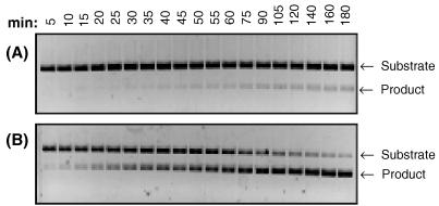
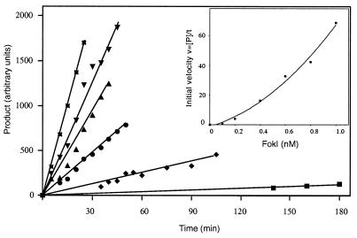
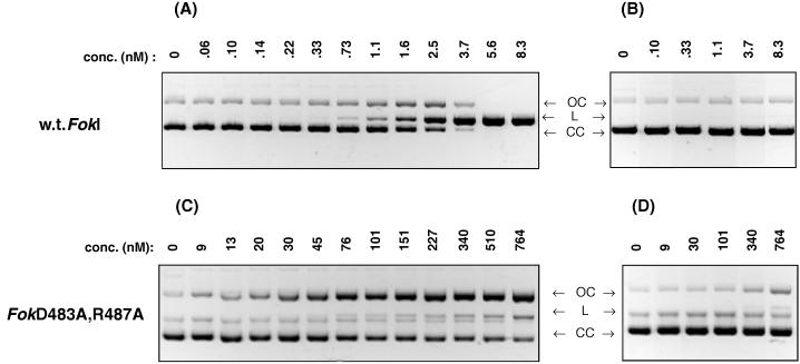
FokI is a type IIs restriction endonuclease comprised of a DNA recognition domain and a catalytic domain. The structural similarity of the FokI catalytic domain to the type II restriction endonuclease BamHI monomer suggested that the FokI catalytic domains may dimerize. In addition, the FokI structure, presented in an accompanying paper in this issue of Proceedings, reveals a dimerization interface between catalytic domains. We provide evidence here that FokI catalytic domain must dimerize for DNA cleavage to occur. First, we show that the rate of DNA cleavage catalyzed by various concentrations of FokI are not directly proportional to the protein concentration, suggesting a cooperative effect for DNA cleavage. Second, we constructed a FokI variant, FokN13Y, which is unable to bind the FokI recognition sequence but when mixed with wild-type FokI increases the rate of DNA cleavage. Additionally, the FokI catalytic domain that lacks the DNA binding domain was shown to increase the rate of wild-type FokI cleavage of DNA. We also constructed an FokI variant, FokD483A, R487A, which should be defective for dimerization because the altered residues reside at the putative dimerization interface. Consistent with the FokI dimerization model, the variant FokD483A, R487A revealed greatly impaired DNA cleavage. Based on our work and previous reports, we discuss a pathway of DNA binding, dimerization, and cleavage by FokI endonuclease.


Figures

Figure 1
Figure 1
Plasmid map of the pTYB4/FokR. The fokIR gene is placed downstream of the T7 promoter, and fokIR is in-frame with the 5′ end of the intein coding sequence. CBD, chitin binding domain.
Figure 2
Figure 2
Time course of the FokI-catalyzed DNA cleavage reaction. Linearized pSKFok1 DNA (6.3 nM) was incubated at 37°C with 0.2 (A) or 0.6 nM (B) FokI restriction endonuclease in 200 μl of reaction buffer (20 mM Tris-acetate, pH 7.9/10 mM magnesium acetate/50 mM potassium acetate/1 mM DTT/100 μg/ml BSA). At the time points indicated, 10-μl samples were withdrawn, immediately quenched by adding 5 μl of stop solution, and subjected to electrophoresis on 1% agarose-gel.
Figure 3
Figure 3
Rate of DNA cleavage with various FokI concentrations. The conditions of DNA cleavage reaction are described in Fig. 2. Samples were withdrawn at the indicated times from the reactions that contained 0.1 nM FokI (■), 0.2 nM FokI (♦), 0.4 nM FokI (•), 0.6 nM FokI (▴), 0.8 nM FokI (▾), and 1.0 nM FokI (∗). The amount of cleaved DNA was determined as in Experimental Procedures. (Inset) Initial velocity vs. FokI concentration.
Figure 4
Figure 4
Stimulation of FokI endonuclease by (A) FokN13Y, (B) FokCD, or (C) FokCD/D450A. Reactions were performed at 37°C in 70 μl of 20 mM Tris-acetate buffer (pH 7.9), 10 mM magnesium acetate, 50 mM potassium acetate, 1 mM DTT, 100 μg/ml BSA, and 6.3 nM linearized pSKFok1 DNA; 10-μl samples were withdrawn at the indicated times from the reactions that contained: (A) either 500 nM FokN13Y, 1.0 nM FokI, or 1.0 nM FokI + 500 nM FokN13Y; (B) either 500 nM FokCD, 0.5 nM FokI, or 0.5 nM FokI + 500 nM FokCD; and (C) either 500 nM FokCD/D450A, 1.0 nM FokI, or 1.0 nM FokI + 500 nM FokCD/D450A. Samples were quenched by adding 5 μl of stop solution and were analyzed by electrophoresis on 1% agarose gel.
Figure 5
Figure 5
Comparison of DNA cleavage by FokI and FokD483A, R487A. The cleavage reactions were performed for 1 h at 37°C in 30 μl of 20 mM Tris-acetate buffer (pH 7.9), 10 mM magnesium acetate, 50 mM potassium acetate, 1 mM DTT, and 100 μg/ml BSA, containing 20 nM of (A and C) supercoiled plasmid pSKFok1 or (B and D) supercoiled plasmid pSKFok0 and either (A and B) FokI or (C and D) FokD483A, R487A. The concentrations of FokI and FokD483A, R487A are indicated. (CC), covalently closed form of plasmid DNA; (OC), open circle form; (L), linear form.

References

    1. Sugisaki H, Kanazawa S. Gene. 1981;16:73–78. - PubMed
    1. Looney M, Moran L S, Jack W E, Feehery G R, Benner J S, Slatko B E, Wilson G G. Gene. 1989;80:193–208. - PubMed
    1. Kita K, Kotani H, Sugisaki H, Takanami M. J Biol Chem. 1989;264:5751–5756. - PubMed
    1. Kaczorowski T, Skowron P, Podhajska A J. Gene. 1989;80:209–216. - PubMed
    1. Skowron P, Kaczorowski T, Tucholski J, Podhajska A. Gene. 1993;125:1–10. - PubMed

Publication types

LinkOut - more resources