. 1998 Sep 1;95(18):10596-601.
doi: 10.1073/pnas.95.18.10596.

The adenomatous polyposis coli-binding protein EB1 is associated with cytoplasmic and spindle microtubules

Affiliations

The adenomatous polyposis coli-binding protein EB1 is associated with cytoplasmic and spindle microtubules

L Berrueta et al. Proc Natl Acad Sci U S A. .

Abstract

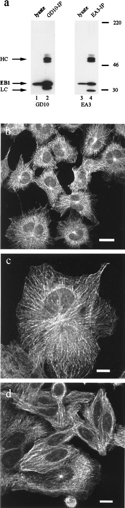
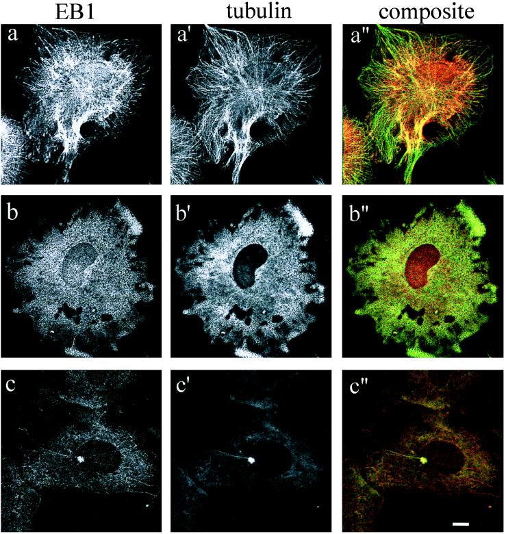
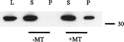
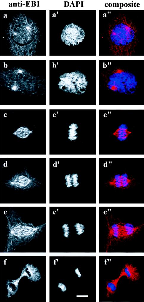
The evolutionarily conserved protein EB1 originally was identified by its physical association with the carboxyl-terminal portion of the adenomatous polyposis coli (APC) tumor suppressor protein, an APC domain commonly mutated in familial and sporadic forms of colorectal neoplasia. The subcellular localization of EB1 in epithelial cells was studied by using immunofluorescence and biochemical techniques. EB1 colocalized both to cytoplasmic microtubules in interphase cells and to spindle microtubules during mitosis, with pronounced centrosome staining. The cytoskeletal array detected by anti-EB1 antibody was abolished by incubation of the cells with nocodazole, an agent that disrupts microtubules; upon drug removal, EB1 localized to the microtubule-organizing center. Immunofluorescence analysis of SW480, a colon cancer cell line that expresses only carboxyl-terminal-deleted APC unable to interact with EB1, demonstrated that EB1 remained localized to the microtubule cytoskeleton, suggesting that this pattern of subcellular distribution is not mediated by its interaction with APC. In vitro cosedimentation with taxol-stabilized microtubules demonstrated that a significant fraction of EB1 associated with microtubules. Recent studies of the yeast EB1 homologues Mal3 and Bim1p have demonstrated that both proteins localize to microtubules and are important in vivo for microtubule function. Our results demonstrate that EB1 is a novel component of the microtubule cytoskeleton in mammalian cells. Associating with the mitotic apparatus, EB1 may play a physiologic role connecting APC to cellular division, coordinating the control of normal growth and differentiation processes in the colonic epithelium.


Figures

Figure 1
Figure 1
Subcellular localization of EB1 in mammalian cells. (a) Specificity of the anti-EB1 mAbs GD10 and EA3 for EB1 in cell lysates (lanes 1 and 3) and immunoprecipitation (lanes 2 and 4). Cell lysates and immunoprecipitated proteins from CV-1 cells, prepared as described, were separated in 10% SDS-polyacrylamide gels, transferred to poly(vinylidene difluoride) membranes, probed with anti-EB1 antibodies (GD10 and EA3), and detected by ECL; heavy chain (HC) and light chain (LC) are indicated. Interphase CV-1 (b and c) and SW480 cells (d) were stained with GD10 antibody after methanol fixation at −20°C. A secondary donkey anti-mouse rhodamine-conjugated antibody was used, and the samples were analyzed by confocal microscopy. [Bars: 25 μm (b) and 10 μm (c and d).]
Figure 2
Figure 2
Colocalization of EB1 with cytoplasmic microtubules and the centrosome. Unsynchronized CV-1 cells were fixed in methanol and processed for dual immunofluorescence with antibodies directed against EB1 (GD10) and α-tubulin (YL1/2), in the presence or absence of the microtubule destabilizing drug nocodazole (10 μM). (a, a′, and a′′) Control untreated cells. (b, b′, and b′′) Cells treated with nocodazole for 1 hr and fixed in methanol at −20°C. (c, c′, and c′′) Cells fixed in methanol 5 min after nocodazole removal. Staining was analyzed by confocal microscopy. (Bar, 10 μm.)
Figure 3
Figure 3
In vitro EB1 microtubule binding. Cell extracts from J77 prepared in buffer A (see Materials and Methods) were incubated with taxol-stabilized microtubules. Supernatants (S) and pellets (P) were collected and separated by SDS-polyacrylamide gel, transferred to poly(vinylidene difluoride), and probed with the anti-EB1 antibody GD10. Microtubule binding was demonstrated by cosedimentation of EB1 with the microtubule pellet. Whole-cell lysate (L) demonstrated endogenous EB1 mobility.
Figure 4
Figure 4
EB1 localizes to the mitotic apparatus during cell division. Unsynchronized CV-1 cells were fixed in methanol and stained with the anti-EB1 antibody GD10 and a secondary donkey anti-mouse rhodamine-conjugated antibody. DNA was counterstained with 4,6-diamidino-2-phenylindole (DAPI). (a, a′, and a′′) Prophase. (b, b′, and b′′) Prometaphase. (c, c′, and c′′) Metaphase. (d, d′, and d′′) Anaphase. (e, e′, and e′′) Telophase. (f, f′, and f′′) Cytokinesis.

Similar articles

Cited by

References

    1. Hyman A A, Karsenti E. Cell. 1996;84:401–410. - PubMed
    1. Merdes A, Cleveland D. J Cell Biol. 1997;138:953–956. - PMC - PubMed
    1. Stearns T. J Cell Biol. 1997;138:957–960. - PMC - PubMed
    1. Barton N R, Goldstein L S. Proc Natl Acad Sci USA. 1996;93:1735–1742. - PMC - PubMed
    1. Sawin K E, Endow S A. BioEssays. 1993;15:399–407. - PubMed

LinkOut - more resources