. 1998 Sep 1;95(18):10602-7.
doi: 10.1073/pnas.95.18.10602.

WW domain-mediated interactions reveal a spliceosome-associated protein that binds a third class of proline-rich motif: the proline glycine and methionine-rich motif

Affiliations

WW domain-mediated interactions reveal a spliceosome-associated protein that binds a third class of proline-rich motif: the proline glycine and methionine-rich motif

M T Bedford et al. Proc Natl Acad Sci U S A. .

Abstract

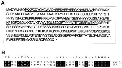
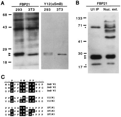
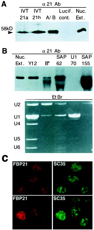
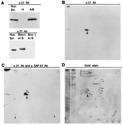
Pre-mRNA splicing requires the bridging of the 5' and 3' ends of the intron. In yeast, this bridging involves interactions between the WW domains in the splicing factor PRP40 and a proline-rich domain in the branchpoint binding protein, BBP. Using a proline-rich domain derived from formin (a product of the murine limb deformity locus), we have identified a family of murine formin binding proteins (FBP's), each of which contains one or more of a special class of tyrosine-rich WW domains. Two of these WW domains, in the proteins FBP11 and FBP21, are strikingly similar to those found in the yeast splicing factor PRP40. We show that FBP21 is present in highly purified spliceosomal complex A, is associated with U2 snRNPs, and colocalizes with splicing factors in nuclear speckle domains. Moreover, FBP21 interacts directly with the U1 snRNP protein U1C, the core snRNP proteins SmB and SmB', and the branchpoint binding protein SF1/mBBP. Thus, FBP21 may play a role in cross-intron bridging of U1 and U2 snRNPs in the mammalian A complex.


Figures

Figure 1
Figure 1
Predicted amino acid sequence of the FBP21 cDNA reveals a zinc finger motif and two WW domains. (A) Amino acid sequence of FBP21. The zinc finger motif is boxed. The two WW domains are underlined with a solid line. The 15-aa spacing between the two WW domains is underlined with a broken line. This spacer distance is conserved between the WW domains of the putative splicing factor FBP11, FBP21, and the yeast U1-associated splicing factor PRP40. (B) A blast search aligning the zinc finger of the splicing factor U1C with the zinc finger of FBP21. The alignment was the best fit obtained by using the blast search.
Figure 2
Figure 2
The WW domain of FBP21 binds SmB as determined by blot overlay. (A) Total cell lysate probed with the 32P-GST-FBP21 WW domain. Total cell lysates from 293 cells (a human cell line) and NIH 3T3 cells (a mouse cell line) were prepared in RIPA buffer. Lysates were run on an SDS/PAGE gel, were blotted onto nitrocellulose, and were incubated with radiolabeled GST-FBP21 WW (see Materials and Methods). A duplicate blot was incubated with the Y12 antibody to visualize the spliceosomal proteins, SmB, and its variant SmB′. The blot was developed by using enhanced chemiluminescence (ECL, Amersham). The upper arrow identifies SmB′; the lower arrow identifies SmB. (B) SmB and SmB′ are detected in HeLa cell nuclear extract (Nuc. ext.) and immunoprecipitates of HeLa cell nuclear extract. Immunoprecipitates were performed with an anti-U170 antibody to “pull down” U1-associated proteins (U1 immunoprecipitates), which were resolved side-by-side on a SDS/PAGE gel, were blotted onto nitrocellulose, and were incubated with radiolabeled GST-FBP21 WW. The upper starred band runs in the expected position of SF1, and the lower starred band runs in the position of U1C. Molecular mass markers (in kDa) are indicated. (C) An alignment of SmB′, U1C, and SF1 reveals a motif rich in proline, glycine, and methionine residues. This motif is repeated three times in SmB′ and SF1 (SF1-HL1) and twice in U1C. The alignment was carried out by using the program megalign (DNAstar, Madison, WI).
Figure 3
Figure 3
FBP21 binds with specificity to the splicing factors SmB, SmB′, and U1C. (A) Diagram of SmB GST fusion proteins. The PGM motif that is repeated three times is underlined. The start of GST-SmB (26 aa) and GST-SmB′ (35 aa) fusion proteins is indicated with an arrow. The fusion protein that includes one repeat unit (GST-SmB13) is underlined in bold. (B) Role of the PGM motif in FBP21 interactions. The indicated purified GST fusion proteins were run on SDS/PAGE gels, and one gel was stained with Coomassie blue whereas the other was blotted to nitrocellulose. The symbols GST, U1A, U1C, B′ (SmB′), and B (SmB) refer to the proteins and peptides described in Materials and Methods and above (A). The term GST represents the fusion peptide alone. ld10 is a GST fusion containing a 10-aa proline-rich peptide from formin (11). The filter was probed with radiolabeled GST-FBP21 WW. (C) The specificity of different WW domains for specific proline-rich sequences. The indicated WW domain-containing GST fusion proteins were purified and were run on SDS/PAGE gels. The lanes were loaded with WW domains from FBP11 (11), FBP21 (21), a longer version of FBP21 containing the same WW domains (21L), the distantly related FBP30 (30) protein (12), and the YES-associated protein (YAP) as described in Materials and Methods. One gel was Coomassie blue-stained, and the other three were blotted to nitrocellulose. The filters were probed with radiolabeled GST-SmB′, GST-Ld10, and GST-WBP1 (14), which are representative of PGM, PPLP, and PPPPY proline-rich motifs, respectively.
Figure 4
Figure 4
FBP21 is a 58-kDa protein that is associated with the U2 snRNP. (A) Analysis of in vitro-translated FBP21. Two different cDNAs (21a and 21b) for FBP21 were transcribed and translated in vitro in reticulocyte lysates as described (Promega). Translated products were run on a SDS/PAGE gel with an A/B complex of spliceosome-specific proteins (A/B), a luciferase in vitro translation product as a control (Lucif. cont.), and HeLa cell nuclear extract (Nuc. Ext.) and were blotted to nitrocellulose. The filter was probed with the α21Ab antibody. The arrowhead shows the detected band of 58 kDa. (B) Association between FBP21 and U2 snRNP’s. The indicated antibodies (Y12, anti-SmB, and SmB′; B", anti-B"; SAP 62, anti-SAP 62; U1–70, anti-U1–70, and SAP 155, anti-SAP 155) were used to perform immunoprecipitations of HeLa cell nuclear extracts under RNase free conditions. The immunoprecipitations were washed three times in a buffer containing 250 mM NaCl and 50 mM Tris (pH8). The immunoprecipitations then were divided in two. One half was run on a SDS/PAGE gel, was blotted to nitrocellulose, and was probed with α21Ab (Upper). The other half of the immunoprecipitation also was fractionated on a SDS/PAGE gel and was stained with EtBr to detect snRNAs (Lower). The large protein band seen in the SAP 155 lane is Ig heavy chain. A light band corresponding to FBP21 can be seen above it. (C) Localization of endogenous FBP21. HeLa cells were fixed, and the cellular localization of FBP21 was determined by indirect immunofluorescence microscopy with αFBP21 antibodies. The FBP21 protein is localized in a speckled nuclear pattern, characteristic of splicing factors. FBP21 colocalizes with SC35, an SR protein that is required for pre-mRNA splicing and is a characterized marker for nuclear speckles. Two different fields are shown. The Texas Red signal represents the endogenous FBP21 protein, and the FITC signal represents SC35 SR protein.
Figure 5
Figure 5
FBP21 is found in an A/B complex of spliceosome-specific proteins. (A) FBP21 is associated with the A/B complex. RNA binding proteins were assembled on AdML pre-mRNA. RNA-protein complexes were size-selected and purified. Two complexes were isolated: the A/B complex [containing spliceosome-specific proteins (snRNPs)] and the H complex (hnRNPs). Nuclear extract and the two complexes were run on an SDS/PAGE gel, were blotted onto nitrocellulose, and were incubated with α21Ab (Upper). The A/B complex isolation was repeated by using biotinylated [Bio (+) A/B] RNA and nonbiotinylated RNA [Bio (−) A/B]. Samples were run on a SDS/PAGE gel, were blotted onto nitrocellulose, and were incubated with α21Ab (Lower). In both upper and lower, the protein band migrates as 58 kDa. (BD) 2D gel analyses of spliceosome-specific proteins. Spliceosome-specific proteins were fractionated by 2D gel electrophoresis and were blotted to nitrocellulose. (B) The filter first was probed with α21Ab. (C) It then was reprobed with an antibody against SAP 61, without removing the α21Ab. All blots were developed by using enhanced chemiluminescence (ECL, Amersham). (D) The same filter then was gold stained. FBP21 is indicated by the closed arrowhead, and SAP 61 is indicated by the open arrowhead. Marker (M) and HeLa cell nuclear extract (NE) were run in the second dimension.

References

    1. Marengere L E, Pawson T. J Cell Sci (Suppl) 1994;18:97–104. - PubMed
    1. Musacchio A, Wilmanns M, Saraste M. Prog Biophys Mol Biol. 1994;61:283–297. - PubMed
    1. Ferguson, K. M., Lemmon, M. A., Sigler, P. B. & Schlessinger, J. Nat. Struct. Biol. 2, 715–718. - PubMed
    1. Bork P, Margolis B. Cell. 1995;80:693–694. - PubMed
    1. Songyang Z, Fanning A S, Fu C, Xu J, Marfatia S M, Chishti A H, Crompton A, Chan A C, Anderson J M, Cantley L C. Science. 1997;275:73–77. - PubMed

Publication types

Associated data