. 1998 Sep 1;95(18):10632-6.
doi: 10.1073/pnas.95.18.10632.

MLL, a mammalian trithorax-group gene, functions as a transcriptional maintenance factor in morphogenesis

Affiliations

MLL, a mammalian trithorax-group gene, functions as a transcriptional maintenance factor in morphogenesis

B D Yu et al. Proc Natl Acad Sci U S A. .

Abstract

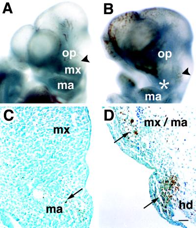
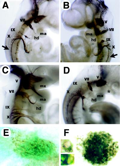
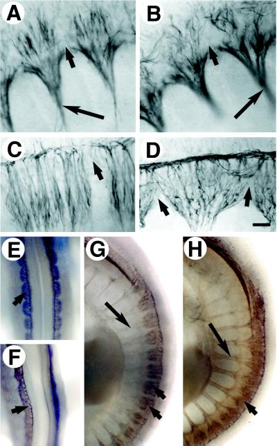
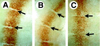
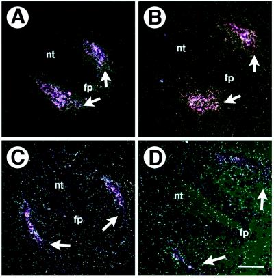
Determinative events in vertebrate embryogenesis appear to require the continuous expression of spatial regulators such as the clustered homeobox genes. The mechanisms that govern long-term patterns of gene expression are not well understood. In Drosophila, active and silent states of developmentally regulated loci are maintained by trithorax and Polycomb group. We have examined the developmental role of a mammalian homolog of trx and putative oncogene, Mll. Knockout mice reveal that Mll is required for maintenance of gene expression early in embryogenesis. Downstream targets of Mll including Hoxa7 are activated appropriately in the absence of Mll but require Mll for sustaining their expression. The Mll-/- phenotype manifests later in development and is characterized by branchial arch dysplasia and aberrant segmental boundaries of spinal ganglia and somites. Thus, Mll represents an essential mechanism of transcriptional maintenance in mammalian development, which functions in multiple morphogenetic processes.


Figures

Figure 1
Figure 1
Defective maintenance of Hoxa7 expression in Mll−/− embryos. Whole mount in situ hybridization of E8.5 and E9 control and Mll−/− embryos (lateral views). (A) Hoxa7 expression in the presomitic mesoderm and neural plate of E8.5 wild-type embryos. (B) Hox-7 expression in E8.5 Mll knockout embryos. Arrows indicate the anterior most extent of Hoxa7 expression. (C) Hoxa7 expression in somites 15–18 and presomitic mesoderm in E9 Mll+/− embryos. (D) Absence of Hoxa7 expression in somites of E9 Mll−/− embryos. Faint expression was detected in Mll−/− embryos proximal to the allantois. Arrows point to the anterior boundary of somite 15 (prospective eleventh vertebrae).
Figure 2
Figure 2
Branchial arch abnormalities in Mll knockout embryos. Gross views and TUNEL staining of control and Mll−/− E10.5 embryos. (A) Wild-type embryo (oblique view), demonstrating normal structures of the first branchial arch, maxillary prominence (MX), the mandibular component (MA), and associated trigeminal ganglia (arrowhead). (B) Mll−/− embryo revealing branchial arch hypoplasia and defects in the development of the maxilla (asterisk). (C) First branchial arch of Mll+/+ embryos. Apoptotic cells are indicated by long arrows. (D) First and second branchial arches of Mll−/− embryos; same magnification as (C). Note the severe hypocellularity and apoptosis in the arch mesenchyme but not the surface ectoderm. op; optic vesicle; HD, hyoid or second branchial arch. (Scale bar = 50 μm.)
Figure 3
Figure 3
Cranial ganglia development in wild-type and mutant embryos at E10 and E10.5. Neurofilament immunostaining of whole embryos and isolated facial ganglia (lateral views). (A) E10 wild-type embryo demonstrating normal morphology of cranial ganglia. (B) E10 Mll−/− embryo displaying condensed morphology of ganglia and present branchial arch structures. Spinal accessory nerves are indicated by arrows. (C) E10.5 Mll+/− embryo showing cranial innervation of the branchial arches. (D) E10.5 Mll−/− cranial ganglia are condensed and innervation of the branchial arches is absent. Defective positioning of the nodose ganglia was noted in only one of six knockout embryos. Note hypoplastic branchial arch development (∗). (E) Flat mount of Mll+/− facial ganglia; blue staining reflects expression of Mll-lacZ marker in cranial ganglia. (F) Mll−/− facial ganglia. (Inset) Magnified view of cells found within the facial ganglia. V, trigeminal; VII, facial; IX, glossopharyngeal; X, vagal nerves.
Figure 4
Figure 4
Defective neurite outgrowth and segmentation of Mll−/− spinal ganglia. Immunohistochemical staining for neurofilament was performed on E10 and E10.5 embryos. (A) Flat mount of E10 Mll+/+ spinal ganglia and motor nerves (long arrow). Short arrows indicate boundaries between somites. (B) E10 Mll−/− spinal ganglia and motor nerves from similar axial level as shown in A. Neurites of knockout spinal ganglia are disorganized but did not cross somite boundaries. (C) E10.5 wild-type spinal ganglia. (D) E10.5 Mll−/− spinal ganglia demonstrating aberrant morphology and loss of segmental restriction. (E) Dorsal view of segmented E10.5 Mll+/+ spinal ganglia at the forelimb level. Short arrows indicate spinal ganglia. (F) Fused E10.5 Mll−/− spinal ganglia. (G) Lateral view of segmented spinal ganglia and spinal nerves in E10.5 Mll+/− embryo. (H) E10.5 Mll−/− embryo displays spinal ganglia fusion. Fusion of spinal ganglia was pronounced less posteriorly. Note the segmented pattern of spinal and motor nerves.
Figure 5
Figure 5
Distribution of EN1-positive cells in E10.5 somites of wild-type and mutant embryos (lateral views). Flat-mounted somites immunostained with EN1/2 antibody. (A) EN1 expression in anterior somites of wild-type embryos; EN1 expression at this stage was in the dermomyotome. Somites shown are at the level of the forelimb. Arrows indicate the somite boundaries. (B) EN1 in posterior somites of Mll−/− embryo. (C) EN1 expression in anterior somites of Mll−/− embryos taken at the level of the forelimb. Note the presence of EN1 positive cells between somites.
Figure 6
Figure 6
Somite architecture and cell death in wild-type and Mll−/− E10.5 embryos. Hematoxylin and eosin staining of lateral sections and TUNEL staining of transverse sections. (A) wild-type somites; note normal morphology of somite epithelium indicated by arrows. (B) Mll−/− somites demonstrating disrupted epithelial boundaries ventrally. (C) Normal cell death in wild-type somites revealed by TUNEL. (D) Cell death in Mll−/− somites; note the relative sparing of spinal ganglia (sg) and neural tube (nt). Arrows indicate areas of apoptosis. (Scale bars = 50 μm.)
Figure 7
Figure 7
Differentiation of somites in wild-type and Mll−/− E10.5 embryos. In situ hybridization of Pax-1 (A and B) and myf-5 (C and D) performed on transverse sections. (A) Pax-1 expression in the sclerotome of wild-type embryo. White arrows show areas of expression. (B) Pax-1 expression in Mll−/− embryo. (C) Myf-5 expression in the dermomyotome of Mll+/+ embryo. (D) Myf-5 expression in Mll−/− embryo. nt, neural tube; fp, floor plate. (Scale bars = 50 μm.)

References

    1. Noden D M. J Neurobiol. 1993;24:248–261. - PubMed
    1. Tam P P L, Trainor P A. Anat Embryol. 1994;189:275–305. - PubMed
    1. Kieny M, Mauger A, Sengel P. Dev Biol. 1972;28:142–161. - PubMed
    1. Beddington R S P, Püschel A W, Rashbass P. In: Postimplantation Development in the Mouse. Chadwick D J, Marsh J, editors. New York: Wiley; 1992. pp. 61–77.
    1. Deschamps J, Wijgerde M. Dev Biol. 1993;156:473–480. - PubMed

Publication types

Substances

LinkOut - more resources