

The optx2 homeobox gene is expressed in early precursors of the eye and activates retina-specific genes
- PMID: 9724757
- PMCID: PMC27948
- DOI: 10.1073/pnas.95.18.10643
The optx2 homeobox gene is expressed in early precursors of the eye and activates retina-specific genes
Abstract
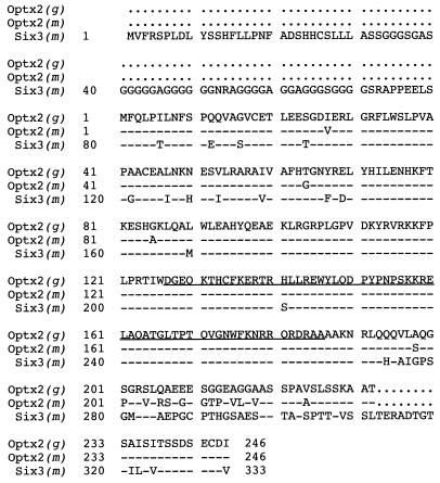
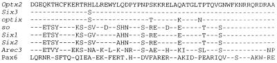
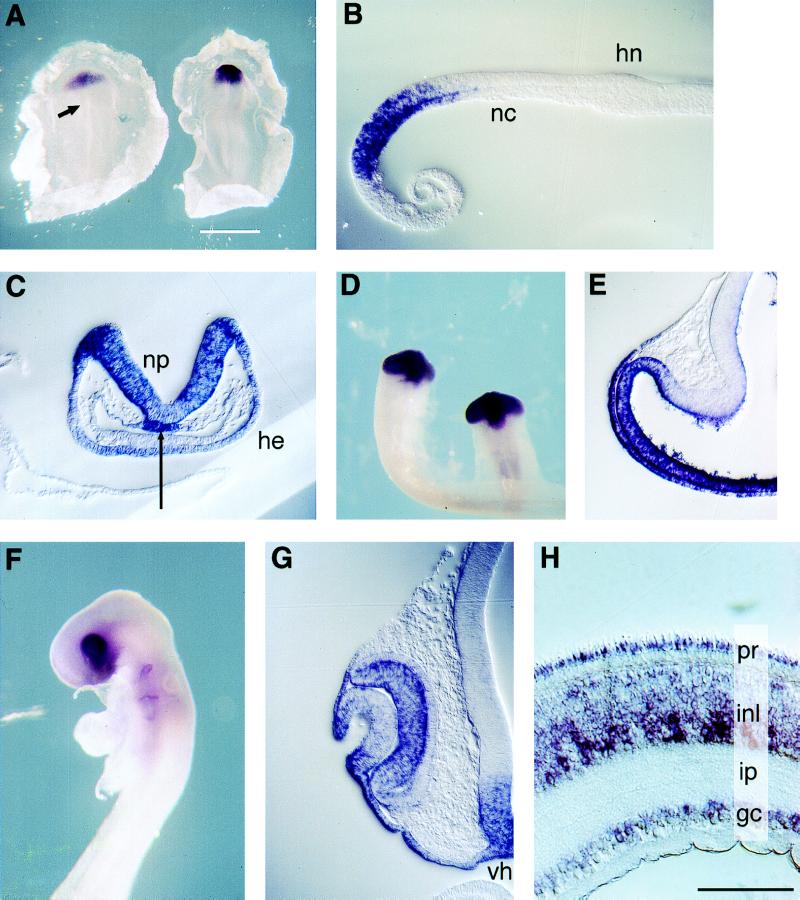
Vertebrate eye development begins at the gastrula stage, when a region known as the eye field acquires the capacity to generate retina and lens. Optx2, a homeobox gene of the sine oculis-Six family, is selectively expressed in this early eye field and later in the lens placode and optic vesicle. The distal and ventral portion of the optic vesicle are fated to become the retina and optic nerve, whereas the dorsal portion eventually loses its neural characteristics and activates the synthesis of melanin, forming the retinal pigment epithelium. Optx2 expression is turned off in the future pigment epithelium but remains expressed in the proliferating neuroblasts and differentiating cells of the neural retina. When an Optx2-expressing plasmid is transfected into embryonic or mature chicken pigment epithelial cells, these cells adopt a neuronal morphology and express markers characteristic of developing neural retina and photoreceptors. One explanation of these results is that Optx2 functions as a determinant of retinal precursors and that it has induced the transdifferentiation of pigment epithelium into retinal neurons and photoreceptors. We also have isolated optix, a Drosophila gene that is the closest insect homologue of Optx2 and Six3. Optix is expressed during early development of the fly head and eye primordia.
Figures
Similar articles
-
Six9 (Optx2), a new member of the six gene family of transcription factors, is expressed at early stages of vertebrate ocular and pituitary development.Mech Dev. 1999 May;83(1-2):155-9. doi: 10.1016/s0925-4773(99)00017-9. Mech Dev. 1999. PMID: 10381575
-
Six3, a medaka homologue of the Drosophila homeobox gene sine oculis is expressed in the anterior embryonic shield and the developing eye.Mech Dev. 1998 Jun;74(1-2):159-64. doi: 10.1016/s0925-4773(98)00055-0. Mech Dev. 1998. PMID: 9651515
-
Six6 (Optx2) is a novel murine Six3-related homeobox gene that demarcates the presumptive pituitary/hypothalamic axis and the ventral optic stalk.Mech Dev. 1999 Jun;84(1-2):31-40. doi: 10.1016/s0925-4773(99)00068-4. Mech Dev. 1999. PMID: 10473118
-
[The myc oncogene and transdifferentiation of the retinal pigment epithelium].J Soc Biol. 2001;195(2):107-13. J Soc Biol. 2001. PMID: 11723821 Review. French.
-
The sine oculis homeobox (SIX) family of transcription factors as regulators of development and disease.Cell Mol Life Sci. 2009 Feb;66(4):565-83. doi: 10.1007/s00018-008-8335-4. Cell Mol Life Sci. 2009. PMID: 18989625 Free PMC article. Review.
Cited by
-
Quantitative Trait Locus Analysis of SIX1-SIX6 With Retinal Nerve Fiber Layer Thickness in Individuals of European Descent.Am J Ophthalmol. 2015 Jul;160(1):123-30.e1. doi: 10.1016/j.ajo.2015.04.001. Epub 2015 Apr 4. Am J Ophthalmol. 2015. PMID: 25849520 Free PMC article.
-
Changes in Rx1 and Pax6 activity at eye field stages differentially alter the production of amacrine neurotransmitter subtypes in Xenopus.Mol Vis. 2007 Jan 26;13:86-95. Mol Vis. 2007. PMID: 17277735 Free PMC article.
-
Comprehensive characterization of the cis-regulatory code responsible for the spatio-temporal expression of olSix3.2 in the developing medaka forebrain.Genome Biol. 2007;8(7):R137. doi: 10.1186/gb-2007-8-7-r137. Genome Biol. 2007. PMID: 17617896 Free PMC article.
-
The homeobox gene Six3 is a potential regulator of anterior segment formation in the chick eye.Dev Biol. 2002 Aug 15;248(2):265-80. doi: 10.1006/dbio.2002.0732. Dev Biol. 2002. PMID: 12167403 Free PMC article.
-
Compartmentalization of vertebrate optic neuroephithelium: external cues and transcription factors.Mol Cells. 2012 Apr;33(4):317-24. doi: 10.1007/s10059-012-0030-5. Epub 2012 Mar 23. Mol Cells. 2012. PMID: 22450691 Free PMC article. Review.
References
Publication types
MeSH terms
Substances
Associated data
- Actions
- Actions
- Actions
Grants and funding
LinkOut - more resources
Full Text Sources
Molecular Biology Databases
Research Materials
Miscellaneous
