. 1998 Sep 1;95(18):10872-7.
doi: 10.1073/pnas.95.18.10872.

Identification of the gene (lgtG) encoding the lipooligosaccharide beta chain synthesizing glucosyl transferase from Neisseria gonorrhoeae

Affiliations

Identification of the gene (lgtG) encoding the lipooligosaccharide beta chain synthesizing glucosyl transferase from Neisseria gonorrhoeae

A Banerjee et al. Proc Natl Acad Sci U S A. .

Abstract

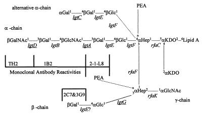
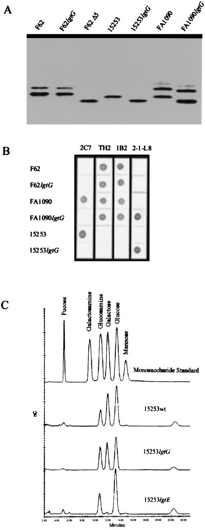
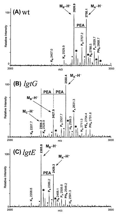
The lipooligosaccharide from Neisseria gonorrhoeae (GC), consists of lipid A, an oligosaccharide core and three branches, alpha, beta, and gamma. We report the cloning of the gene (lgtG, lipooligosaccharide glycosyl transferase G) encoding the glucosyl transferase of GC that initiates the beta chain which consists of a lactosyl moiety. This gene contains a homopolymeric tract of cytidine [poly(C)] and we demonstrate that changes in the number of Cs in poly(C) account for the variation of beta chain expression in different GC strains. Biochemical analyses and mass spectrometry clearly attribute the reactivity of mAb 2C7 to the presence of the lactosyl beta chain. In addition, we demonstrate that in the absence of the lactosyl group, a phosphoethanolamine is added to generate a new antigenic epitope as evidenced by the gain of reactivity to mAb 2-L1-8. These results show that, like the alpha chain, the beta chain of lipooligosaccharide is subject to antigenic variation.


Figures

Figure 1
Figure 1
Structure of LOS from GC. Alternative LOS structures are indicated by dotted lines. The genes that show phase variation are underlined. The positions of bond formation on the sugars are given in small font. “n” denotes a yet undetermined position of bond formation. The epitopes for mAbs used in this study are also shown. The “?” next to lgtE indicates that it is not known whether it plays a direct or indirect role in that particular galactosyl bond formation.
Figure 2
Figure 2
DNA sequence of lgtG locus from GC strain 15253. RBS 1 and RBS 2 indicate two possible ribosome-binding sites (marked by double underlines) corresponding to two alternative translational starts (thick underlines), START 1 and START 2, respectively. poly(C) (black box) indicates the phase variable 11 consecutive Cs. The FA1090 sequence has 11 Cs also. The number of Cs in F62 and 1291 is 12 and the ORF terminates at STOP 2 instead of STOP 1, the putative translational terminator (white box) in 15253/FA1090. In MS11, 10 Cs are present and translation terminates at STOP 3. The locations of the relevant PCR primers are underlined by arrows. The ermC′ cassette was introduced into the indicated BstBI restriction enzyme site (italic font) for insertional inactivation of lgtG ORF. The NdeI sites were used to create pK18-LgtG18ΔNdeI. −10? and −30?, possible −10 consensus and −30 consensus of σ70-binding site (gray boxes).
Figure 3
Figure 3
Involvement of lgtG and its poly(C) tract on LOS biosynthesis of GC. (A) SDS-PAGE analysis of LOS isolated from F62Δ8–1 and its derivatives. The lanes represent LOS isolated from: 1, F62Δ8–1; 2, F62Δ8–1(3G9+); 3, F62Δ8–1(3G9+)lgtG; and 4, F62Δ8–1(3G9+)lgtG transformed with pK18-LgtG18 (Table 1). (B) DNA sequencing gel containing the C lanes of the three derivatives of pK18-LgtG18. (C) Photoreproduction of the region of a GC-agar plate containing spots of various F62Δ8–1-derived cells. The small dots inside the spots represent actual colonies after 24 hr of growth. Rows 1 through 3 represent cells that were transformed with constructs that contained 10 Cs, 12 Cs, and 11 Cs, respectively, in poly(C). Row 4 represents cells that were transformed with N. gonorrhoeae strain 4318 (Table 1) chromosomal DNA. (D) Colony immunoblot of the cells shown in C using mAb 3G9.
Figure 4
Figure 4
Comparison of size, immunoreactivity, and monosaccharide composition of LOSs from lgtG mutants with that from their parent strains and other mutants. (A) Silver-stained SDS-PAGE of different LOS preparations as indicated. (B) Immunoblot of LOSs from wild-type GC strains and their lgtG mutants. mAbs are in columns and GC strains are in rows. The epitopes for the mAbs are indicated in Fig. 1. (C) Monosaccharide analysis of GC LOS by HPAE-PAD. The top chromatogram is of a standard mixture of monosaccharides containing 2 nmol of each component. The other chromatograms are hydrolysates of indicated GC LOSs.
Figure 5
Figure 5
Negative ion MALDI-TOF-MS spectra of O-deacylated LOS from wild-type (A), lgtG knockout (B), and 1gtE (C) mutants of GC 15253. Measured masses (m/z) for all of the major peaks are given in the spectra. Peaks corresponding to major intact O-deacylated LOS glycoforms are denoted as their deprotonated ions (M-H). Several less abundant glycoforms were also observed in the spectra and they have either one less ketodeoxyoctulosonate (Kn), one more phosphate group (Pn), one more hexose (Hn), one more phospho-PEA (PEn), or addition of both phosphate and hexose moieties (PHn) to the major glycoforms. Peaks labeled with (•) and (∗) are water loss peaks and sodium-cationized peaks, respectively.

Similar articles

Cited by

References

    1. World Health Organization. World Health Report 1997. Geneva: W.H.O.; 1997.
    1. Institute of Medicine. Committee on Prevention and Control of Sexually Transmitted Diseases. The Hidden Epidemic: Confronting Sexually Transmitted Diseases. Washington, DC: National Academy Press; 1997.
    1. Schwan E T, Robertson B D, Brade H, van Putten J P M. Mol Microbiol. 1995;15:267–275. - PubMed
    1. van Putten J P M. Mol Microbiol. 1995;16:847–853. - PubMed
    1. Yamasaki R, Kerwood D E, Schneider H, Quinn K P, Griffiss J M, Mandrell R E. J Biol Chem. 1994;269:30345–30351. - PubMed

Publication types

MeSH terms

Substances

Associated data

LinkOut - more resources