. 1998 Sep 1;95(18):10890-5.
doi: 10.1073/pnas.95.18.10890.

Characterization of a calmodulin kinase II inhibitor protein in brain

Affiliations

Characterization of a calmodulin kinase II inhibitor protein in brain

B H Chang et al. Proc Natl Acad Sci U S A. .

Abstract

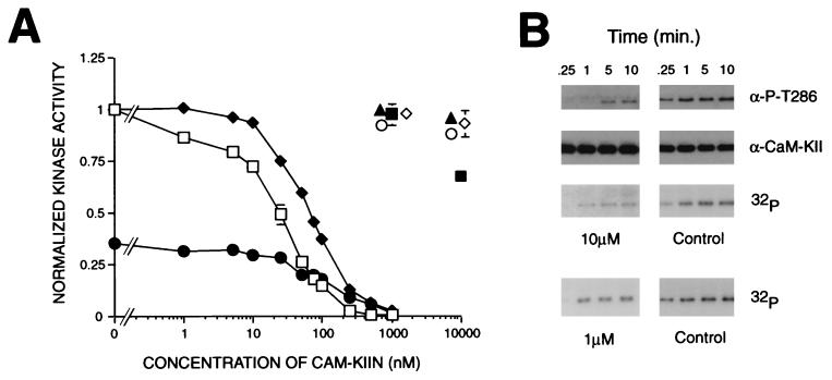
Ca2+/calmodulin-dependent protein kinase II (CaM-KII) regulates numerous physiological functions, including neuronal synaptic plasticity through the phosphorylation of alpha-amino-3-hydroxy-5-methyl-4-isoxazolepropionic acid-type glutamate receptors. To identify proteins that may interact with and modulate CaM-KII function, a yeast two-hybrid screen was performed by using a rat brain cDNA library. This screen identified a unique clone of 1.4 kb, which encoded a 79-aa brain-specific protein that bound the catalytic domain of CaM-KII alpha and beta and potently inhibited kinase activity with an IC50 of 50 nM. The inhibitory protein (CaM-KIIN), and a 28-residue peptide derived from it (CaM-KIINtide), was highly selective for inhibition of CaM-KII with little effect on CaM-KI, CaM-KIV, CaM-KK, protein kinase A, or protein kinase C. CaM-KIIN interacted only with activated CaM-KII (i. e., in the presence of Ca2+/CaM or after autophosphorylation) by using glutathione S-transferase/CaM-KIIN precipitations as well as coimmunoprecipitations from rat brain extracts or from HEK293 cells cotransfected with both constructs. Colocalization of CaM-KIIN with activated CaM-KII was demonstrated in COS-7 cells transfected with green fluorescent protein fused to CaM-KIIN. In COS-7 cells phosphorylation of transfected alpha-amino-3-hydroxy-5-methyl-4-isoxazolepropionic acid-type glutamate receptors by CaM-KII, but not by protein kinase C, was blocked upon cotransfection with CaM-KIIN. These results characterize a potent and specific cellular inhibitor of CaM-KII that may have an important role in the physiological regulation of this key protein kinase.


Figures

Figure 1
Figure 1
Cloning and expression of CaM-KIIN. (A) DNA sequence of CaM-KIIN and deduced amino acid sequence. The underlined region denotes the inhibitory domain. Only part of the 3′ noncoding sequence is shown. (B) Tissue expression of CaM-KIIN message. A Northern blot was performed on poly(A) RNA (Left) and on total RNA (Right). The estimated size of the band is 1.4 kb (arrow). (C) Protein expression of CaM-KIIN. Western blot was performed by using a polyclonal antibody produced to a peptide within the underlined region of CaM-KIIN. (Upper Left) Rat tissues were harvested, and 50 μg of total protein was run per lane. (Upper Right) COS-7 cells were transiently transfected with CaM-KIIN or vector alone and harvested, and 20 μg of soluble protein was run per lane. (Lower) Same as Upper except the antibody was preadsorbed by preincubation with 100 μM of peptide antigen.
Figure 2
Figure 2
Binding of CaM-KII to CaM-KIIN. (A) GST/CaM-KIIN precipitation of CaM-KII. Either recombinant CaM-KIV, CaM-KII, or soluble brain extract was incubated with GST/CaM-KIIN in the presence of Ca2+/CaM or EGTA, precipitated with glutathione-Sepharose, washed, and separated by SDS/PAGE. Samples then were visualized by Coomassie staining or by Western blot with anti-CaM-KII antibody. The positions of CaM-KII (upper arrow) and GST/CaM-KIIN (lower arrow) are indicated. The Western blot of the soluble brain extract precipitation (Right) visualized both the α and β subunits of CaM-KII. (B) Coimmunoprecipitation of CaM-KII and CaM-KIIN. Rat forebrain supernatant was split into 200-μl aliquots and incubated with either protein A Sepharose alone (first two lanes) or with a mAb CaM-KIIα in the presence of 3 mM CaCl2 or 1 mM EGTA. After centrifugation the immune complex was separated by SDS/PAGE and immunoblotted with the CaM-KIIN antibody (Upper Left) or with preadsorption with the antigenic peptide (Upper Right). The membrane then was immunoblotted with the polyclonal CaM-KII Ab showing both the α and β isoforms (Lower). (C) Coimmunoprecipitation of CaM-KIIN with CaM-KII from transfected cells. HEK 293 cells were transiently transfected with CaM-KIIN, CaM-KII (His-282–Arg), or together and subsequently lysed. CaM-KII was immunoprecipitated from the soluble cell extract with 3 mM Ca2+ and separated by SDS/PAGE. The samples then were immunoblotted with anti-CaM-KIIN. (D) Colocalization of active CaM-KII and CaM-KIIN with transfected cells. COS-7 cells were transiently transfected with the indicated combinations of GFP, GFP/CaM-KIIN fusion protein (GFP-KIIN), wild-type CaM-KII, or activated CaM-KII (H282R). Cells were visualized for GFP (Upper) or for activated CaM-KII by using a Thr286 phosphospecific-antibody (Lower).
Figure 3
Figure 3
Inhibition of CaM-KII by CaM-KIIN. (A) Concentration response curves of inhibition of kinase activity by CaM-KIIN and CaM-KIINtide. Autophosphorylated CaM-KII was assayed in the presence of Ca2+/CaM and varying concentrations of GST/CaM-KIIN (⧫) or CaM-KIINtide (□) for its ability to phosphorylate the peptide substrate syntide 2. Ca2+-independent activity (normalized to total activity) also was assayed in the presence of EGTA and CaM-KIINtide (•). Other kinases were assayed with their substrates by using either 0, 1, or 10 μM CaM-KIINtide and normalized to 0 μM CaM-KIINtide: PKC (1 nM, ▴), EGF receptor peptide (Sigma); PKA (100 nM, ○), RII peptide; CaM-KI (20 nM, ◊), and CaM-KIV (40 nM, ■) were activated by CaM-KK and then assayed for their abilities to phosphorylate syntide 2. (B) Effect of CaM-KIINtide on autophosphorylation of CaM-KII. Autophosphorylation reactions were performed at 5°C with 0 (Control), 1 or 10 μM CaM-KIINtide containing either cold ATP or [γ 32P]-ATP, stopped at the indicated times, and blotted with either antiphosphospecific Thr286 antibody (gift from L. Griffith, Brandeis University, Waltham, MA) or polyclonal CaM-KII antibody, or visualized by 32P incorporation, respectively.
Figure 4
Figure 4
Effect of CaM-KIIN on GluR1 phosphorylation by CaM-KII and PKC. (A) Kinase reactions, performed at 30°C for 20 min with the indicated mixtures of 0.2 mM [γ 32P]-ATP, 20 nM PKC catalytic fragment (PKM, Calbiochem) or 20 nM CaM-KII, 4 μM GST or 4 μM GST/GluR1816–889 with or without 1 μM CaM-KIINtide, were separated by SDS/PAGE and visualized by autoradiography. GST/GluR1816–889 migrated as a doublet. (B) HEK 293 cells, transfected with GluR1 without or with cotransfected CaM-KII (H282R) were 32P-labeled. Some cells without CaM-KII transfection were stimulated with phorbol 12-tetradecanoate 13-acetate to activate PKC for 20 min. Cells were harvested, and GluR1 was immunoprecipitated, analyzed by SDS/PAGE/autoradiography (Upper), and subjected to GluR1 Western blot (Lower).

References

    1. Braun A P, Schulman H. Annu Rev Physiol. 1995;57:417–445. - PubMed
    1. Barria A, Muller D, Derkach V, Griffith L C, Soderling T R. Science. 1997;276:2042–2045. - PubMed
    1. Pettit D L, Perlman S, Malinow R. Science. 1994;266:1881–1885. - PubMed
    1. Lledo P M, Hjelmstad G O, Mukherji S, Soderling T R, Malenka R C, Nicoll R A. Proc Natl Acad Sci USA. 1995;92:11175–11179. - PMC - PubMed
    1. Soderling T R. Biochem Biophys Acta. 1996;1297:131–138. - PubMed

Publication types

MeSH terms

Substances

Associated data