. 1998 Sep 1;95(18):10902-7.
doi: 10.1073/pnas.95.18.10902.

Mental chronometry using latency-resolved functional MRI

Affiliations

Mental chronometry using latency-resolved functional MRI

R S Menon et al. Proc Natl Acad Sci U S A. .

Abstract

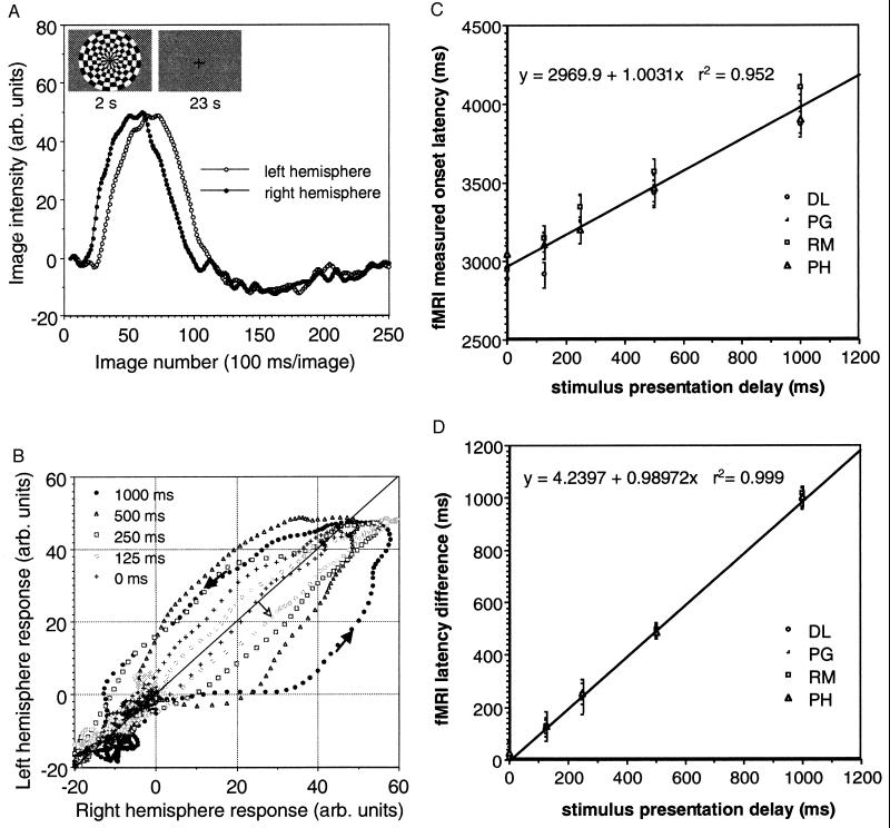
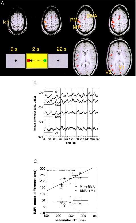
Vascular responses to neural activity are exploited as the basis of a number of brain imaging techniques. The vascular response is thought to be too slow to resolve the temporal sequence of events involved in cognitive tasks, and hence, imaging studies of mental chronometry have relied on techniques such as the evoked potential. Using rapid functional MRI (fMRI) of single trials of two simple behavioral tasks, we demonstrate that while the microvascular response to the onset of neural activity is delayed consistently by several seconds, the relative timing between the onset of the fMRI responses in different brain areas appears preserved. We examined a number of parameters that characterize the fMRI response and determined that its onset time is best defined by the inflection point from the resting baseline. We have found that fMRI onset latencies determined in this manner correlate well with independently measurable parameters of the tasks such as reaction time or stimulus presentation time and can be used to determine the origin of processing delays during cognitive or perceptual tasks with a temporal accuracy of tens of milliseconds and spatial resolution of millimeters.


Figures

Figure 1
Figure 1
Hemifield experiment. (A) Typical fMRI response from left and right hemispheres of V1 because of a 500-ms offset between left and right side checkerboard presentation of a single subject (Inset and see Materials and Methods). (B) Lissajous plot of left-hemisphere fMRI signal vs. right-hemisphere fMRI signal in V1 for a single subject and all presentation delays. Time evolution is indicated by the dark arrowheads. Phase delays during onset appear on or below the diagonal (right hemisphere leads left since left hemifield appears first), while phase delays on the falling side of the response appear above the diagonal. (C) Plot of fMRI onset delay for left hemisphere (see Materials and Methods) vs. actual presentation delay. (D) Plot of fMRI latency between hemispheres referenced to the onset of the right-hemisphere activity (see Materials and Methods) vs. actual presentation delay.
Figure 2
Figure 2
Visuomotor reaction-time experiment. (A) Activation maps generated by using the cross-correlation approach of the areas invoked by the visuomotor task in a single subject. The activation maps, derived from EPI images with 1-cm slice thicknesses, have been interpolated onto the much thinner anatomic slices. preM, premotor area; M1, primary motor area; SMA, supplementary motor area; V5, motion-sensitive area of visual cortex; V1, primary visual cortex. These areas have been determined anatomically. (B) fMRI responses for 10 trials after bandpass filtering and Fourier interpolation in the four areas chosen for analysis (see Materials and Methods). The visual stimulus that cued subject motor response is shown as an Inset along with the direction of cursor movement. (C) Plot of fMRI onset delay between V1 and SMA (see Materials and Methods) vs. measured reaction time from kinematic trace as well as the onset differences between SMA and M1 vs. RT. Note there are eight points, as two of the six subjects were repeated to assess consistency. In order of increasing kinematic RT, the subjects are LS(2), BG, JD, ML, DB(2), LS(1), DB(1), and ET. The number in parentheses denotes the session number for those scanned twice. Note that RT improved on the second session in both these subjects, demonstrating some practice effect.

Similar articles

Cited by

References

    1. Posner M I. Chronometric Explorations of Mind. London: Oxford Univ. Press; 1978.
    1. Georgopoulos A P, Pellizzer G. Neuropsychologia. 1995;33:1531–1547. - PubMed
    1. Kok A. Biol Psychol. 1997;45:19–56. - PubMed
    1. Regan D. Human Brain Electrophysiology: Evoked Potentials and Evoked Magnetic Fields in Science and Medicine. Amsterdam: Elsevier; 1989.
    1. Celesia G G, Bodis-Wollner I, Chatrian G E, Harding G F, Sokol S, Spekreijse H. Electroencephalograph Clin Neurophysiol. 1993;87:421–436. - PubMed

Publication types