

Evidence for the presence of 5S rRNA in mammalian mitochondria
- PMID: 9725900
- PMCID: PMC25503
- DOI: 10.1091/mbc.9.9.2375
Evidence for the presence of 5S rRNA in mammalian mitochondria
Abstract
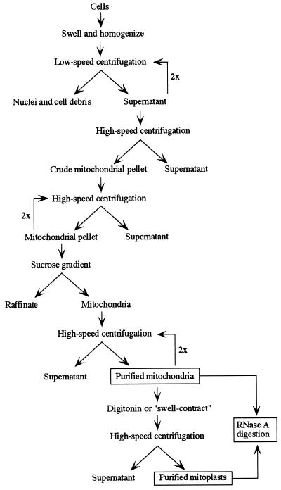
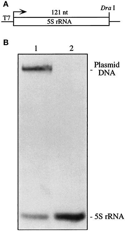
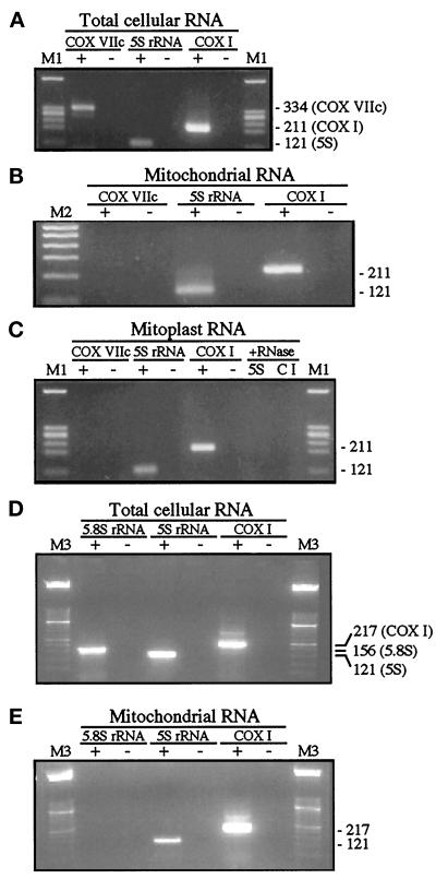
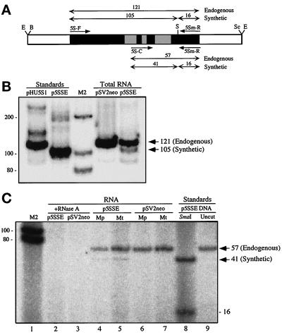
Mammalian mitochondrial ribosomes contain two prokaryotic-like rRNAs, 12S and 16S, both encoded by mitochondrial DNA. As opposed to cytosolic ribosomes, however, these ribosomes are not thought to contain 5S rRNA. For this reason, it has been unclear whether 5S rRNA, which can be detected in mitochondrial preparations, is an authentic organellar species imported from the cytosol or is merely a copurifying cytosol-derived contaminant. We now show that 5S rRNA is tightly associated with highly purified mitochondrial fractions of human and rat cells and that 5S rRNA transcripts derived from a synthetic gene transfected transiently into human cells are both expressed in vivo and present in highly purified mitochondria and mitoplasts. We conclude that 5S rRNA is imported into mammalian mitochondria, but its function there still remains to be clarified.
Figures
Similar articles
-
Biological significance of 5S rRNA import into human mitochondria: role of ribosomal protein MRP-L18.Genes Dev. 2011 Jun 15;25(12):1289-305. doi: 10.1101/gad.624711. Genes Dev. 2011. PMID: 21685364 Free PMC article.
-
Existence of nuclear-encoded 5S-rRNA in bovine mitochondria.FEBS Lett. 1994 Jan 31;338(2):137-42. doi: 10.1016/0014-5793(94)80351-x. FEBS Lett. 1994. PMID: 7508404
-
Abundant 5S rRNA-like transcripts encoded by the mitochondrial genome in amoebozoa.Eukaryot Cell. 2010 May;9(5):762-73. doi: 10.1128/EC.00013-10. Epub 2010 Mar 19. Eukaryot Cell. 2010. PMID: 20304999 Free PMC article.
-
Epitranscriptomics of Mammalian Mitochondrial Ribosomal RNA.Cells. 2020 Sep 27;9(10):2181. doi: 10.3390/cells9102181. Cells. 2020. PMID: 32992603 Free PMC article. Review.
-
Mitochondrial noncoding RNA transport.BMB Rep. 2017 Apr;50(4):164-174. doi: 10.5483/bmbrep.2017.50.4.013. BMB Rep. 2017. PMID: 28115039 Free PMC article. Review.
Cited by
-
Mitochondrial enzyme rhodanese is essential for 5 S ribosomal RNA import into human mitochondria.J Biol Chem. 2010 Oct 1;285(40):30792-803. doi: 10.1074/jbc.M110.151183. Epub 2010 Jul 27. J Biol Chem. 2010. PMID: 20663881 Free PMC article.
-
5S ribosomal RNA database Y2K.Nucleic Acids Res. 2000 Jan 1;28(1):166-7. doi: 10.1093/nar/28.1.166. Nucleic Acids Res. 2000. PMID: 10592212 Free PMC article.
-
Isolation of mitochondria from cells and tissues.Methods Cell Biol. 2020;155:3-31. doi: 10.1016/bs.mcb.2019.10.002. Epub 2019 Dec 10. Methods Cell Biol. 2020. PMID: 32183964 Free PMC article.
-
Biological significance of 5S rRNA import into human mitochondria: role of ribosomal protein MRP-L18.Genes Dev. 2011 Jun 15;25(12):1289-305. doi: 10.1101/gad.624711. Genes Dev. 2011. PMID: 21685364 Free PMC article.
-
Correction of the consequences of mitochondrial 3243A>G mutation in the MT-TL1 gene causing the MELAS syndrome by tRNA import into mitochondria.Nucleic Acids Res. 2011 Oct;39(18):8173-86. doi: 10.1093/nar/gkr546. Epub 2011 Jun 30. Nucleic Acids Res. 2011. PMID: 21724600 Free PMC article.
References
-
- Adhya S, Ghosh T, Das A, Bera SK, Mahapatra S. Role of an RNA-binding protein in import of tRNA into Leishmania mitochondria. J Biol Chem. 1997;272:21396–21402. - PubMed
-
- Anderson S, et al. Sequence and organization of the human mitochondrial genome. Nature. 1981;290:457–465. - PubMed
-
- Attardi B, Cravioto B, Attardi G. Membrane bound ribosomes in HeLa cells. I. Their proportion to total cell ribosomes and their association with messenger RNA. J Mol Biol. 1969;44:47–70. - PubMed
-
- Attardi G, Montoya J. Analysis of human mitochondrial RNA. Methods Enzymol. 1983;97:435–469. - PubMed
Publication types
MeSH terms
Substances
Grants and funding
LinkOut - more resources
Full Text Sources
Other Literature Sources
Research Materials
