. 1998 Sep;9(9):2383-92.
doi: 10.1091/mbc.9.9.2383.

Binding of insulin-like growth factor (IGF)-binding protein-5 to smooth-muscle cell extracellular matrix is a major determinant of the cellular response to IGF-I

Affiliations
Free PMC article

Binding of insulin-like growth factor (IGF)-binding protein-5 to smooth-muscle cell extracellular matrix is a major determinant of the cellular response to IGF-I

A Parker et al. Mol Biol Cell. 1998 Sep.
Free PMC article

Abstract

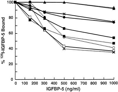
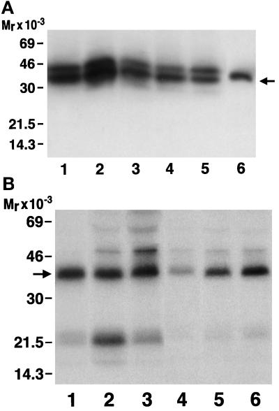
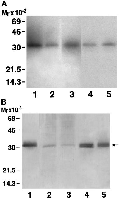
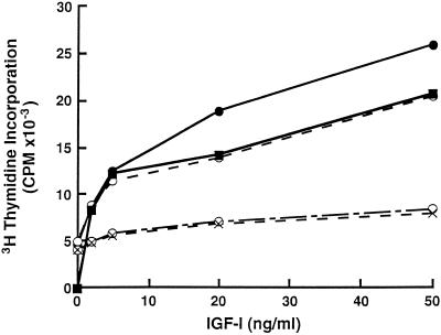
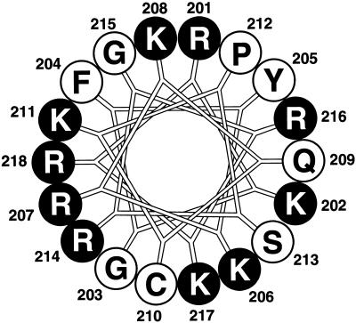
Insulin-like growth factor-binding protein-5 (IGFBP-5) has been shown to bind to fibroblast extracellular matrix (ECM). Extracellular matrix binding of IGFBP-5 leads to a decrease in its affinity for insulin-like growth factor-I (IGF-I), which allows IGF-I to better equilibrate with IGF receptors. When the amount of IGFBP-5 that is bound to ECM is increased by exogenous addition, IGF-I's effect on fibroblast growth is enhanced. In this study we identified the specific basic residues in IGFBP-5 that mediate its binding to porcine smooth-muscle cell (pSMC) ECM. An IGFBP-5 mutant containing alterations of basic residues at positions 211, 214, 217, and 218 had the greatest reduction in ECM binding, although three other mutants, R214A, R207A/K211N, and K202A/R206N/R207A, also had major decreases. In contrast, three other mutants, R201A/K202N/R206N/R208A, and K217N/R218A and K211N, had only minimal reductions in ECM binding. This suggested that residues R207 and R214 were the most important for binding, whereas alterations in K211 and R218, which align near them, had minimal effects. To determine the effect of a reduction in ECM binding on the cellular replication response to IGF-I, pSMCs were transfected with the mutant cDNAs that encoded the forms of IGFBPs with the greatest changes in ECM binding. The ECM content of IGFBP-5 from cultures expressing the K211N, R214A, R217A/R218A, and K202A/R206N/R207A mutants was reduced by 79.6 and 71.7%, respectively, compared with cells expressing the wild-type protein. In contrast, abundance of the R201A/K202N/R206N/R208A mutant was reduced by only 14%. Cells expressing the two mutants with reduced ECM binding had decreased DNA synthesis responses to IGF-I, but the cells expressing the R201A/K202N/R206N/R208A mutant responded well to IGF-I. The findings suggest that specific basic amino acids at positions 207 and 214 mediate the binding of IGFBP-5 to pSMC/ECM. Smooth-muscle cells that constitutively express the mutants that bind weakly to ECM are less responsive to IGF-I, suggesting that ECM binding of IGFBP-5 is an important variable that determines cellular responsiveness.


Figures

Figure 1
Figure 1
Competition for binding to the ECM between native IGFBP-5 and IGFBP-5 mutants. The IGFBP-5 mutants listed in Table 1 were added in increasing concentrations with 125I-IGFBP-5 and incubated with Immobilon membranes that contained the ECM extracts. The binding reaction was carried out as described in MATERIALS AND METHODS. Specific binding was determined by subtracting the counts bound in the presence of 50 μg/ml IGFBP-5 from the total counts per minute bound. This was consistently <15% of total binding. Each point represents the mean of quadruplicate determinations from three independent experiments. The individual mutants that were added were as follows: K211N/R214A/K217/R218A (▴- - -▴); K202A/K206A/R207N (▴—▴); K201A/K202A/R206A/K208N (○- - -○); R217A/R218A (▪—▪); R214A (×—×); native IGFBP-5 (×- - -×); K211N (□—□); R207A/K211N (•—•); R201A/K202N (▪- - -▪).
Figure 2
Figure 2
Relative abundance of IGFBP-5 in the cell lysates (A) and conditioned medium (B) of pSMCs that are constitutively expressing IGFBP-5 or IGFBP-5 mutants. (A) pSMC cell lysates (50 μl) from each cell line were electrophoresed and transferred to filters that were analyzed by ligand blotting using 125I-IGF-I. Lane 1, R202A/R206A/R207A; lane 2, wild-type IGFBP-5; lane 3, R201A/K202N/R206A/R208A; lane 4, K211N/R214A/K217A/R218A; lane 5, mock-transfected cells; lane 6, pure IGFBP-5 standard (10 ng). The arrow denotes the position of intact, purified IGFBP-5. (B) Radiolabeled conditioned medium was collected from the cultures expressing mutants shown in A for 6 h. One milliliter was analyzed by immunoprecipitation using anti-IGFBP-5 antiserum. The lane positions are as follows: lane 1, K202A/R206A/R207A; lane 2, native IGFBP-5; lane 3, K211N/R214A/K217A/R218A; lane 4, nontransfected; lane 5, R201A/K202N/R206A/R208A; lane 6, mock transfected. The arrow denotes the position of intact IGFBP-5. The 22-kDa band represents an IGFBP-5 fragment.
Figure 3
Figure 3
(A) Ligand blot of IGFBPs in the ECM. The cultures that were secreting the various forms of IGFBP-5 shown in Figure 2 were used. ECM extracts were prepared from confluent cultures as described previously and analyzed by SDS-PAGE followed by Western ligand blotting. The amount of ECM that was loaded in each lane was determined by the relative abundance of IGFBP-5 that was produced by each cell line shown in Figure 2A. Lane 1, wild-type IGFBP-5; lane 2, R201A/K202A/R207A; lane 3, R201A/K202A/R206N/R208A; lane 4, K211N/R214A/K217A/R218A; lane 5, mock transfected culture. The 31-kDa band that is shown represents intact IGFBP-5. No fragment is detected because IGFBP-5 within ECM is protected from proteolysis (Jones et al., 1993a). The experiment was repeated three times with similar results. (B) Immunoblot of IGFBP-5 in the ECM. The cultures that were analyzed in A were also analyzed by immunoblotting as described in MATERIALS AND METHODS. Lane 1, wild-type IGFBP-5; lane 2, K211N/R214A/K217A/R218A; lane 3, R201A/K202A/R207A; lane 4, mock transfected; lane 5, R201A/K202A/R206N/R208A. The arrow denotes the position of IGFBP-5.
Figure 4
Figure 4
DNA synthesis response of cells expressing mutant forms of IGFBP-5. To determine whether the cells that were transfected with mutant forms of IGFBP-5 that resulted in less IGFBP-5 in their extracellular matrix would respond similarly to IGF-I, confluent, quiescent cultures were exposed to increasing concentrations of IGF-I for 36 h in DMEM containing 0.2% PPP. After 36 h, [3H]thymidine incorporation into DNA was determined as described in MATERIALS AND METHODS. Each point represents the mean of triplicate determinations. The cultures expressing the wild-type IGFBP-5 (•—•) responded to IGF-I better than the mock-transfected cultures (▪—▪). Those synthesizing the R201A/K202N/K206N/K208N (○—○) mutant responded equally well. In contrast, the cultures synthesizing the K202A/K206A/K207A (○- · -○) or K211N/R214A/K217N/R218A (×- - -×) mutants had attenuated responses to IGF-I. The experiment was repeated three times with similar results.
Figure 5
Figure 5
Helical wheel alignment of the region of IGFBP-5 between amino acids 201 and 218. The basic residues are shown in black.

Similar articles

Cited by

References

    1. Arai A, Busby WH, Clemmons DR. Binding of insulin-like growth factor I or II to IGF binding protein 2 enables it to bind to heparin or extracellular matrix. Endocrinology. 1996a;137:4571–4575. - PubMed
    1. Arai T, Clarke J, Parker A, Busby WH, Clemmons DR. Effect of substitution of specific amino acids in insulin-like growth factor binding protein-5 on heparin binding and its change in affinity for IGF-I in response to heparin. J Biol Chem. 1996b;271:6099–6106. - PubMed
    1. Blat C, Villaudy J, Binoux M. In vivo proteolysis of serum insulin-like growth factor binding protein-3 results in minimal availability of IGF to target cells. J Clin Invest. 1994;93:2286–2290. - PMC - PubMed
    1. Borstein P, Sage EH. Thrombospondins. Methods Enzymol. 1994;245:62–85. - PubMed
    1. Camacho-Hubner C, Busby WH, McCusker RH, Wright G, Clemmons DR. Identification of the forms of insulin-like growth factor binding proteins produced by human fibroblasts and the mechanisms that regulate their secretion. J Biol Chem. 1992;267:11949–11956. - PubMed

Publication types

MeSH terms

Substances

LinkOut - more resources