. 1998 Sep;9(9):2407-22.
doi: 10.1091/mbc.9.9.2407.

Partially processed pre-rRNA is preserved in association with processing components in nucleolus-derived foci during mitosis

Affiliations
Free PMC article

Partially processed pre-rRNA is preserved in association with processing components in nucleolus-derived foci during mitosis

M Dundr et al. Mol Biol Cell. 1998 Sep.
Free PMC article

Abstract

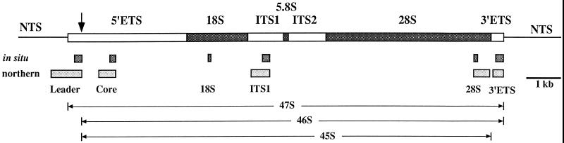
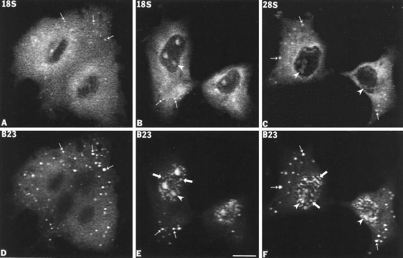
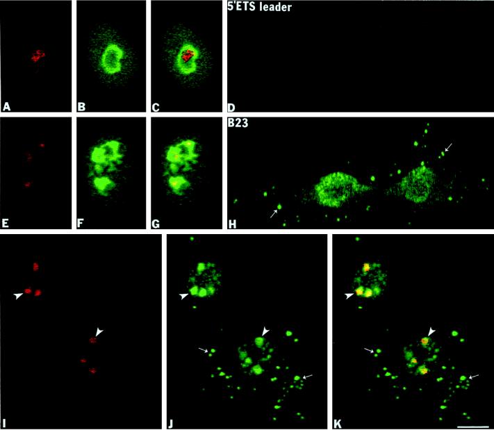
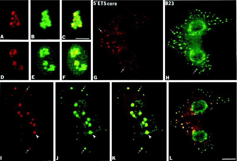
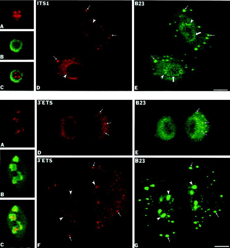
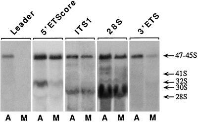
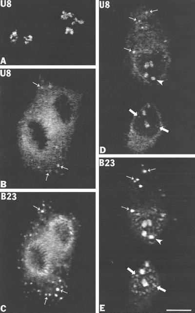
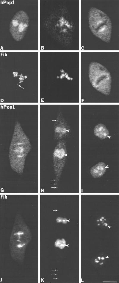
Previous studies showed that components implicated in pre-rRNA processing, including U3 small nucleolar (sno)RNA, fibrillarin, nucleolin, and proteins B23 and p52, accumulate in perichromosomal regions and in numerous mitotic cytoplasmic particles, termed nucleolus-derived foci (NDF) between early anaphase and late telophase. The latter structures were analyzed for the presence of pre-rRNA by fluorescence in situ hybridization using probes for segments of pre-rRNA with known half-lives. The NDF did not contain the short-lived 5'-external transcribed spacer (ETS) leader segment upstream from the primary processing site in 47S pre-rRNA. However, the NDF contained sequences from the 5'-ETS core, 18S, internal transcribed spacer 1 (ITS1), and 28S segments and also had detectable, but significantly reduced, levels of the 3'-ETS sequence. Northern analyses showed that in mitotic cells, the latter sequences were present predominantly in 45S-46S pre-rRNAs, indicating that high-molecular weight processing intermediates are preserved during mitosis. Two additional essential processing components were also found in the NDF: U8 snoRNA and hPop1 (a protein component of RNase MRP and RNase P). Thus, the NDF appear to be large complexes containing partially processed pre-rRNA associated with processing components in which processing has been significantly suppressed. The NDF may facilitate coordinated assembly of postmitotic nucleoli.


Figures

Figure 1
Figure 1
Diagram of human 47S pre-rRNA transcription region (boxed) and adjacent nontranscribed spacers (NTS; single lines). The coding regions for 18S, 5.8S, and 28S rRNAs are designated by stippled areas, and the transcribed spacer regions are designated by open areas. The arrow indicates the position of the human primary processing site (+414). The sequence locations of the biotin-labeled antisense riboprobes for in situ hybridization (dark stippled boxes) and fluorescein-labeled DNA probes for Northern analysis (gray stippled boxes) are shown below the 47S diagram. The sequence locations of the major high molecular weight pre-rRNA precursors (45S–47S) are shown at the bottom of the diagram.
Figure 2
Figure 2
Sequences from 18S and 28S rRNAs are present in the NDF in CMT3 cells. 18S and 28S rRNAs were detected by fluorescence in situ hybridization using a biotin-labeled antisense riboprobe. (A) Anaphase cells labeled with an anti-18S rRNA probe. The in situ hybridization signal is diffusely distributed in the cytoplasm, but distinct bright foci predominate over the general cytoplasmic labeling. These bright foci (A, small arrows) colocalize with signal for protein B23 in the NDF (D, small arrows). (B) Late telophase cells labeled with the anti-18S rRNA probe. The signal is present in reforming postmitotic nucleoli (B, arrowheads) and in the cytoplasm with detectable labeling of NDF (B, small arrows) where it colocalizes with protein B23 (E, small arrows). (C) In early telophase cells, the signal for 28S rRNA is present predominantly over the cytoplasm with detectable NDF (C, small arrows), which colocalized with protein B23 (F, small arrows) as well as in postmitotic nucleoli (C and F, arrowheads). PNBs were also detectable with the anti-B23 antibody (E and F, large arrows). Bar, 10 μm.
Figure 3
Figure 3
The 5′-ETS leader pre-rRNA is not found in the NDF of CMT3 cells. The 5′-ETS leader pre-rRNA was detected by fluorescence in situ hybridization using an antisense biotinylated riboprobe (nucleotides +195/+424). (A–C) Interphase cells labeled with the 5′-ETS probe and anti-B23 antibody. In the nucleoli of these cells the 5′-ETS leader is localized to a restricted area (A) compared with the region labeled with anti-B23 antibody (B), which is clearly shown on the overlay (C). (E–G) Early prophase cells. In these cells the 5′-ETS leader segment is detectable in restricted areas in the disintegrating nucleoli in early (E) compared with protein B23 labeling (F) in the superimposed picture (G). (D and H) Anaphase cells colabeled with the 5′-ETS leader probe and anti-B23 antibody. In these cells the signal from the 5′-ETS leader was reduced to background levels (D) but showing the presence of protein B23 in the perichromosomal region and in NDF (H, small arrows). (I–K) Late telophase cells colabeled with the 5′-ETS leader probe and anti-B23 antibody. The 5′-ETS leader was detected only in postmitotic transcriptionally reactivated nucleoli (I, arrowheads) where it colocalized with protein B23 (J, arrowheads; K, yellow in superimposed picture). Bar, 10 μm.
Figure 4
Figure 4
The 5′-ETS core of pre-rRNA is present in the NDF of CMT3 cells. The 5′-ETS core pre-rRNA was detected by fluorescence in situ hybridization using a biotinylated antisense riboprobe (nucleotides +1272/+1446). (A–C) Interphase cells probed with the 5′-ETS core probe and with anti-B23 antibody. The 5′-ETS core segment is localized in the central regions of interphase nucleoli (A) compared with overall nucleolar labeling of protein B23 (B) and yellow areas of the superimposed picture (C). (D–F) In early prophase a similar labeling pattern of 5′-ETS signal (D) is seen when colocalized with protein B23 (E) and in the superimposed picture (F). (G and H) During anaphase the 5′-ETS core segment is enriched in the perichromosomal region and in NDF (G, small arrows) where it colocalizes with protein B23 (H, small arrows; L, yellow in overlay). Note that more NDF appeared to be labeled by immunofluorescence than by in situ hybridization because the signal faded more rapidly in the latter case. (I–K) In late telophase the 5′-ETS core segment is present mostly in reformed nucleoli (I, arrowhead) and in persisting cytoplasmic NDF (I, small arrows) where it colocalizes with protein B23 (J, arrowhead, small arrows; K, yellow in superimposed picture). Bar, 10 μm.
Figure 6
Figure 6
The 3′-ETS pre-rRNA is present in the NDF of CMT3 cells. The 3′-ETS pre-rRNA segment was detected by fluorescence in situ hybridization with a biotinylated antisense riboprobe (nucleotides +13,111/+13,343). (A–C) Labeling of interphase cells with the 3′-ETS probe and the anti-B23 antibody. The 3′-ETS signal was present in restricted intranucleolar areas (A) in contrast to general nucleolar staining with anti-B23 antibody as seen in panel B and in the overlay (C). (D and E) In anaphase cells the 3′-ETS signal is detectable throughout the cytoplasm with several brighter foci (D, small arrows) that were identified as the NDF by colocalization with protein B23 (E, small arrows). (F and G) In late telophase cells the 3′-ETS signal reappeared in the reformed nucleoli (F, arrowheads), but the signal is also present in persisting cytoplasmic NDF (F, small arrows) where it colocalizes with protein B23 (G, small arrows). Bar, 10 μm.
Figure 7
Figure 7
Northern blot analysis of pre-rRNA transcripts. Equal amounts of total RNA from asynchronous (A) or mitotic (M) CMT3 cells were separated on multiple lanes of 0.6% agarose formaldehyde gels. The RNA was transferred to nylon membranes and probed with the fluorescein-labeled probes complementary to segments of 47S pre-rRNA as indicated above each pair of lanes (see Figure 1). The positions of major pre-rRNA species and 28S rRNA are indicated on the right.
Figure 8
Figure 8
U8 snoRNA is present in nucleolus-derived foci (NDF) in CMT3 cells during mitosis. U8 snoRNA was detected by fluorescence in situ hybridization using a biotin-labeled antisense riboprobe. (A) Interphase cells hybridized with anti-U8 snoRNA probe. Labeling is observed exclusively in intranucleolar subdomains. (B and C). Labeling with U8 probe and with anti-B23 antibody in anaphase cells. The U8 snoRNA in situ hybridization signal is dispersed throughout the cytoplasm with some enrichment in the perichromosomal region (B) and in NDF (B, small arrows) where it colocalizes with protein B23 (C, small arrows). (D and E) During late telophase U8 snoRNA is visible inside of reforming nucleoli (D, arrowheads) but is also weakly detectable in prenucleolar bodies (D, large arrows) and in several cytoplasmic NDF (D, small arrows) that colocalize with protein B23 (E, small arrows). Bar, 10 μm.
Figure 9
Figure 9
The hPop1 protein subunit of human RNase P and RNase MRP is present in NDF in HeLa cells during mitosis. (A and D) Labeling of interphase cell with anti-hPop1 and the S4 autoimmune antifibrillarin serum. The hPop1 protein was predominantly localized in nucleoli with weak nucleoplasmic staining whereas fibrillarin was present in the nucleolar DFC and in a coiled body (D, long arrow). (B and E) In early prophase the hPop1 protein (B) and fibrillarin (E) disperse from disintegrating nucleolar bodies. (C and F) In metaphase the hPop1 protein is found highly concentrated at the external central portion of chromosome periphery (C), whereas fibrillarin is concentrated in the entire perichromosomal region of the metaphase plate (F). (G and J) In late anaphase the hPop1 protein was seen associated with chromatids (G) where it colocalizes with fibrillarin (J), but hPop1 was also seen inside of mitotic spindle (G). (H and K) In late telophase when the nuclear envelope is formed, hPop1 protein is concentrated in the postmitotic nuclei but it is not detectable in newly reformed nucleoli (H, arrowheads) where fibrillarin is prominantly localized (K, arrowheads). In addition, both proteins colocalized in NDF in the cytoplasm (H, K, small arrows). (I and L) In late telophase/early G1 phase the hPop1 protein reenters nucleoli (I) and is colocalized with fibrillarin (L). Bar, 10 μm.

Similar articles

Cited by

References

    1. Beltrame M, Tollervey D. Base pairing between U3 and the pre-ribosomal RNA is required for 18S rRNA synthesis. EMBO J. 1995;14:4350–4356. - PMC - PubMed
    1. Beven AF, Lee R, Razaz M, Leader DJ, Brown JWS, Shaw PJ. The organization of ribosomal RNA processing correlates with the distribution of nucleolar snRNAs. J Cell Sci. 1996;109:1241–1251. - PubMed
    1. Bootsma D, Budke L, Vos O. Studies on synchronous division of tissue culture cells initiated by excess thymidine. Exp Cell Res. 1964;33:301–309. - PubMed
    1. Chamberlain J, Pagan-Ramos E, Kindelberger DW, Engelke D. An RNase P RNA subunit mutation affects ribosomal RNA processing. Nucleic Acids Res. 1996;24:3158–3166. - PMC - PubMed
    1. Chu S, Archer RH, Zengel JM, Lindahl L. The RNA of RNase MRP is required for normal processing of ribosomal RNA. Proc Natl Acad Sci USA. 1994;91:659–663. - PMC - PubMed

Publication types

LinkOut - more resources