. 1998 Sep;9(9):2423-37.
doi: 10.1091/mbc.9.9.2423.

A novel synaptobrevin/VAMP homologous protein (VAMP5) is increased during in vitro myogenesis and present in the plasma membrane

Affiliations
Free PMC article

A novel synaptobrevin/VAMP homologous protein (VAMP5) is increased during in vitro myogenesis and present in the plasma membrane

Q Zeng et al. Mol Biol Cell. 1998 Sep.
Free PMC article

Abstract

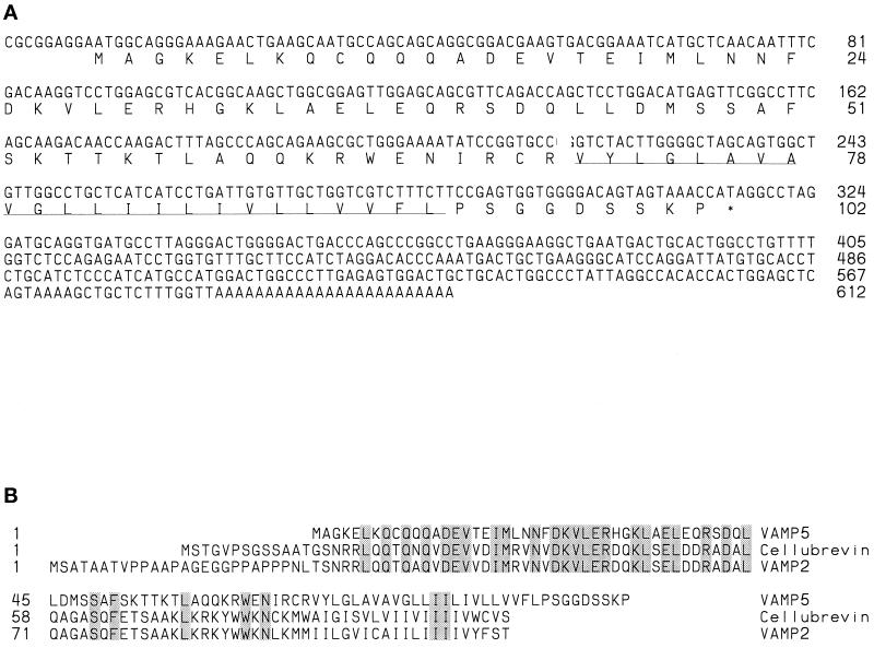
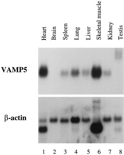
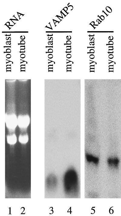
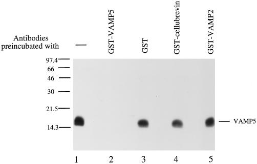
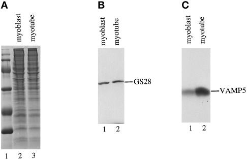
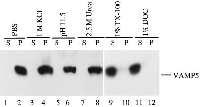
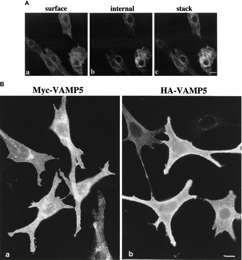
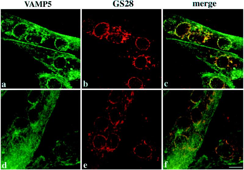
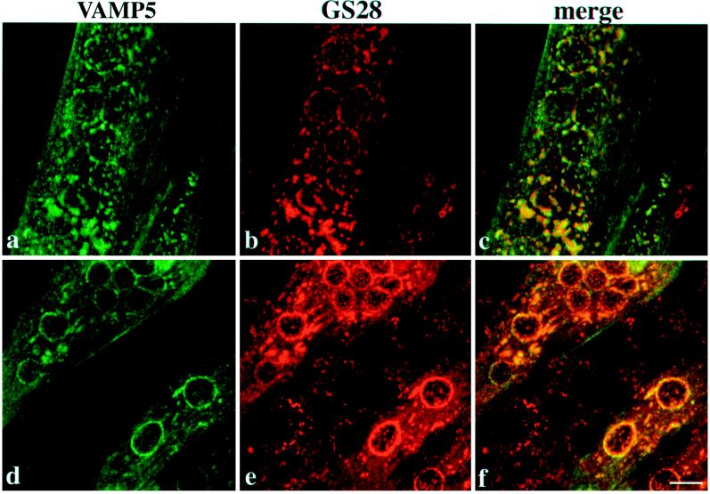
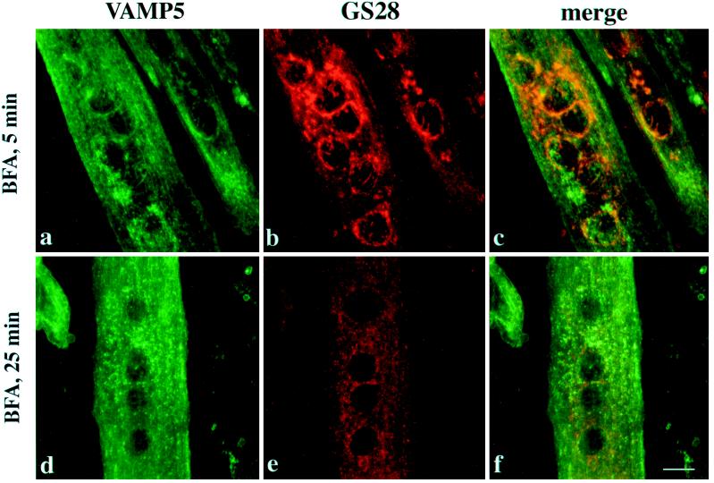
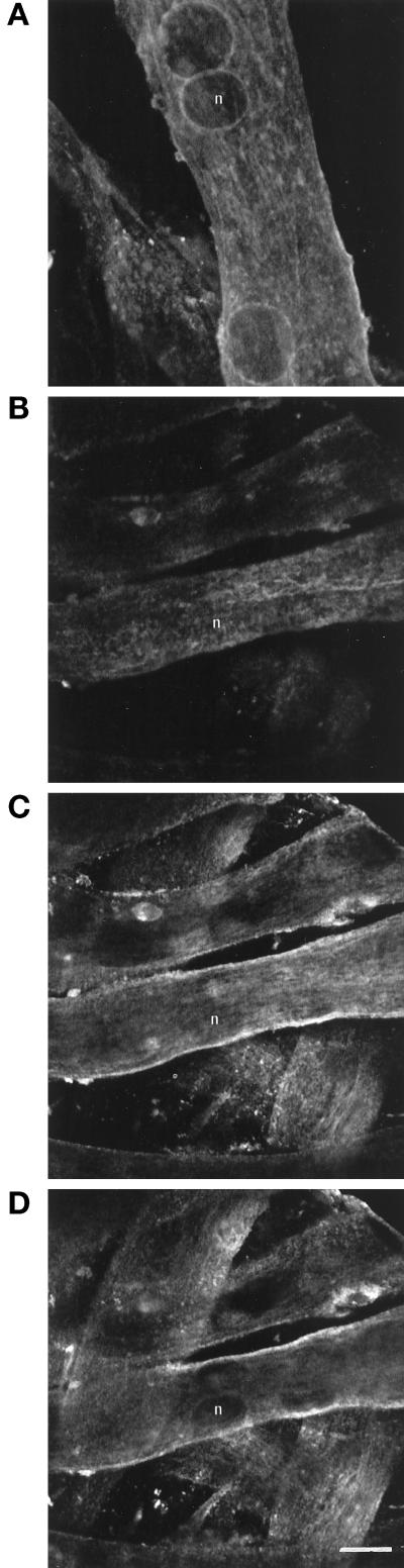
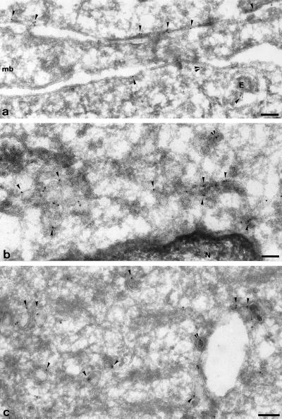
cDNA clones encoding a novel protein (VAMP5) homologous to synaptobrevins/VAMPs are detected during database searches. The predicted 102-amino acid VAMP5 harbors a 23-residue hydrophobic region near the carboxyl terminus and exhibits an overall amino acid identity of 33% with synaptobrevin/VAMP1 and 2 and cellubrevin. Northern blot analysis reveals that the mRNA for VAMP5 is preferentially expressed in the skeletal muscle and heart, whereas significantly lower levels are detected in several other tissues but not in the brain. During in vitro differentiation (myogenesis) of C2C12 myoblasts into myotubes, the mRNA level for VAMP5 is increased approximately 8- to 10-fold. Immunoblot analysis using antibodies specific for VAMP5 shows that the protein levels are also elevated approximately 6-fold during in vitro myogenesis of C2C12 cells. Indirect immunofluorescence microscopy and immunoelectron microscopy reveal that VAMP5 is associated with the plasma membrane as well as intracellular perinuclear and peripheral vesicular structures of myotubes. Epitope-tagged versions of VAMP5 are similarly targeted to the plasma membrane.


Figures

Figure 1
Figure 1
(A) Nucleotide and the derived amino acid sequence of mouse VAMP5. The C-terminal hydrophobic region is underlined. (B) Alignment of amino acid sequences of mouse VAMP5, cellubrevin, and VAMP2/synaptobrevin 2. Residues identical in the three proteins are shaded.
Figure 2
Figure 2
Northern blot analysis of VAMP5 transcript. A mouse multiple-tissue blot containing 2 μg of poly(A)+ mRNA from the indicated tissues was probed with 32P-labeled VAMP5 cDNA probe (top panel) and then subsequently with 32P-labeled β-actin probe (bottom panel). Highest levels of VAMP5 transcript were detected in the heart and the skeletal muscle.
Figure 3
Figure 3
Increase in VAMP5 transcript during in vitro myogenesis. Thirty micrograms of total RNA isolated from myoblasts (lanes 1, 3, and 5) and 15 μg of total RNA isolated from myotubes (lanes 2, 4, and 6) were analyzed by agarose gel electrophoresis. The RNAs were stained with ethidium bromide and photographed (lanes 1 and 2) or transferred to a filter and probed with 32P-labeled VAMP5 probe (lanes 3 and 4) or Rab10 probe (lanes 5 and 6).
Figure 4
Figure 4
Characterization of VAMP5 antibodies. Total cellular extracts of myotubes were resolved by SDS-PAGE and transferred to a filter. The blot was probed with affinity-purified rabbit antibodies against VAMP5, and a 16-kDa protein was specifically detected (lane 1). The detection of this protein is abolished by preincubation of antibodies with recombinant GST-VAMP5 (lane 2) but not by GST (lane 3), GST-cellubrevin (lane 4), or GST-VAMP2 (lane 5).
Figure 5
Figure 5
Increase of VAMP5 protein during myogenesis. One hundred micrograms of total protein extracts derived from growing myoblasts (A, lane 2; B, lane 1; C, lane 1) and myotubes after growing in differentiation medium for 5 d (A, lane 3; B, lane 2; C, lane 2) were resolved by SDS-PAGE and stained with Coommasie blue (A) or processed for immunoblot to detect Golgi SNARE GS28 (B) or VAMP5 (C). Although GS28 is present at comparable levels in both myoblasts and myotubes, VAMP5 is present at much higher levels in myotubes.
Figure 6
Figure 6
VAMP5 is an integral membrane protein. Total membranes derived from myotubes were extracted by different reagents as indicated. The extracts (S) and the membrane pellets (P) were then analyzed by immunoblot to detect VAMP5. As shown, VAMP5 is not extracted by PBS (lanes 1 and 2), 1 M KCl (lanes 3 and 4), 150 mM sodium bicarbonate (pH 11.5) (lanes 5 and 6), or 2.5 M urea (lanes 7 and 8) but is extracted effectively by detergents such as 1% Triton X-100 (lanes 9 and 10) and 1% DOC (lanes 11 and 12).
Figure 7
Figure 7
(A) Labeling of VAMP5 in few myoblasts in differentiated C2C12 cultures by indirect immunofluorescence microscopy. Cells were fixed, permeabilized, and incubated with affinity-purified rabbit antibodies against VAMP5 followed by FITC-conjugated anti-rabbit IgG. The cells were mounted, and images captured with the Bio-Rad MRC1024 confocal system. Although the majority of the unfused myoblasts did not exhibit any specific labeling, a small fraction of unfused myoblasts had strong labeling on the cell surface (A, a) and intracellular vesicular structures (A, b) when viewed at the cell surface and internal focal planes, respectively. Shown in c (A) is the combined image of 0.3-μm optical sections, and it is again obvious that VAMP5 is associated with the plasma membrane and intracellular vesicular structures. (B) Epitope-tagged versions of VAMP5 are targeted to the cell surface. Pooled transfectants of C2C12 cells stably transfected with expression vector expressing myc-tagged VAMP5 (a) or HA-tagged VAMP5 (b) were fixed and labeled with monoclonal antibody against myc or HA followed by FITC-conjugated anti-mouse IgG. Cells were viewed and photographed. Bars, 10 μm.
Figure 8
Figure 8
Differentiated C2C12 cells were double-labeled with rabbit antibodies against VAMP5 and a mouse monoclonal antibody against GS28 and then with FITC-conjugated anti-rabbit IgG and rhodamine-conjugated anti-mouse IgG. Cells were viewed with confocal microscopy, and combined images are shown. In myotubes, both surface and intracellular labelings are detected, and VAMP5 is thus associated with the plasma membrane and intracellular vesicular structures of myotubes. The broken-ring labeling of VAMP5 around the nuclei overlaps well with that of GS28, as does some of the peripheral labeling. However, the labeling on the plasma membrane and the majority of peripheral vesicular structures labeled by VAMP5 are devoid of GS28 As shown, most unfused myoblasts (except for the one on the right bottom corners of a–c) do not exhibit specific labeling of VAMP5, although their Golgi apparatuses are labeled by the GS28 monoclonal antibody. Bar, 10 μm.
Figure 9
Figure 9
Differentiated C2C12 cells were incubated with 10 μg/ml nocodazole for 60 min and then analyzed by indirect immunofluorescence microscopy to detect VAMP5 and GS28. The top and bottom panels represent views from different myotubes. Note that in the bottom panel, unfused myoblasts are labeled by anti-GS28 antibody (e and f) but are negative for VAMP5 labeling (d and f). Although the Golgi apparatus marked by GS28 in unfused myoblasts is clearly fragmented, the Golgi apparatus marked by GS28 in myotubes is less fragmented. Under this condition, the Golgi-like labeling of VAMP5 is significantly segregated from that of GS28. Bar, 10 μm.
Figure 10
Figure 10
Differentiated C2C12 cells were incubated with BFA for 5 or 25 min and then processed for indirect immunofluorescence microscopy to detect VAMP5 and GS28. After a 5-min treatment with BFA, the perinuclear Golgi-like labeling for VAMP5 is essentially dispersed (a and c), although the perinuclear Golgi labeling for GS28 remains essentially intact (b and c), suggesting that structures marked by VAMP5 are more quickly affected by BFA. After 25 min of BFA treatment, both VAMP5 (d) and GS28 (e) are redistributed into fine dotted structures. However, the majority of VAMP5-positive structures do not colocalize with that of GS28, suggesting that VAMP5 and GS28 are segregated into distinct structures. Bar, 10 μm.
Figure 11
Figure 11
Immunolabeling of VAMP5 in young and mature C2C12 myotube cultures. C2C12 cells were grown in culture snd studied at either day 3 (a) or day 9 (b–d) after differentiation. Cells were fixed and labeled with anti-VAMP5 antibodies. In young cultures, both intracellular and surface labelings were evident. In mature cultures, surface labeling became more predominant. Panels b–d represent sequential serial sections using a confocal laser microscope through the same field of a mature culture and illustrate the relative absence of intracellular labeling compared with that observed in panel a. Representative nuclei in both preparations are indicated (n). Bar, 10 μm.
Figure 12
Figure 12
Ultrastructural localization of VAMP5. Ultrathin frozen sections of differentiated C2C12 cells were labeled with anti-VAMP5 antibodies followed by swine anti-rabbit antibodies and then 10 nm protein A-gold. Panel a shows plasma membrane regions of three closely apposed cells; a region of myoblast (mb) is unlabeled, whereas the neighboring myotubes (above and below) show significant labeling close to the plasma membrane (arrowheads). Note the labeling on a small vesicle (double arrowheads) and on a multivesicular endosome (E). Panel b shows the juxtanuclear area of a myotube; labeling is concentrated on uncoated vesicular and tubular profiles. The ER surrounding the nucleus (N) is unlabeled. Panel c shows characteristic labeling of large uncoated vesicles (arrowheads), some of which are associated with putative endosomal structure. The double arrowheads in b and c indicate a labeled clathrin coated buds. Bars, 100 nm.

References

    1. Altschul SF, Gish W, Miller W, Myers EW, Lipman DJ. Basic local alignment search tool. J Mol Biol. 1990;215:403–410. - PubMed
    1. Baumert M, Maycox PR, Navone F, De Camilli P, Jahn R. Synaptobrevin: an integral membrane protein of 18,000 daltons present in small synaptic vesicles of rat brain. EMBO J. 1989;8:379–384. - PMC - PubMed
    1. Bennett MK, Calakos N, Scheller RH. Syntaxin: a synaptic protein implicated in docking of synaptic vesicles at presynaptic active zones. Science. 1992;257:255–259. - PubMed
    1. Bennett MK, Garcia-Arraras JE, Elferink LA, Peterson K, Fleming AM, Hazuka CD, Scheller RH. The syntaxin family of vesicular transport receptors. Cell. 1993;74:863–873. - PubMed
    1. Bock JB, Klumperman J, Davanger S, Scheller RH. Syntaxin 6 functions in trans-Golgi network vesicle trafficking. Mol Biol Cell. 1997;8:1261–1271. - PMC - PubMed