. 1998 Sep;9(9):2439-61.
doi: 10.1091/mbc.9.9.2439.

A novel fluorescence-based genetic strategy identifies mutants of Saccharomyces cerevisiae defective for nuclear pore complex assembly

Affiliations
Free PMC article

A novel fluorescence-based genetic strategy identifies mutants of Saccharomyces cerevisiae defective for nuclear pore complex assembly

M Bucci et al. Mol Biol Cell. 1998 Sep.
Free PMC article

Abstract

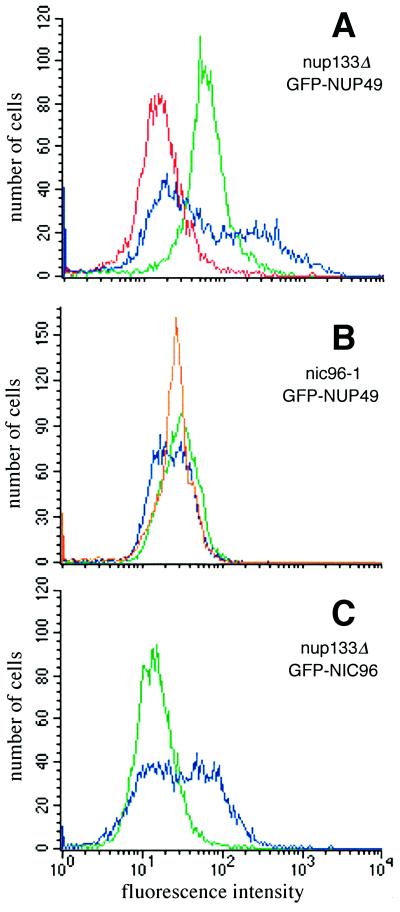
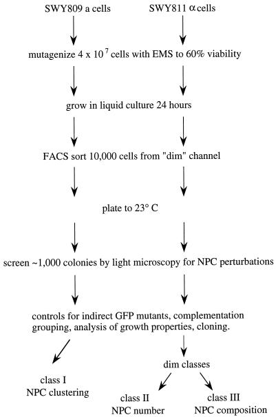
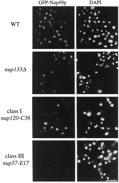
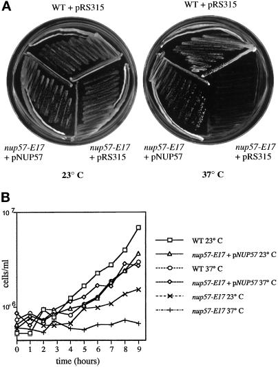
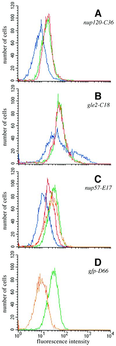
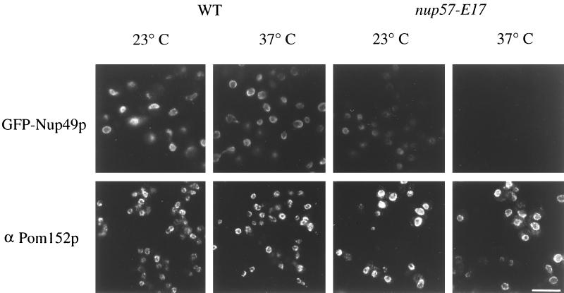
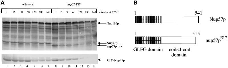
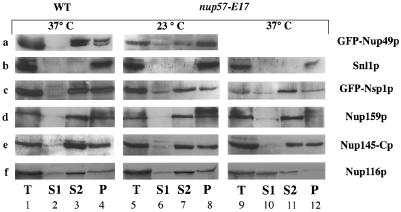
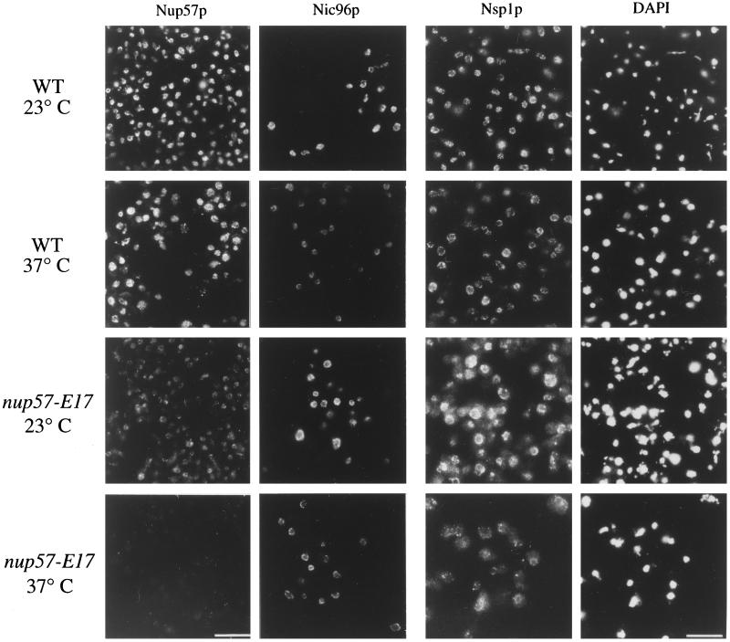
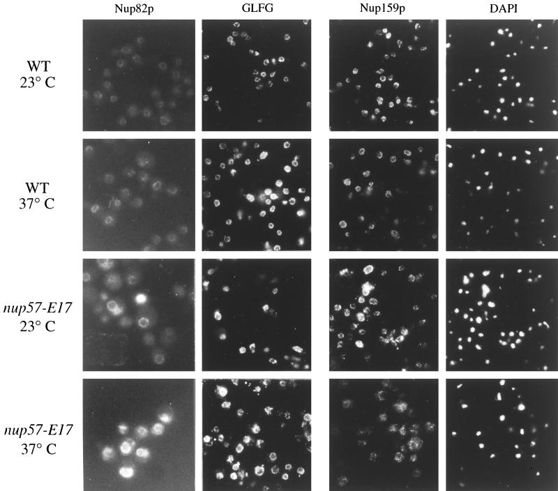
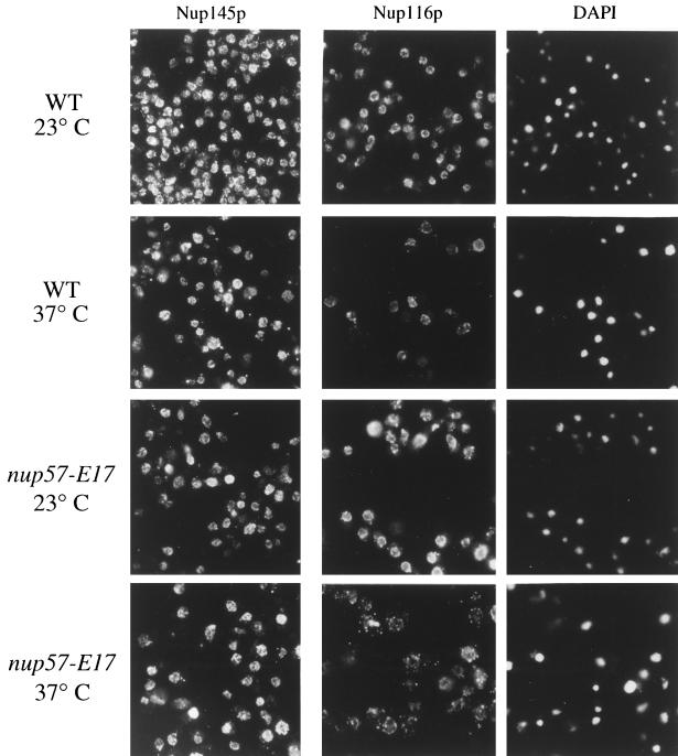
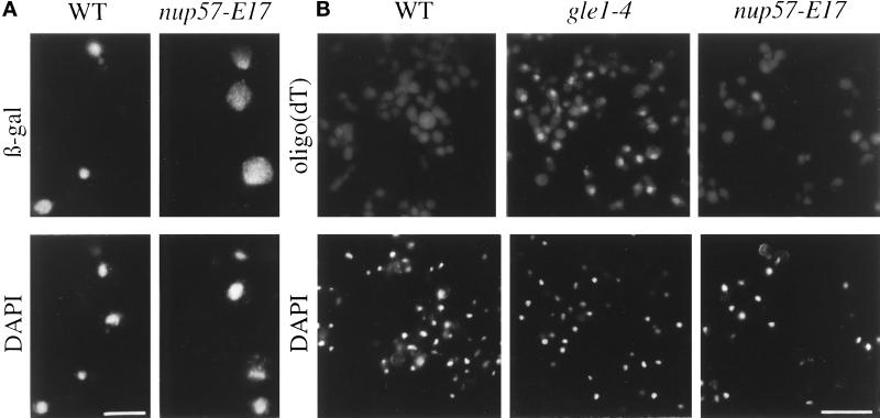
Nuclear pore complexes (NPCs) are large proteinaceous portals for exchanging macromolecules between the nucleus and the cytoplasm. Revealing how this transport apparatus is assembled will be critical for understanding the nuclear transport mechanism. To address this issue and to identify factors that regulate NPC formation and dynamics, a novel fluorescence-based strategy was used. This approach is based on the functional tagging of NPC proteins with the green fluorescent protein (GFP), and the hypothesis that NPC assembly mutants will have distinct GFP-NPC signals as compared with wild-type (wt) cells. By fluorescence-activated cell sorting for cells with low GFP signal from a population of mutagenized cells expressing GFP-Nup49p, three complementation groups were identified: two correspond to mutant nup120 and gle2 alleles that result in clusters of NPCs. Interestingly, a third group was a novel temperature-sensitive allele of nup57. The lowered GFP-Nup49p incorporation in the nup57-E17 cells resulted in a decreased fluorescence level, which was due in part to a sharply diminished interaction between the carboxy-terminal truncated nup57pE17 and wt Nup49p. Interestingly, the nup57-E17 mutant also affected the incorporation of a specific subset of other nucleoporins into the NPC. Decreased levels of NPC-associated Nsp1p and Nup116p were observed. In contrast, the localizations of Nic96p, Nup82p, Nup159p, Nup145p, and Pom152p were not markedly diminished. Coincidentally, nuclear import capacity was inhibited. Taken together, the identification of such mutants with specific perturbations of NPC structure validates this fluorescence-based strategy as a powerful approach for providing insight into the mechanism of NPC biogenesis.


Figures

Figure 1
Figure 1
NPC mutants expressing GFP-Nup49p have distinct fluorescence properties compared with wt cells expressing GFP-Nup49p. Flow cytometry analysis was performed on cells grown to logarithmic phase in SD lacking leucine (488 nm ex, 530 nm em). wt strains expressing GFP-Nup49p (SWY894, green profiles in panels A and B) have a fluorescence emission level nearly five times as great as the endogenous fluorescence of cells not expressing GFP (SWY1601, red profile in panel A). (A) nup133Δ cells expressing GFP-Nup49p (SWY1667, blue) display a bimodal distribution reflecting two populations of GFP-labeled cells with different fluorescence levels. (B) nic96–1 cells expressing GFP-Nup49p (SWY1860) show a broad, left-shifted peak after growth at 23°C (blue) and a sharp symmetrical peak after growth for 5 h at 37°C (orange). (C) nup133Δ cells expressing GFP-Nic96p (SWY1824, blue) have a distinct profile compared with wt cells expressing GFP-Nic96p (green).
Figure 2
Figure 2
Schematic diagram for a fluorescence-based strategy to isolate NPC assembly mutants. Strains expressing GFP-Nup49p were selected by FACS and screened by microscopic inspection of GFP-NPC fluorescence patterns. Candidate mutants were tested for plasmid-dependent complementation and for growth phenotypes. We predicted that at least three different classes of mutants would be identified: Class I, NPC clustering; Class II, fewer total NPCs per nucleus, and Class III, wt NPC number, but each NPC with a decreased amount of GFP-Nup incorporated.
Figure 3
Figure 3
Two classes of NPC mutants were isolated in the GFP-Nup49p fluorescence-based strategy: NPC clustering mutants and dim mutants. Direct fluorescence microscopy analysis of GFP-Nup49p localization in wt, nup133Δ, and two representative mutant strains isolated in the FACS-based screen. The strains were grown in SD media lacking leucine: wt (SWY894), nup133Δ clustering (SWY1667), clustering-Class I (nup120-C36 mutant, SWY1710), Dim-Class III (nup57-E17 mutant, SWY1708). Within each column, photographs were exposed and printed for identical times. DAPI staining for each respective field is shown on the left. Bar, 10 μm.
Figure 4
Figure 4
Expression of NUP57 from a plasmid complements the temperature sensitivity of the nup57-E17 mutant. (A) Strains were grown on SD plates lacking leucine for 4 d at 23 or 37°C: wt (SWY894), nup57-E17 with pRS315 (SWY1708), nup57-E17 + pSW806/NUP57 (SWY1709). NUP57 expression restores growth to the nup57-E17 strain at 37°C. (B) Growth analysis of wt, nup57-E17, and nup57-E17 + NUP57 cells at 23 and 37°C. Aliquots of cells growing at early logarithmic phase were counted at various time points after shift to 37°C or continued growth at 23°C. Cell counts are expressed as cell number per ml of culture.
Figure 5
Figure 5
FACS analysis of mutant strains shows that expression of NUP120, GLE2, or NUP57 rescues the GFP phenotypes of the respective mutants. The mutant strains transformed with CEN/LEU plasmids were analyzed by flow cytometry as in Figure 1. In all panels, the GFP-Nup49p profile in wt cells is shown in green, the mutant transformed with an empty CEN/LEU vector is shown in blue, and the mutant transformed with a complementing plasmid is shown in red. (A) nup120-C36 at 23°C (blue, SWY1710; red, SWY1711). (B) gle2-C18 at 23°C (blue, SWY1705; red, SWY1706). (C) nup57-E17 at 23°C (blue, SWY1708; red, SWY1709) and nup57-E17 + NUP57 at 37°C (orange, SWY1709). (D) gfp-D66 (orange, SWY1707).
Figure 6
Figure 6
Localization of GFP-Nup49p at the NPC is decreased in nup57-E17 cells, whereas Pom152p localization is not perturbed. Within each row, images were photographed and printed for identical times for direct comparison of fluorescence intensities. Top row, direct fluorescence microscopy was performed on wt and nup57-E17 cells expressing GFP-Nup49p (SWY809 and SWY1586, respectively), grown in SD lacking tryptophan. At 23°C, the nup57-E17 strain has less GFP fluorescence signal localized at the NE than wt cells. After 4 h of growth at 37°C, the nup57-E17 cells lack any detectable GFP-Nup49p staining. Bottom row, Indirect immunofluorescence microscopy was performed with monoclonal anti-Pom152p antibodies (mAb 118C3) on wt (SWY519) and nup57-E17 (SWY1587) strains grown in YPD. The anti-Pom152p staining is localized at the NE/NPC, and the intensity level is not notably altered in nup57-E17 cells at either 23 or 37°C compared with wt. Bar, 10 μm.
Figure 7
Figure 7
The nup57-E17 allele results in a C-terminal truncation of nup57pE17 and temperature-dependent degradation of nup57pE17 and GFP-Nup49p. (A) Immunoblot analysis reveals the truncated nup57pE17 protein is unstable at 37°C. Total cell lysates prepared from wt (SWY809) and nup57-E17 (SWY1586) cells were separated on 8% SDS-polyacrylamide gels, transferred to nitrocellulose, and analyzed by immunoblotting with the indicated antibody (upper, affinity-purified polyclonal anti-GLFG; lower, polyclonal anti-GFP). The samples were prepared from cells grown at 23°C (lanes 1 and 8) or shifted to growth at 37°C for the indicated times (lanes 2–7 and 9–14). For each sample, identical cell number equivalents were loaded in each well making the protein levels directly comparable. Lysates from wt cells have similar levels of Nup116p, Nup57p, and GFP-Nup49p at all time points. In nup57-E17 cells, the levels of nup57pE17 and GFP-Nup49p decrease with increasing time at 37°C. (B) Diagram of wt Nup57p and mutated nup57pE17. Sequencing of the nup57-E17 allele revealed a single point mutation resulting in a stop codon after the codon for residue 515 and truncation of 26 residues from the C terminus. Numbers reflect amino acid positions within Nup57p. Vertical boxes designate individual GLFG repeats.
Figure 8
Figure 8
Subcellular fractionation of wt and nup57-E17 cells. Spheroplasts of the respective strains were osmotically lysed and fractionated into soluble (S1) and pellet (P1) fractions. The P1 fraction was further extracted with 1 M NaCl and separated into a supernatant (S2) and insoluble pellet (P2) fractions. Samples of each fraction and an aliquot of total spheroplast lysate (T) were separated by SDS-PAGE and analyzed by immunoblotting with the designated antibodies. For each antibody, only the full-length protein product is shown. In cases where some degradation of the full-length product was observed, the levels of degradation between the wt and mutant cells were equivalent (e.g., Nup116p and Nup159p). For each individual strain under the given growth conditions, identical cell number equivalents were analyzed for each S1, S2, and P2 fraction. The amount of each given protein in the S1, S2, and P2 fractions was quantified as described in the MATERIALS AND METHODS. (A) GFP-Nup49p (SWY809, SWY1586). (B) Snl1p (SWY809, SWY1586). (C) GFP-Nsp1p (SWY1728, SWY1736). (D) Nup159p (SWY809, SWY1586). (E) Nup145-Cp (SWY809, SWY1586). (F) Nup116p (SWY809, SWY1586).
Figure 9
Figure 9
Microscopic analysis of NPC/NE association of nup57pE17, Nic96p, and Nsp1p in nup57-E17 cells reveals decreased staining for nup57pE17 and Nsp1p. Indirect immunofluorescence microscopy was performed on wt (SWY519) and nup57-E17 (SWY1587) strains using antibodies recognizing the C-terminal region of Nup57p (left column). Direct fluorescence microscopy was performed on wt (SWY1695, SWY1728) and nup57-E17 (SWY1722, SWY1736) strains expressing GFP-tagged Nic96p or Nsp1p, respectively. Cells were shifted to growth at 37°C for 4 h. Within each column, images were photographed and printed for identical times for direct comparison of fluorescence intensities. The DAPI photographs in the right column correspond to the fields of Nsp1p cells. Bars, 10 μm, with Nup57p and Nic96p images at identical magnifications.
Figure 10
Figure 10
NPC/NE association of Nup82p, a GLFG nucleoporin(s), and Nup159p are not markedly diminished in nup57-E17 cells. Direct fluorescence microscopy was performed on wt (SWY1411) and nup57-E17 (SWY1744) strains expressing GFP-tagged Nup82p (left column). Indirect immunofluorescence microscopy was performed on wt (SWY519) and nup57-E17 (SWY1587) strains using antibodies recognizing either the GLFG family of nucleoporins (middle column) or Nup159p (right column), with DAPI staining shown for the anti-Nup159p fields. Cells were shifted to growth at 37°C for 4 h. Within each column, images were photographed and printed for identical times for direct comparison of fluorescence intensities.
Figure 11
Figure 11
Localization of the GLFG nucleoporin Nup116p at the NPC/NE is perturbed in temperature-arrested nup57-E17 cells, whereas Nup145-Cp remains predominantly at the NPC/NE. Indirect immunofluorescence microscopy was performed on wt (SWY519) and nup57-E17 (SWY1587) strains using antibodies recognizing either the C-terminal region of Nup145p (left column) or the C-terminal region of Nup116p (middle column). Coincident DAPI staining for the anti-Nup116p field is shown. Cells were shifted to growth at 37°C for 4 h. Within each column, images were photographed and printed for identical times for direct comparison of fluorescence intensities.
Figure 12
Figure 12
Nuclear import capacity is decreased in temperature-arrested nup57-E17 cells. (A) To assay nuclear import, wt (SWY519) and nup57-E17 (SWY1587) cells transformed with a plasmid expressing NLS-β-galactosidase under GAL10 control (pNLS-E1) were analyzed. Cells grown in 2% raffinose media at 37°C for 2.5 h were induced for NLS-β-galactosidase expression by addition of 2% galactose, and growth at 37°C was continued for 3.5 h. Cells were fixed and processed for indirect immunofluorescence microscopy. Localization of the reporter was determined using mAbs against β-galactosidase as described in MATERIALS AND METHODS. (B) Export of poly(A)+ RNA was not strongly inhibited. wt (SWY809), gle1–4 (SWY1186), and nup57-E17 (SWY1586) cells were grown at 23°C, shifted to 37°C for 4 h, and processed for in situ hybridization with a digoxigenin-oligo(dT)30 probe. Rhodamine-conjugated anti-digoxigenin antibodies were used to localize probe binding. Exposure and printing times are identical for wt and mutant cells in a given experiment. Coincident DAPI staining is shown. Bar, 5 μm (A); 15 μm (B).
Figure 13
Figure 13
Examination by electron microscopy of NE and NPC morphology in wt and nup57-E17 cells. NUP57 (SWY809) and nup57-E17 (SWY1586) cells were grown at either 23°C or shifted to growth at 37°C for 4 h before processing for thin section electron microscopy. Arrows point to NPCs with apparently wt structure; black arrowheads point to NPC-like electron dense masses that may not span the NE lumen between the inner and outer membranes; open arrowheads point to NE herniations. (A) nup57-E17 at 23°C. (B–D, G, H) nup57-E17 cells at 37°C. (E) wt at 23°C. (F) wt at 37°C. n, nucleus; c, cytoplasm. Bars, 500 nm (A–F); 250 nm (G and H).

Similar articles

Cited by

References

    1. Aitchison JD, Blobel G, Rout MP. Nup120p: a yeast nucleoporin required for NPC distribution and mRNA transport. J Cell Biol. 1995;131:1659–1675. - PMC - PubMed
    1. Akey CW. Structural plasticity of the nuclear pore complex. J Mol Biol. 1995;248:273–293. - PubMed
    1. Akey CW, Radermacher M. Architecture of the Xenopus nuclear pore complex revealed by three-dimensional cryo-electron microscopy. J Cell Biol. 1993;122:1–19. - PMC - PubMed
    1. Baudin A, Ozier KO, Denouel A, Lacroute F, Cullin C. A simple and efficient method for direct gene deletion in Saccharomyces cerevisiae. Nucleic Acids Res. 1993;21:3329–3330. - PMC - PubMed
    1. Bogerd AM, Hoffman JA, Amberg DC, Fink GR, Davis LI. nup1 mutants exhibit pleiotropic defects in nuclear pore complex function. J Cell Biol. 1994;127:319–332. - PMC - PubMed

Publication types

MeSH terms