. 1998 Sep;9(9):2463-76.
doi: 10.1091/mbc.9.9.2463.

Real-time imaging of the axonal transport of granules containing a tissue plasminogen activator/green fluorescent protein hybrid

Affiliations
Free PMC article

Real-time imaging of the axonal transport of granules containing a tissue plasminogen activator/green fluorescent protein hybrid

J E Lochner et al. Mol Biol Cell. 1998 Sep.
Free PMC article

Abstract

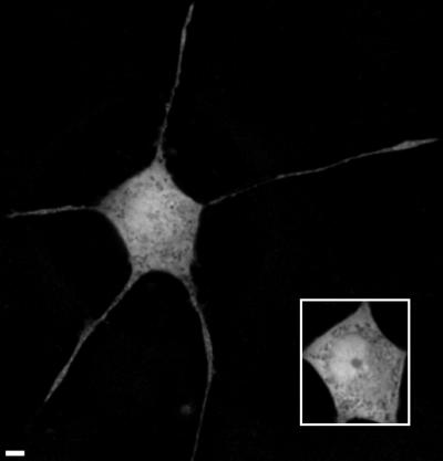
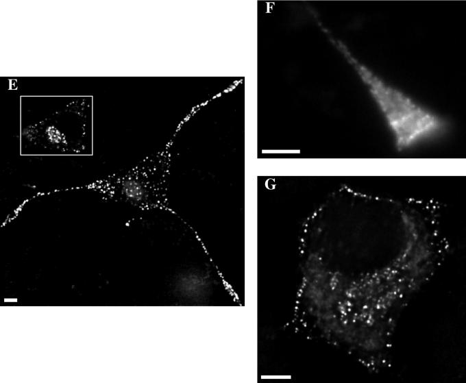
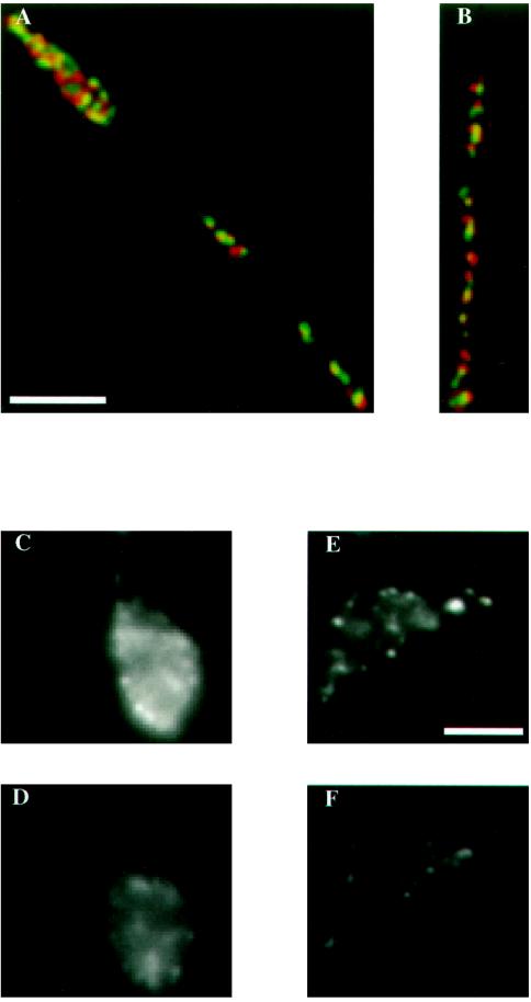
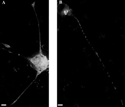
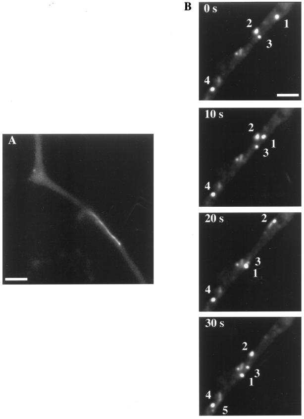
A hybrid protein, tPA/GFP, consisting of rat tissue plasminogen activator (tPA) and green fluorescent protein (GFP) was expressed in PC12 cells and used to study the distribution, secretory behavior, and dynamics of secretory granules containing tPA in living cells with a neuronal phenotype. High-resolution images demonstrate that tPA/GFP has a growth cone-biased distribution in differentiated cells and that tPA/GFP is transported in granules of the regulated secretory pathway that colocalize with granules containing secretogranin II. Time-lapse images of secretion reveal that secretagogues induce substantial loss of cellular tPA/GFP fluorescence, most importantly from growth cones. Time-lapse images of the axonal transport of granules containing tPA/GFP reveal a surprising complexity to granule dynamics. Some granules undergo canonical fast axonal transport; others move somewhat more slowly, especially in highly fluorescent neurites. Most strikingly, granules traffic bidirectionally along neurites to an extent that depends on granule accumulation, and individual granules can reverse their direction of motion. The retrograde component of this bidirectional transport may help to maintain cellular homeostasis by transporting excess tPA/GFP back toward the cell body. The results presented here provide a novel view of the axonal transport of secretory granules. In addition, the results suggest that tPA is targeted for regulated secretion from growth cones of differentiated cells, strategically positioning tPA to degrade extracellular barriers or to activate other barrier-degrading proteases during axonal elongation.


Figures

Figure 1
Figure 1
Deblurred images of a fixed GFP-expressing PC12 cell. The two images (including the inset) were obtained by optically sectioning the sample and correspond to two different depths through the cell. Bar, 4 μm.
Figure 2
Figure 2
Images of tPA/GFP-expressing PC12 cells. The fluorescence images in E and G were deblurred, whereas the fluorescence images in B, D, and F were not. Phase (A and C) and fluorescence (B and D) images of a fixed, differentiated cell and a neurite taken with a 40× objective. Images of a fixed, differentiated cell (E), a bright growth cone from a living differentiated cell (F), and a fixed, undifferentiated cell (G). Note that the Matrigel is a source of background in the phase images. Bar, 4 μm.
Figure 2
Figure 2
Images of tPA/GFP-expressing PC12 cells. The fluorescence images in E and G were deblurred, whereas the fluorescence images in B, D, and F were not. Phase (A and C) and fluorescence (B and D) images of a fixed, differentiated cell and a neurite taken with a 40× objective. Images of a fixed, differentiated cell (E), a bright growth cone from a living differentiated cell (F), and a fixed, undifferentiated cell (G). Note that the Matrigel is a source of background in the phase images. Bar, 4 μm.
Figure 3
Figure 3
Deblurred image of the distribution of endogenous tPA in a fixed, immunostained PC12 cell showing the similarity in distribution between endogenous tPA and tPA/GFP. Some surface staining is visible in the immunofluorescence image, which may reflect endogenous tPA that is bound to its membrane receptor. Bar, 4 μm.
Figure 4
Figure 4
Images demonstrating that tPA/GFP traffics through the regulated secretory pathway (A and B) and is secreted from growth cones after stimulation with carbachol (C–F). The images in panels A and B were deblurred, whereas the images in C–F were not. Representative two-color images (A and B) of regions of neurites from two different cells that demonstrate extensive colocalization of tPA/GFP and SgII. The distributions of tPA/GFP and SgII are shown in green and red, respectively. Areas of extensive overlap appear yellow in the images. Most green granules overlap with a red granule, although in some cases the sizes of the green and red spots differ; also, the centers of some spots are shifted slightly. Lack of perfect overlap could reflect antibody accessibility, wavelength-dependent image shifts, or differences in protein concentration in individual granules, especially in granules produced before transfection. Images (C–F) of two growth cones from living cells before (C and E) and 20 min after (D and F) the addition of 10 mM carbachol. Panels A and B and C–F share a scale bar. Bar, 4 μm.
Figure 5
Figure 5
Deblurred images of fixed (A) tPA/GFP(SIG−) and (B) GFP(SIG+)-expressing PC12 cells. One significant difference in distribution between tPA/GFP and GFP(SIG+) is that the staining in growth cones and neurites is often so faint in GFP(SIG+)-expressing cells that these structures are almost invisible when viewed in fluorescence mode. The GFP(SIG+) labeling in the neurite and growth cone shown here is somewhat more intense than is typical, permitting these structures to be seen in the image. Differences in growth cone brightness exhibited by tPA/GFP and GFP(SIG+) are not due to a lower level of expression in GFP(SIG+)-expressing cells. In fact, GFP(SIG+)-expressing cells on average are brighter than tPA/GFP-expressing cells. Bar, 4 μm.
Figure 6
Figure 6
Time-lapse images of the axonal transport of secretory granules in PC12 cells. The granule in panel A is streaking in the anterograde direction along the neurite of a PC12 cell at a speed of ∼1.6 μm/s. This granule covered the entire length of the neurite in one long, unidirectional excursion. The sequential images in panel B exhibit a spectrum of generally less rapid granule motions. Granule 1 moves in the anterograde direction at a speed of ∼0.4 μm/s, slowing somewhat during the last frame. Granule 2 is motionless at first; it then starts moving in the retrograde direction at a speed of ∼0.4 μm/s, leaving a streak as it achieves an instantaneous speed of at least 0.9 μm/s. Granule 2 then reverses direction and finally moves in the anterograde direction at a speed of ∼0.7 μm/s. Granule 3 at first moves slowly in the anterograde direction; it then moves slightly out of the focal plane, reappearing more clearly in the last image. Granule 4 is essentially immobile during the entire observation period. Finally, granule 5 appears in the last frame near granule 4, streaking along the neurite at ∼1.3 μm/s. Bar, 4 μm.

References

    1. Alberts B, Bray D, Lewis J, Raff M, Roberts K, Watson JD. Molecular Biology of the Cell. 3rd ed. New York: Garland; 1994.
    1. Allen RD, Metuzals J, Tasaki I, Brady ST, Gilbert SP. Fast axonal transport in squid giant axon. Science. 1982;218:1127–1129. - PubMed
    1. Allen RD, Weiss DG, Hayden JH, Brown DT, Fujiwake H, Simpson M. Gliding movement of and bidirectional transport along single native microtubules from squid axoplasm: evidence for an active role of microtubules in cytoplasmic transport. J Cell Biol. 1985;100:1736–1752. - PMC - PubMed
    1. Bottenstein JE, Sato GH. Growth of a rat neuroblastoma cell line in serum-free supplemented medium. Proc Natl Acad Sci USA. 1979;76:514–517. - PMC - PubMed
    1. Brady ST. A novel brain ATPase with properties expected for the fast axonal transport motor. Nature. 1985;317:73–75. - PubMed

Publication types

MeSH terms

Substances

LinkOut - more resources