. 1998 Sep;9(9):2509-25.
doi: 10.1091/mbc.9.9.2509.

Differential expression and functions of cortical myosin IIA and IIB isotypes during meiotic maturation, fertilization, and mitosis in mouse oocytes and embryos

Affiliations
Free PMC article

Differential expression and functions of cortical myosin IIA and IIB isotypes during meiotic maturation, fertilization, and mitosis in mouse oocytes and embryos

C Simerly et al. Mol Biol Cell. 1998 Sep.
Free PMC article

Abstract

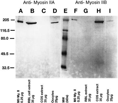
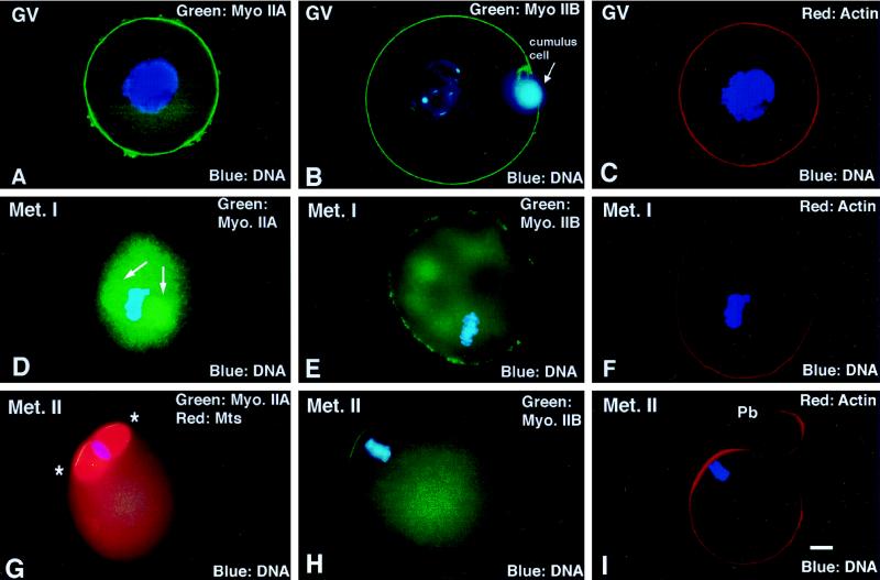
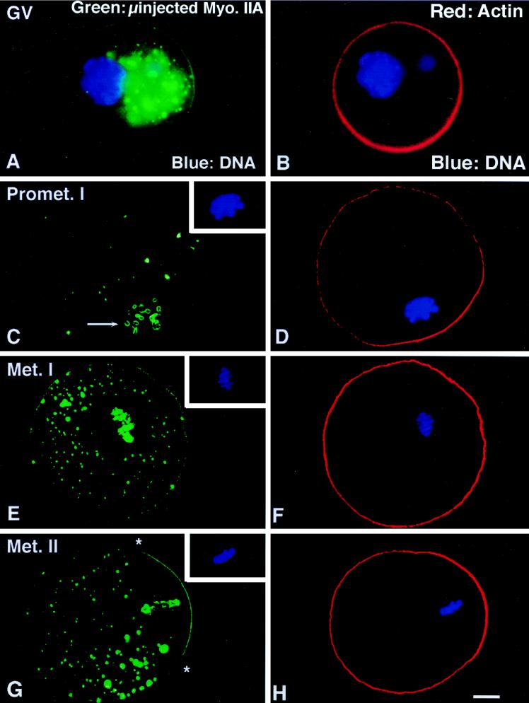
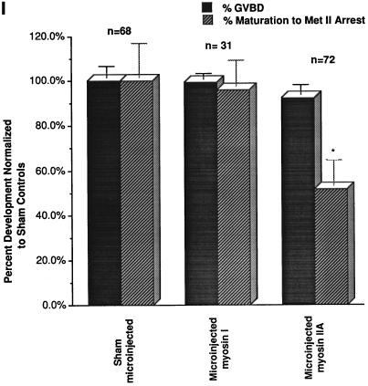
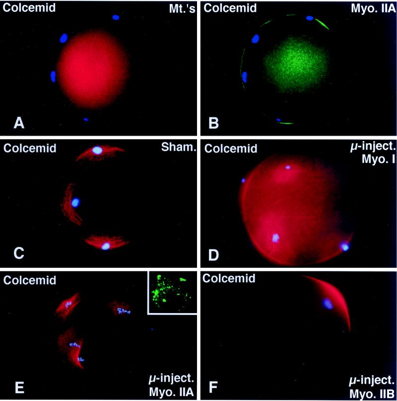
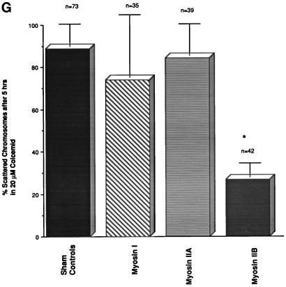
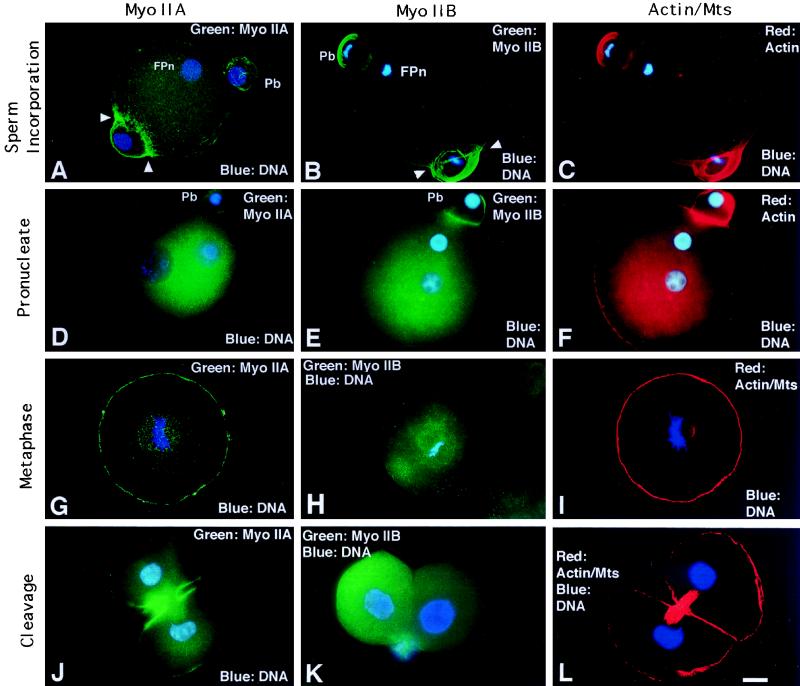
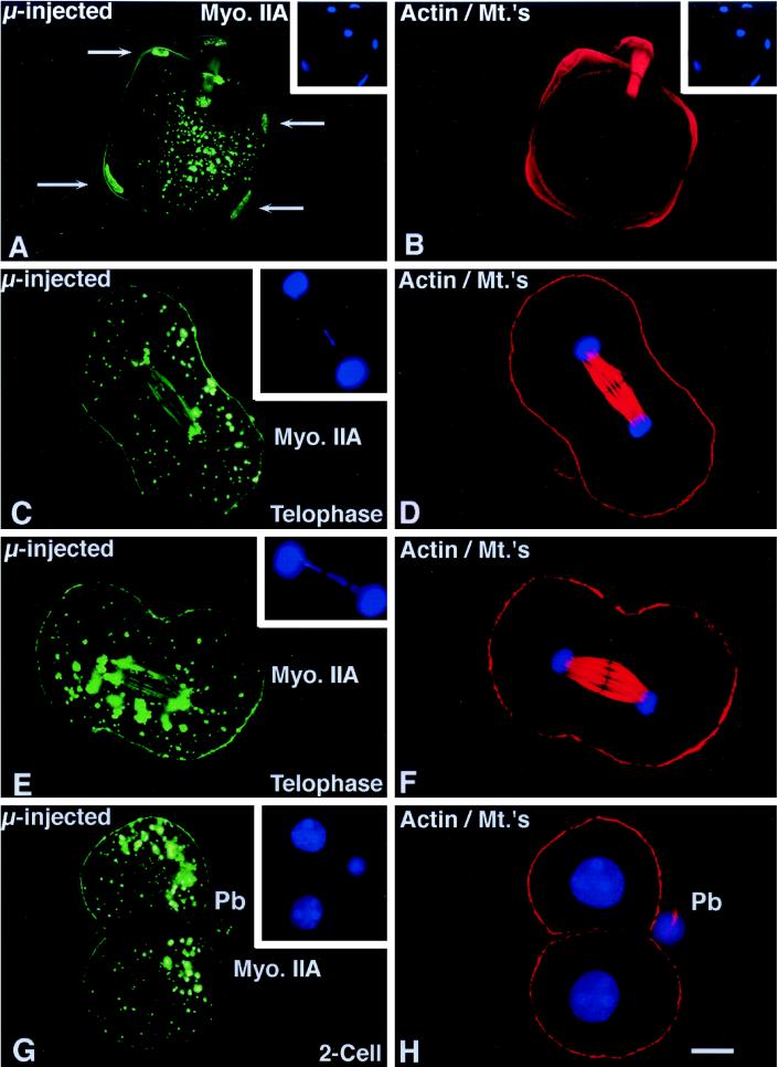
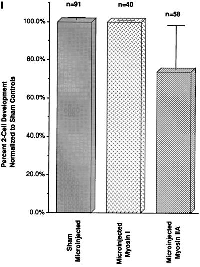
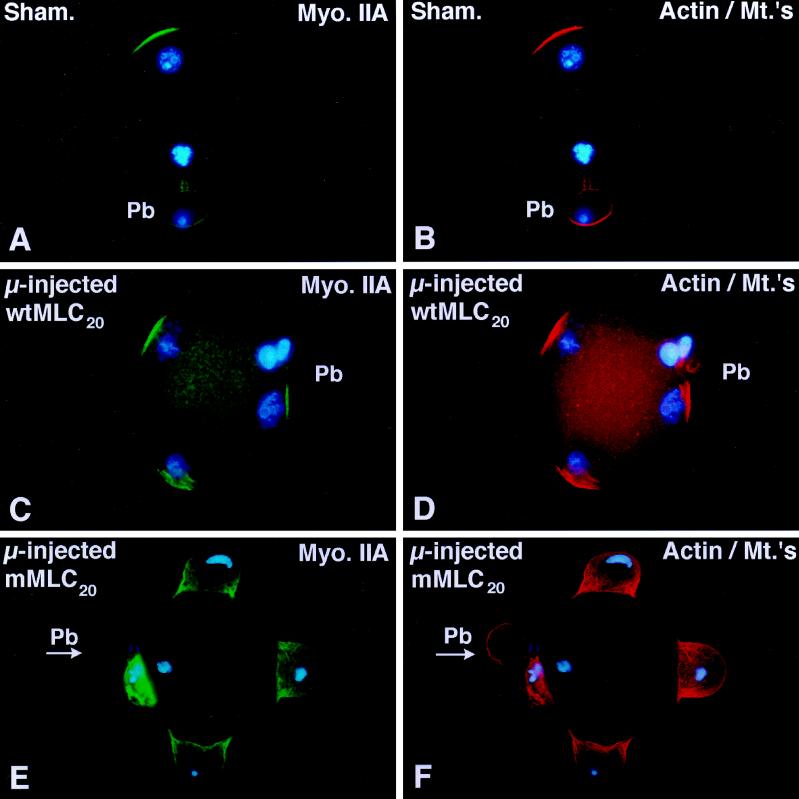
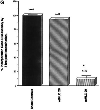
To explore the role of nonmuscle myosin II isoforms during mouse gametogenesis, fertilization, and early development, localization and microinjection studies were performed using monospecific antibodies to myosin IIA and IIB isotypes. Each myosin II antibody recognizes a 205-kDa protein in oocytes, but not mature sperm. Myosin IIA and IIB demonstrate differential expression during meiotic maturation and following fertilization: only the IIA isoform detects metaphase spindles or accumulates in the mitotic cleavage furrow. In the unfertilized oocyte, both myosin isoforms are polarized in the cortex directly overlying the metaphase-arrested second meiotic spindle. Cortical polarization is altered after spindle disassembly with Colcemid: the scattered meiotic chromosomes initiate myosin IIA and microfilament assemble in the vicinity of each chromosome mass. During sperm incorporation, both myosin II isotypes concentrate in the second polar body cleavage furrow and the sperm incorporation cone. In functional experiments, the microinjection of myosin IIA antibody disrupts meiotic maturation to metaphase II arrest, probably through depletion of spindle-associated myosin IIA protein and antibody binding to chromosome surfaces. Conversely, the microinjection of myosin IIB antibody blocks microfilament-directed chromosome scattering in Colcemid-treated mature oocytes, suggesting a role in mediating chromosome-cortical actomyosin interactions. Neither myosin II antibody, alone or coinjected, blocks second polar body formation, in vitro fertilization, or cytokinesis. Finally, microinjection of a nonphosphorylatable 20-kDa regulatory myosin light chain specifically blocks sperm incorporation cone disassembly and impedes cell cycle progression, suggesting that interference with myosin II phosphorylation influences fertilization. Thus, conventional myosins break cortical symmetry in oocytes by participating in eccentric meiotic spindle positioning, sperm incorporation cone dynamics, and cytokinesis. Although murine sperm do not express myosin II, different myosin II isotypes may have distinct roles during early embryonic development.


Figures

Figure 1
Figure 1
Antibodies prepared against isoform-specific regions of myosin heavy chains A and B are monospecific and detect 205-kDa proteins in unfertilized mouse oocytes. Lanes A–D, antimyosin IIA antibody. (A) 0.25 μg of purified macrophage (Mφ) myosin II protein; (B) 35 μg of RBL cell extract; (C) 35 μg of COS cell extract; and (D) 25 μg of mouse unfertilized oocyte extract. Lanes F–I, antimyosin IIB antibody. (F) 0.25 μg of purified (Mφ) myosin II protein; (G) 35 μg of RBL cell extract; (H) 35 μg of COS cell extract; and (I) 25 μg of mouse unfertilized oocytes extract. Lane E, molecular mass standards, in kDa. Myosin IIA antibody recognizes a 205-kDa myosin IIA isoform in RBL cell extracts, but not extracts from COS cells which lack myosin IIA. In contrast, the 205-kDa myosin IIB isoform is detected in COS cell extracts, but is absent in RBL cell extracts which lack the IIB protein. Myosin IIA and IIB antibodies detect 205-kDa proteins in mature oocytes. The IIB isoform appears to be less prevalent in oocytes than myosin IIA.
Figure 2
Figure 2
Detection of myosin IIA and IIB isotypes during meiotic maturation and in the unfertilized oocyte arrested at second meiotic metaphase. (A–C) In immature GV stage oocytes, myosin IIA, myosin IIB, and actin are uniform in the cortex. (D–F) By the first meiotic metaphase, myosin IIA decreases in the cortical region and increases in the cytoplasm, associating prominently with the metaphase spindle (D, arrows). Myosin IIB is discontinuous in the cortex and detected within the cytoplasm, but no association with the meiotic spindle or chromosomes is observed. Cortical actin filament detection remains uniform. (G–I). In the unfertilized oocyte, myosins IIA and IIB as well as actin filaments are enhanced in the region overlying the metaphase-arrested second meiotic spindle (G, ∗, second meiotic spindle region). (A, C, D, and F) Triple-labeled images for myosin IIA (green), actin (red), and DNA (blue). (B, E, and H) Double labeled for myosin IIB (green) and DNA (blue). (G) Triple labeled for myosin IIA (green), microtubules (red), and DNA (blue). (I) Double labeled for actin (red) and DNA (blue). Bar, 10 μm.
Figure 3
Figure 3
Microinjection of myosin IIA antibody blocks the eccentric positioning of the metaphase I spindle during meiotic maturation. (A and B) Microinjection of myosin IIA antibody into GV-arrested oocytes reduces myosin IIA, but not actin filaments, in the cortex and results in the formation of large immunofluorescent aggregates within the cytoplasm. (C and D) By 16 h after microinjection of myosin IIA antibody, oocytes arrested at prometaphase I (C, inset) demonstrate cytoplasmic myosin aggregates, a reduced cortical intensity of myosin IIA, and peripheral staining of chromosomal surfaces (C, arrow). Cortical actin enhancement overlying the peripheral chromosomes is not influenced after microinjection of the myosin IIA antibody (D). (E and F) In E, a GV stage oocyte microinjected with myosin IIA antibody has arrested at metaphase I (inset, DNA). Myosin IIA is not detected in the meiotic spindle region, except at surfaces of the aligned equatorial chromosomes. The large cytoplasmic immunofluorescent aggregates which form after microinjection of myosin IIA antibody are also excluded from the meiotic spindle region. Actin filaments remain uniform in the cortex of metaphase I-blocked oocytes. (G and H) In G, a GV stage oocyte microinjected with myosin IIA antibody has arrested at metaphase of second meiosis (inset, DNA). Both cortical myosin IIA and actin filaments are enhanced in the region overlying the spindle (∗, spindle region). Microinjection of myosin IIA antibody eliminates myosin IIA detection in the meiotic spindle but labels the peripheries of the meiotic chromosomes. (I) An analysis of the impact of microinjected myosin antibodies on meiotic maturation in vitro demonstrates that GVBD is not prevented in the presence of either myosin I or myosin IIA antibodies (solid bars), but completion of meiotic maturation to metaphase II arrest is significantly reduced by the IIA antibody (stippled bars). ∗, significant difference compared with microinjected sham controls (p < 0.001). All images triple labeled for myosin IIA (green), actin (red), and DNA (blue). (A and B) epifluorescence. (C–H) Laser scanning confocal microscopy. (I) Graph of the mean ± SD. Bar, 10 μm.
Figure 3
Figure 3
Microinjection of myosin IIA antibody blocks the eccentric positioning of the metaphase I spindle during meiotic maturation. (A and B) Microinjection of myosin IIA antibody into GV-arrested oocytes reduces myosin IIA, but not actin filaments, in the cortex and results in the formation of large immunofluorescent aggregates within the cytoplasm. (C and D) By 16 h after microinjection of myosin IIA antibody, oocytes arrested at prometaphase I (C, inset) demonstrate cytoplasmic myosin aggregates, a reduced cortical intensity of myosin IIA, and peripheral staining of chromosomal surfaces (C, arrow). Cortical actin enhancement overlying the peripheral chromosomes is not influenced after microinjection of the myosin IIA antibody (D). (E and F) In E, a GV stage oocyte microinjected with myosin IIA antibody has arrested at metaphase I (inset, DNA). Myosin IIA is not detected in the meiotic spindle region, except at surfaces of the aligned equatorial chromosomes. The large cytoplasmic immunofluorescent aggregates which form after microinjection of myosin IIA antibody are also excluded from the meiotic spindle region. Actin filaments remain uniform in the cortex of metaphase I-blocked oocytes. (G and H) In G, a GV stage oocyte microinjected with myosin IIA antibody has arrested at metaphase of second meiosis (inset, DNA). Both cortical myosin IIA and actin filaments are enhanced in the region overlying the spindle (∗, spindle region). Microinjection of myosin IIA antibody eliminates myosin IIA detection in the meiotic spindle but labels the peripheries of the meiotic chromosomes. (I) An analysis of the impact of microinjected myosin antibodies on meiotic maturation in vitro demonstrates that GVBD is not prevented in the presence of either myosin I or myosin IIA antibodies (solid bars), but completion of meiotic maturation to metaphase II arrest is significantly reduced by the IIA antibody (stippled bars). ∗, significant difference compared with microinjected sham controls (p < 0.001). All images triple labeled for myosin IIA (green), actin (red), and DNA (blue). (A and B) epifluorescence. (C–H) Laser scanning confocal microscopy. (I) Graph of the mean ± SD. Bar, 10 μm.
Figure 4
Figure 4
Microinjected myosin IIB antibody blocks microfilament-mediated chromosome scattering following Colcemid-induced disassembly of the second meiotic spindle. (A and B) Unfertilized oocytes incubated in 20 μM Colcemid for 5 h disassemble the meiotic spindle (A, red), resulting in meiotic chromosome scattering (A, blue). Meiotic chromosome scattering induces an increase in cortical myosin IIA (B, green) at sites adjacent to, but often not over, each set of chromosomes (B, blue). (C–E) Mature unfertilized oocytes which were sham-treated (C) and microinjected with myosin I (D) or myosin IIA (E) antibodies. After 5 h in 20 μM Colcemid, all three sets of oocytes demonstrate dispersed cortical chromosomes and an increase in actin accumulation (red) in the plasma membrane regions adjacent to the scattered chromosome masses (blue). Inset in E, microinjected myosin IIA labeling of the scattered meiotic chromosomes. (F) Microinjection of myosin IIB antibody blocks chromosomal dispersion in Colcemid-treated unfertilized oocytes. A single region of enhanced cortical actin overlies the intact chromosomes. (G) To quantify the effects of myosin antibodies on meiotic chromosome scattering following Colcemid-induced spindle disassembly, unfertilized oocytes were microinjected with myosin I, myosin IIA, or myosin IIB antibodies before placement into Colcemid for 5 h. The percentage of scattered chromosomes with overlying enhanced cortical actin was then recorded. The graph demonstrates that chromosome scattering is significantly reduced in the presence of myosin IIB antibody but not by the microinjection of myosin I or IIA antibodies. ∗, significant difference with sham-microinjected oocytes (p < 0.01). (A and B) Triple labeled for myosin IIA (green), microtubules (red), and DNA (blue). (C–F) Double labeled images for actin (red) and DNA (blue). Bar, 10 μm.
Figure 4
Figure 4
Microinjected myosin IIB antibody blocks microfilament-mediated chromosome scattering following Colcemid-induced disassembly of the second meiotic spindle. (A and B) Unfertilized oocytes incubated in 20 μM Colcemid for 5 h disassemble the meiotic spindle (A, red), resulting in meiotic chromosome scattering (A, blue). Meiotic chromosome scattering induces an increase in cortical myosin IIA (B, green) at sites adjacent to, but often not over, each set of chromosomes (B, blue). (C–E) Mature unfertilized oocytes which were sham-treated (C) and microinjected with myosin I (D) or myosin IIA (E) antibodies. After 5 h in 20 μM Colcemid, all three sets of oocytes demonstrate dispersed cortical chromosomes and an increase in actin accumulation (red) in the plasma membrane regions adjacent to the scattered chromosome masses (blue). Inset in E, microinjected myosin IIA labeling of the scattered meiotic chromosomes. (F) Microinjection of myosin IIB antibody blocks chromosomal dispersion in Colcemid-treated unfertilized oocytes. A single region of enhanced cortical actin overlies the intact chromosomes. (G) To quantify the effects of myosin antibodies on meiotic chromosome scattering following Colcemid-induced spindle disassembly, unfertilized oocytes were microinjected with myosin I, myosin IIA, or myosin IIB antibodies before placement into Colcemid for 5 h. The percentage of scattered chromosomes with overlying enhanced cortical actin was then recorded. The graph demonstrates that chromosome scattering is significantly reduced in the presence of myosin IIB antibody but not by the microinjection of myosin I or IIA antibodies. ∗, significant difference with sham-microinjected oocytes (p < 0.01). (A and B) Triple labeled for myosin IIA (green), microtubules (red), and DNA (blue). (C–F) Double labeled images for actin (red) and DNA (blue). Bar, 10 μm.
Figure 5
Figure 5
Myosin IIA and IIB localization during sperm incorporation and first mitosis. (A–C) Myosin IIA (A), IIB (B), and actin (C) assemble in the second polar bodies (Pb) and the sperm incorporation cones, where both isoforms ensheathe the sperm penetration site (A and B, arrowheads). (D–F) In early interphase oocytes, cytoplasmic myosin IIA (D) and IIB (E) increases as cortical detection decreases, except in the region of the second polar body (Pb). Cortical actin staining remains uniform (F). (G–I) At first mitotic metaphase, cortical and spindle-associated myosin IIA staining is prominent (G). Myosin IIB appears diffuse in the cytoplasm, is absent cortically, and is not detected in the mitotic spindle (H). Actin filaments remain uniform in the cortex (I, spindle poles colabeled with acetylated α-tubulin antibody). (J) In telophase zygotes, myosin IIA strongly localizes to the cleavage furrow and opposing plasma membranes of daughter blastomeres; the midbody is weakly detected. (K) Assembly of myosin IIB is not detected in cleaving zygotes or daughter cells following division. (L) A newly formed two-cell embryo double labeled for microfilaments with antiactin antibody and for midbody microtubules with acetylated α-tubulin antibody. (A, D, G, I, J, and L) Quadrupled labeled for myosin IIA (green), actin, acetylated α-tubulin (red), and DNA (blue). (B, C, E, F, and H) Double labeled for myosin IIB (green) and DNA (blue). (K) Double labeled for myosin IIB (green) and DNA (blue). Fpn, female pronucleus; Pb, second polar body. Confocal images: A, B, C, G, I, and L. Epifluorescence: D, E, F, H, J, and K. Bar, 10 μm.
Figure 6
Figure 6
Effects of microinjected myosin IIA antibody on first mitosis. (A and B) Sperm incorporation in zona-free in vitro fertilized mature oocytes is not blocked in the presence of microinjected myosin IIA antibody. The assembly of cortical myosin IIA at the sperm incorporation cones appears reduced (A, arrows; compare with Figure 5A). Immunoreactive cytoplasmic aggregates are observed and both the paternal (A, arrows) as well as maternal chromatin bind the microinjected myosin IIA antibody. (C–F) Microinjection of myosin IIA into interphase zygotes does not impair chromosome congression, segregation, and cleavage furrow formation during mitosis. Cortical myosin IIA is not uniform and the intensity of antibody staining is reduced, especially within the forming cleavage furrow. Numerous cytoplasmic aggregates are observed and the labeling of mitotic chromosome is also evident. An occasional lagging chromosome is observed during late anaphase within the midbody region (insets). (D and F) Simultaneous antiactin and acetylated α-tubulin labeling demonstrates midbody and cortical microfilaments labeling in the same zygotes. (G and H) Cleavage of zygotes microinjected with myosin IIA appears to be normal and at the correct time for division. Detection of cortical myosin IIA in sister blastomeres is reduced in the cell–cell contact regions. No myosin IIA is found within daughter cell nuclei after cytokinesis, suggesting a transient association of myosin IIA with the condensed chromosomal surfaces during mitosis. H is the corresponding antiactin and acetylated α-tubulin image of the oocyte in G. (I) To quantify the effects of myosin II antibodies on cell division, pronucleate stage oocytes were microinjected with myosin I (I, middle column) or myosin IIA antibody (I, right column) and allowed to develop in vitro. Neither myosin antibody significantly impacts cytokinesis and two-cell formation following mitosis. Similar observations were made following microinjection of myosin IIB antibody, either injected alone or coinjection with the IIA antibody (our unpublished results). Left column, sham controls. Confocal images quadruple labeled for microinjected myosin IIA (green), actin, and acetylated α-tubulin (red) and DNA (blue). Bar, 10 μm.
Figure 6
Figure 6
Effects of microinjected myosin IIA antibody on first mitosis. (A and B) Sperm incorporation in zona-free in vitro fertilized mature oocytes is not blocked in the presence of microinjected myosin IIA antibody. The assembly of cortical myosin IIA at the sperm incorporation cones appears reduced (A, arrows; compare with Figure 5A). Immunoreactive cytoplasmic aggregates are observed and both the paternal (A, arrows) as well as maternal chromatin bind the microinjected myosin IIA antibody. (C–F) Microinjection of myosin IIA into interphase zygotes does not impair chromosome congression, segregation, and cleavage furrow formation during mitosis. Cortical myosin IIA is not uniform and the intensity of antibody staining is reduced, especially within the forming cleavage furrow. Numerous cytoplasmic aggregates are observed and the labeling of mitotic chromosome is also evident. An occasional lagging chromosome is observed during late anaphase within the midbody region (insets). (D and F) Simultaneous antiactin and acetylated α-tubulin labeling demonstrates midbody and cortical microfilaments labeling in the same zygotes. (G and H) Cleavage of zygotes microinjected with myosin IIA appears to be normal and at the correct time for division. Detection of cortical myosin IIA in sister blastomeres is reduced in the cell–cell contact regions. No myosin IIA is found within daughter cell nuclei after cytokinesis, suggesting a transient association of myosin IIA with the condensed chromosomal surfaces during mitosis. H is the corresponding antiactin and acetylated α-tubulin image of the oocyte in G. (I) To quantify the effects of myosin II antibodies on cell division, pronucleate stage oocytes were microinjected with myosin I (I, middle column) or myosin IIA antibody (I, right column) and allowed to develop in vitro. Neither myosin antibody significantly impacts cytokinesis and two-cell formation following mitosis. Similar observations were made following microinjection of myosin IIB antibody, either injected alone or coinjection with the IIA antibody (our unpublished results). Left column, sham controls. Confocal images quadruple labeled for microinjected myosin IIA (green), actin, and acetylated α-tubulin (red) and DNA (blue). Bar, 10 μm.
Figure 7
Figure 7
Microinjection of a mutated 20-kDa myosin light chain which cannot be phosphorylated delays the timing of fertilization cone disassembly. (A and B) Sham-microinjected oocytes disassemble the fertilization cone 5 h after insemination in vitro. As male and female pronuclei form, myosin IIA (A) and actin (B) are detected in the second polar body and at the cortex overlying the site of insemination cone disassembly. (C and D) Microinjection of unfertilized oocytes with bacterially expressed wtMLC20 has no effects on second polar body formation, sperm penetration, or fertilization cone disassembly 5 h after insemination. Myosin IIA (C) and actin filament (D) costain similar cortical regions overlying the developing male and female pronuclei. (E and F) A polyspermic zygote derived from an oocyte microinjected with the mMLC20 and fertilized in vitro demonstrates the failure to disassemble the fertilization cone by 5 h after insemination. Second polar body formation is not prevented (E, arrow, PB) and extensive myosin IIA (E) and actin (F) organization is detected within each fully formed insemination cone. The paternal and maternal DNA remains condensed. (G) Analysis of unfertilized oocytes microinjected with either wtMLC20 or mMLC20 protein and subsequently fertilized in vitro demonstrates that fertilization cone disassembly is not affected in the presence of wtMLC20 protein (middle column), but is significantly impaired in the presence of mMLC20 (right column). Sham controls, left column. p < 0.01. (G) mean ± SD of three independent trials. Confocal images are triple labeled for myosin IIA (green), actin (red), and DNA (blue). Bar, 10 μm.
Figure 7
Figure 7
Microinjection of a mutated 20-kDa myosin light chain which cannot be phosphorylated delays the timing of fertilization cone disassembly. (A and B) Sham-microinjected oocytes disassemble the fertilization cone 5 h after insemination in vitro. As male and female pronuclei form, myosin IIA (A) and actin (B) are detected in the second polar body and at the cortex overlying the site of insemination cone disassembly. (C and D) Microinjection of unfertilized oocytes with bacterially expressed wtMLC20 has no effects on second polar body formation, sperm penetration, or fertilization cone disassembly 5 h after insemination. Myosin IIA (C) and actin filament (D) costain similar cortical regions overlying the developing male and female pronuclei. (E and F) A polyspermic zygote derived from an oocyte microinjected with the mMLC20 and fertilized in vitro demonstrates the failure to disassemble the fertilization cone by 5 h after insemination. Second polar body formation is not prevented (E, arrow, PB) and extensive myosin IIA (E) and actin (F) organization is detected within each fully formed insemination cone. The paternal and maternal DNA remains condensed. (G) Analysis of unfertilized oocytes microinjected with either wtMLC20 or mMLC20 protein and subsequently fertilized in vitro demonstrates that fertilization cone disassembly is not affected in the presence of wtMLC20 protein (middle column), but is significantly impaired in the presence of mMLC20 (right column). Sham controls, left column. p < 0.01. (G) mean ± SD of three independent trials. Confocal images are triple labeled for myosin IIA (green), actin (red), and DNA (blue). Bar, 10 μm.

Similar articles

Cited by

References

    1. Alexandre H, Van Cauwenberge A, Mulnard J. Involvement of microtubules and microfilaments in the control of the nuclear movement during maturation of mouse oocyte. Dev Biol. 1989;136:311–320. - PubMed
    1. Amsterdam A, Lindner AR, Groschel-Stewart U. Localization of actin and myosin in the rat oocyte and follicular wall by immunofluorescence. Anat Rec. 1976;187:311–328. - PubMed
    1. Battaglia DE, Gaddum-Rosse P. The distribution of polymerized actin in the rat egg and its sensitivity to cytochalasin B during fertilization. J Exp Zool. 1986;237:97–105. - PubMed
    1. Bavister BD. A consistently successful procedure for in vitro fertilization of golden hamster eggs. Gamete Res. 1989;23:139–158. - PubMed
    1. Campanella C, Gabbiani G, Baccetti B, Burrini AG, Pallini V. Actin and myosin in the vertebrate acrosomal region. J Submicrosc Cytol. 1979;11:53–71.

Publication types

MeSH terms