. 1998 Sep;9(9):2545-60.
doi: 10.1091/mbc.9.9.2545.

Human and yeast cdk-activating kinases (CAKs) display distinct substrate specificities

Affiliations
Free PMC article

Human and yeast cdk-activating kinases (CAKs) display distinct substrate specificities

P Kaldis et al. Mol Biol Cell. 1998 Sep.
Free PMC article

Abstract

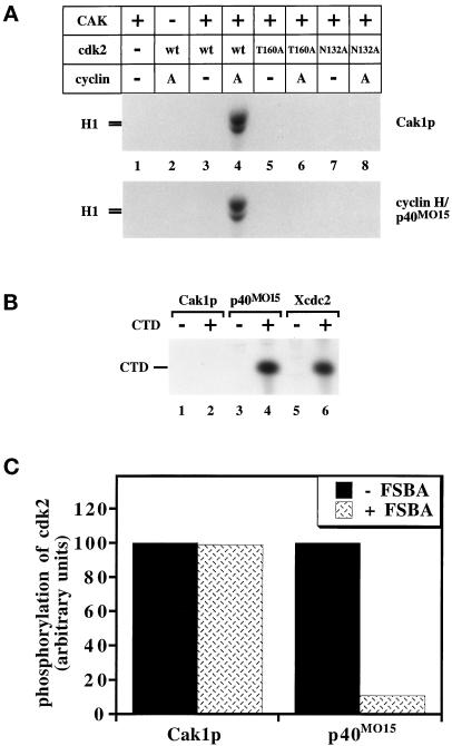
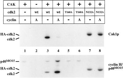
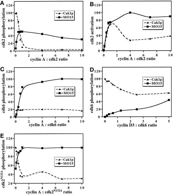
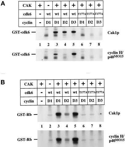
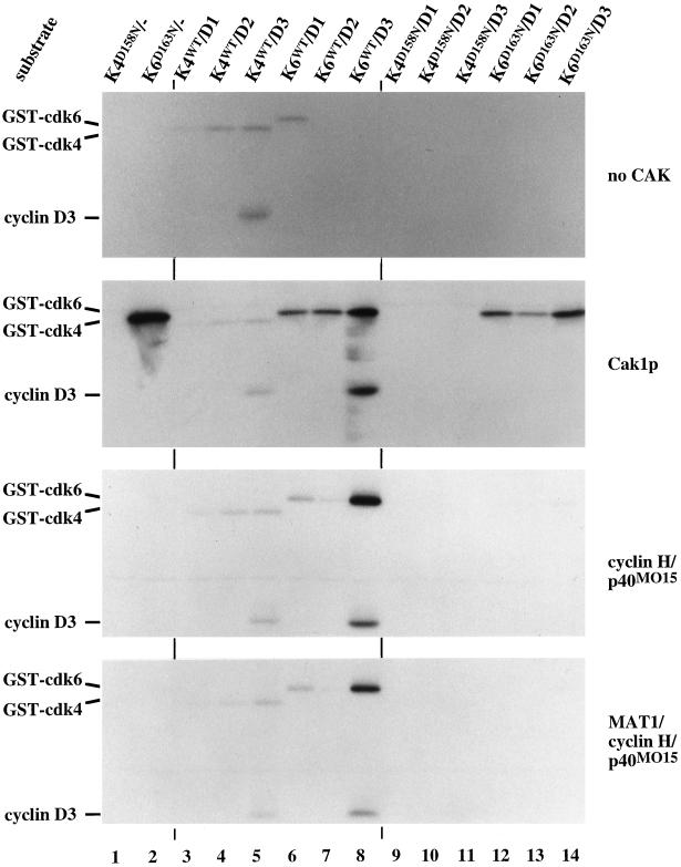
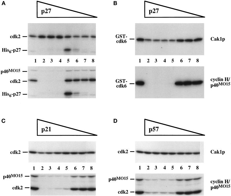
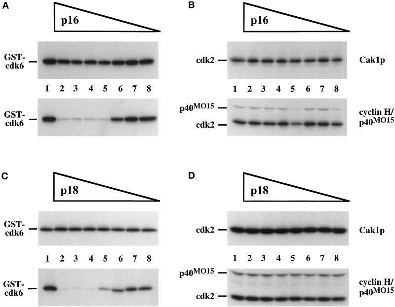
Cell cycle progression is controlled by the sequential functions of cyclin-dependent kinases (cdks). Cdk activation requires phosphorylation of a key residue (on sites equivalent to Thr-160 in human cdk2) carried out by the cdk-activating kinase (CAK). Human CAK has been identified as a p40(MO15)/cyclin H/MAT1 complex that also functions as part of transcription factor IIH (TFIIH) where it phosphorylates multiple transcriptional components including the C-terminal domain (CTD) of the large subunit of RNA polymerase II. In contrast, CAK from budding yeast consists of a single polypeptide (Cak1p), is not a component of TFIIH, and lacks CTD kinase activity. Here we report that Cak1p and p40(MO15) have strikingly different substrate specificities. Cak1p preferentially phosphorylated monomeric cdks, whereas p40(MO15) preferentially phosphorylated cdk/cyclin complexes. Furthermore, p40(MO15) only phosphorylated cdk6 bound to cyclin D3, whereas Cak1p recognized monomeric cdk6 and cdk6 bound to cyclin D1, D2, or D3. We also found that cdk inhibitors, including p21(CIP1), p27(KIP1), p57(KIP2), p16(INK4a), and p18(INK4c), could block phosphorylation by p40(MO15) but not phosphorylation by Cak1p. Our results demonstrate that although both Cak1p and p40(MO15) activate cdks by phosphorylating the same residue, the structural mechanisms underlying the enzyme-substrate recognition differ greatly. Structural and physiological implications of these findings will be discussed.


Figures

Figure 1
Figure 1
Basic properties of yeast and human CAKs. (A) The activation of wild-type cdk2 expressed in insect cells and of mutant forms of cdk2 expressed in bacteria by CAK in the presence or absence of cyclin A173–432 was monitored by the ability of cdk2 to phosphorylate histone H1. The T160A mutation alters the site of activating phosphorylation, whereas the N132A mutation renders cdk2 catalytically inactive. (B) Phosphorylation of the CTD peptide by GST-Cak1p (lanes 1 and 2), p40MO15/cyclin H (lanes 3 and 4), and Xenopus cdc2/cyclin B complexes (lanes 5 and 6). The CTD peptide was present in the even-numbered lanes. The same amounts of Cak1p and of p40MO15 were used as in panel A. (C) GST-Cak1p or p40MO15/cyclin H was preincubated with the irreversible inhibitory ATP-analog 5′-p-fluorosulfonylbenzoyladenosine (FSBA) before assaying for phosphorylation of cdk2/cyclin A173–432 (mass ratio, 1:0.75). As a control, FSBA was inactivated by incubation with DTT before addition to CAK (−FSBA lanes). Lanes represent the averages of three and five measurements for GST-Cak1p and p40MO15/cyclin H, respectively.
Figure 2
Figure 2
Phosphorylation of cdk2. Wild-type cdk2 expressed in insect cells and mutant forms of cdk2 expressed in bacteria were incubated in the presence of radiolabeled ATP with GST-Cak1p or p40MO15/cyclin H. Samples were analyzed on 10% SDS-PAGE for incorporation of radiolabeled phosphate by autoradiography. T160A contains a mutation of the activating phosphorylation site (lanes 5 and 6), whereas N132A renders cdk2 catalytically inactive (lanes 7 and 8). Cyclin A173–432 was present in the indicated lanes in a threefold mass excess over cdk2. The mobility of cdk2N132A is decreased due to the presence of an N-terminal influenza hemagglutinin (HA) epitope tag (lanes 7 and 8). A low level of phosphorylation of p40MO15 by active cdk2 complexes is apparent in lane 4 (see also Fisher et al., 1995).
Figure 3
Figure 3
Effect of cyclins on phosphorylation of cdks by Cak1p and p40MO15/cyclin H. Phosphorylation (A) and activation (B, measured as the phosphorylation of histone H1 by cdk2) of wild-type cdk2 or phosphorylation of cdk2N132A (E) by GST-Cak1p (triangles) and by p40MO15/cyclin H (squares) were determined in the presence of increasing concentrations of cyclin A173–432. Phosphorylation of cdk6 expressed in insect cells was analyzed in a similar manner in the presence of various concentrations of cyclin A173–432 (C), which binds cdk6 very weakly, or of His6-cyclin D3 (D). Note that His6-cyclin D3 binds less tightly to cdk6 in vitro (Kato et al., 1994a) than does cyclin A173–432 to cdk2, resulting in less pronounced effects on cdk phosphorylation. Data were analyzed by phosphorimaging and are expressed in arbitrary units. The cyclin:cdk ratios are mass ratios of the indicated proteins.
Figure 4
Figure 4
Cdk6 phosphorylation and activation by CAK. (A) Wild-type and mutant forms of GST-cdk6 expressed in bacteria were incubated in the presence of radiolabeled ATP with GST-Cak1p (upper panel) or p40MO15/cyclin H (lower panel). T177A contains a mutation of the activating phosphorylation site (lanes 6, 7, and 8). Purified His6-cyclin D1, D2, or D3 was present in the indicated lanes. (B) Activation of cdk6 by CAKs. Wild-type and mutant forms of GST-cdk6 expressed in bacteria were incubated with the indicated D-type cyclins and GST-Cak1p (upper panel) or p40MO15/cyclin H (lower panel) in the presence of unlabeled ATP. The reaction was then incubated with GST-Rb605–928 as a substrate in the presence of radiolabeled ATP. Samples were analyzed by 10% SDS-PAGE and autoradiography. His6-cyclin D1, D2, and D3 proteins were expressed in insect cells and purified via their His6-tags, whereas GST-cdk6 was expressed in E. coli.
Figure 5
Figure 5
Phosphorylation of cdk4 and cdk6-cyclin D complexes by CAK. Monomeric and cyclin-bound GST-cdk4 and GST-cdk6 complexes were purified from baculovirus-infected insect cells and used as substrates for autophosphorylation (upper panel) or for direct phosphorylation by GST-Cak1p (second panel), p40MO15/cyclin H (third panel), and p40MO15/cyclin H/MAT1 (bottom panel) in the presence of radiolabeled ATP. Samples were analyzed by 10% SDS-PAGE and autoradiography. Lanes 3–8 contained wild-type cdks whereas lanes 1, 2, and 9–14 contained catalytically inactive enzymes. The positions of the phosphorylated cdks and of cyclin D3 are indicated at the left. Note that the GST-cdk4/cyclin D complexes undergo significant autophosphorylation (top panel, lanes 3–5) and display Rb kinase activity (our unpublished results).
Figure 6
Figure 6
Cdk inhibitors from the CIP/KIP family block phosphorylation of cdks by p40MO15 but not by Cak1p. Cdk2 (A, C, and D) or GST-cdk6 (B) was phosphorylated directly by GST-Cak1p (upper panels) or by p40MO15/cyclin H (lower panels) in the presence of various concentrations of His6-p27 (A and B), p21 (C), or p57 (D), respectively. Lanes 1 contained no inhibitors. Mass ratios of inhibitor:cdk are 10:1 (lanes 2), 5:1 (lanes 3), 2.5:1 (lanes 4), 1.2:1 (lanes 5), 0.3:1 (lanes 6), 0.09:1 (lanes 7), and 0.03:1 (lanes 8). In all experiments the cdk:cyclin A173–432 ratio was chosen to be 1:0.75. In panel A, the position of His6-p27 that has been phosphorylated by cdk2/cyclin A173–432 is indicated.
Figure 7
Figure 7
Inhibitors of the INK4 family prevent phosphorylation of cdk6 by p40MO15 but not by Cak1p. GST-cdk6 (A and C) or cdk2 (B and D) was phosphorylated directly by GST-Cak1p (upper panels) or by p40MO15/cyclin H (lower panels) in the presence of various concentrations of p16 (A and B) or GST-p18 (C and D). Lanes 1 contained no inhibitors. Mass ratios of cdk:inhibitor are as in Figure 6. In all experiments the cdk:cyclin A173–432 ratio was chosen to be 1:0.75.

Similar articles

Cited by

References

    1. Adamczewski JP, Rossignol M, Tassan J-P, Nigg EA, Moncollin V, Egly J-M. MAT1, cdk7 and cyclin H form a kinase complex which is UV light-sensitive upon association with TFIIH. EMBO J. 1996;15:1877–1884. - PMC - PubMed
    1. Aprelikova O, Xiong Y, Liu ET. Both p16 and p21 families of cyclin-dependent kinase (CDK) inhibitors block the phosphorylation of cyclin-dependent kinases by the CDK-activating kinase. J Biol Chem. 1995;270:18195–18197. - PubMed
    1. Blain SW, Montalvo E, Massagué J. Differential interaction of the cyclin-dependent kinase (cdk) inhibitor p27Kip1 with cyclin A-cdk2 and cyclin D2-cdk4. J Biol Chem. 1997;272:25863–25872. - PubMed
    1. Canagarajah BJ, Khokhlatchev A, Cobb MH, Goldsmith EJ. Activation mechanism of the MAP kinase ERK2 by dual phosphorylation. Cell. 1997;90:859–869. - PubMed
    1. Chan FKM, Zhang J, Chen L, Shapiro DN, Winoto A. Identification of human and mouse p19, a novel CDK4 and CDK6 inhibitor with homology to p16ink4. Mol Cell Biol. 1995;15:2682–2688. - PMC - PubMed

Publication types