. 1998 Sep;9(9):2561-75.
doi: 10.1091/mbc.9.9.2561.

Rho and Rab small G proteins coordinately reorganize stress fibers and focal adhesions in MDCK cells

Affiliations
Free PMC article

Rho and Rab small G proteins coordinately reorganize stress fibers and focal adhesions in MDCK cells

H Imamura et al. Mol Biol Cell. 1998 Sep.
Free PMC article

Abstract

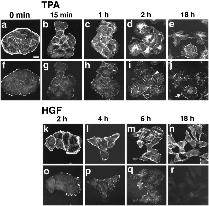
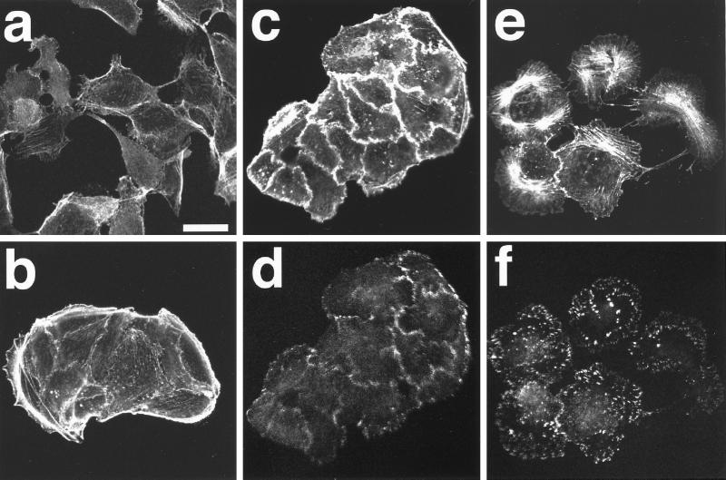
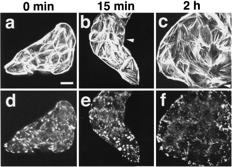
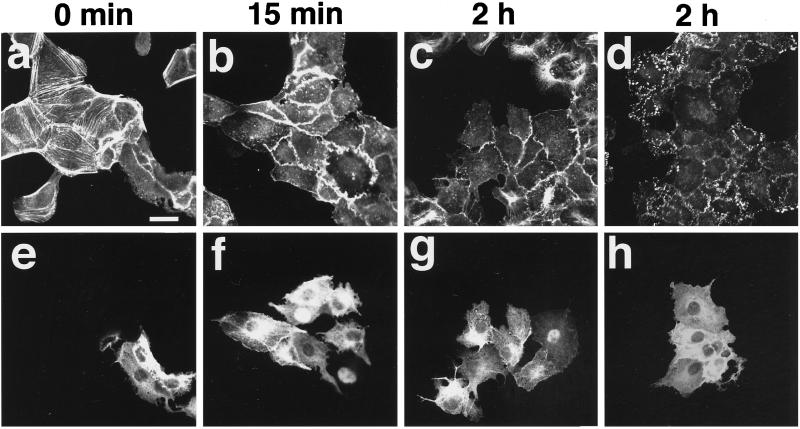
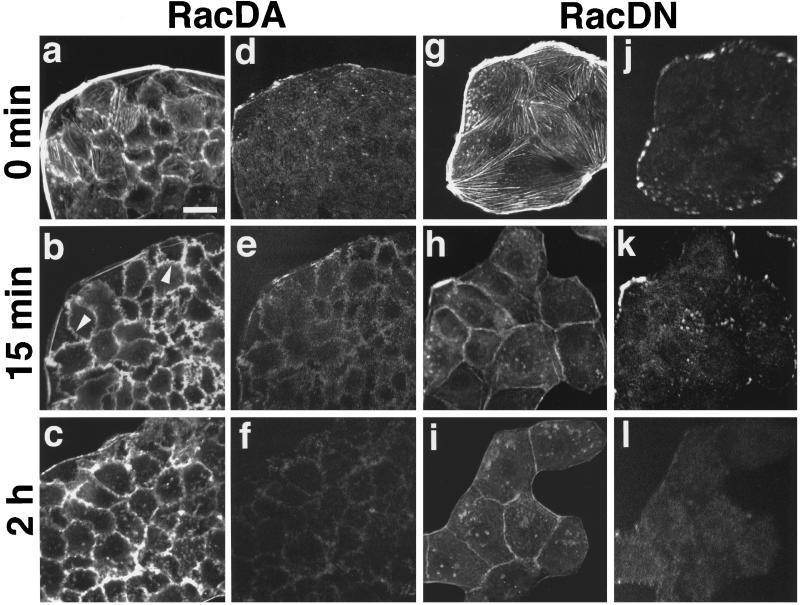
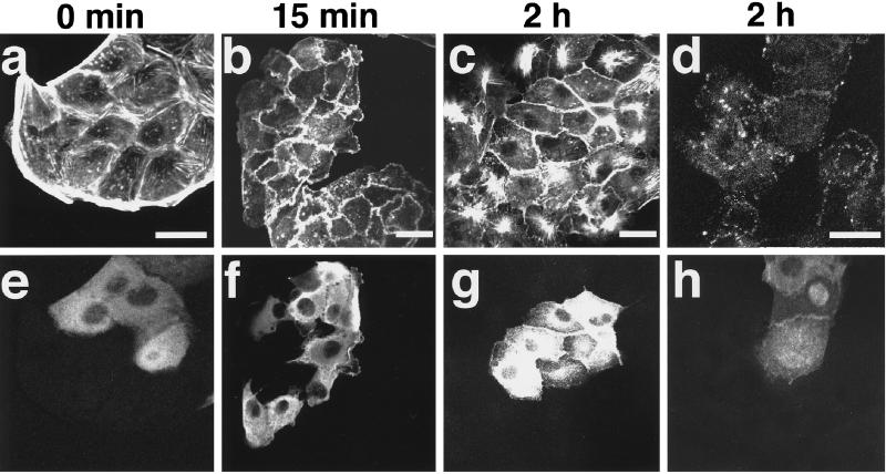
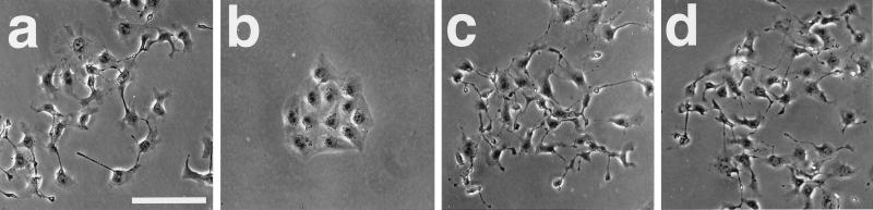
The Rho subfamily of the Rho small G protein family (Rho) regulates formation of stress fibers and focal adhesions in many types of cultured cells. In moving cells, dynamic and coordinate disassembly and reassembly of stress fibers and focal adhesions are observed, but the precise mechanisms in the regulation of these processes are poorly understood. We previously showed that 12-O-tetradecanoylphorbol-13-acetate (TPA) first induced disassembly of stress fibers and focal adhesions followed by their reassembly in MDCK cells. The reassembled stress fibers showed radial-like morphology that was apparently different from the original. We analyzed here the mechanisms of these TPA-induced processes. Rho inactivation and activation were necessary for the TPA-induced disassembly and reassembly, respectively, of stress fibers and focal adhesions. Both inactivation and activation of the Rac subfamily of the Rho family (Rac) inhibited the TPA-induced reassembly of stress fibers and focal adhesions but not their TPA-induced disassembly. Moreover, microinjection or transient expression of Rab GDI, a regulator of all the Rab small G protein family members, inhibited the TPA-induced reassembly of stress fibers and focal adhesions but not their TPA-induced disassembly, indicating that, furthermore, activation of some Rab family members is necessary for their TPA-induced reassembly. Of the Rab family members, at least Rab5 activation was necessary for the TPA-induced reassembly of stress fibers and focal adhesions. The TPA-induced, small G protein-mediated reorganization of stress fibers and focal adhesions was closely related to the TPA-induced cell motility. These results indicate that the Rho and Rab family members coordinately regulate the TPA-induced reorganization of stress fibers and focal adhesions that may cause cell motility.


Figures

Figure 1
Figure 1
TPA- and HGF/SF-induced reorganization of the actin cytoskeleton in wt MDCK cells. wt MDCK cells were incubated in DMEM containing 10% FCS for 24 h. After the incubation, the cells were stimulated with none (a and f), 100 nM TPA (b–e and g–j), or 20 ng/ml HGF/SF (k–r). The cells were fixed at 15 min (b and g), 1 h (c and h), 2 h (d and i), or 18 h (e and j) after TPA stimulation, and at 2 h (k and o), 4 h (l and p), 6 h (m and q), or 18 h (n and r) after HGF/SF stimulation, double stained with rhodamine-phalloidin (a–e and k–n) or the V115 anti-vinculin mAb (f–j and o–r), and analyzed by confocal microscopy. Confocal images are shown at the basal levels. The results shown are representative of three independent experiments. Bar, 10 μm. An arrowhead in panel i indicates the staining of vinculin at the newly formed focal adhesions. An arrow in panel j indicates the dot-like staining of vinculin that is located at the sites different from the focal adhesions.
Figure 2
Figure 2
Effect of cycloheximide on the TPA- and HGF/SF-induced reorganization of actin filaments in wt MDCK cells. wt MDCK cells were incubated in DMEM containing 10% FCS for 24 h. After the incubation, the cells were stimulated with 20 ng/ml HGF/SF (a and b) or 100 nM TPA (c–f) in the absence (a) or presence (b–f) of 10 μg/ml cycloheximide. Cycloheximide was added 30 min before HGF/SF or TPA stimulation. At 18 h after HGF/SF stimulation, the cells were fixed and stained with rhodamine-phalloidin (a and b). At 15 min (c and d) or 2 h (e and f) after TPA stimulation, the cells were fixed, double stained with rhodamine-phalloidin (c and e) or the V115 anti-vinculin mAb (d and f), and analyzed by confocal microscopy. Confocal images are shown at the basal levels. The results shown are representative of three independent experiments. Bar, 10 μm.
Figure 3
Figure 3
Inhibition of the TPA-induced disassembly of stress fibers and focal adhesions in sMDCK-RhoDA cells. sMDCK-RhoDA cells were incubated in DMEM containing 10% FCS for 24 h. After the incubation, the cells were stimulated with none (a and d) or 100 nM TPA (b, c, e, and f). The cells were fixed at 15 min (b and e) or 2 h (c and f) after TPA stimulation, double stained with rhodamine-phalloidin (a–c) or the V115 anti-vinculin mAb (d–f), and analyzed by confocal microscopy. Confocal images are shown at the basal levels. The results shown are representative of three independent experiments. Bar, 10 μm. Arrowheads in panels b and c indicate the reduced peripheral bundles.
Figure 4
Figure 4
Inhibition by C3 of the TPA-induced reassembly of stress fibers and focal adhesions. wt MDCK cells were incubated in DMEM containing 10% FCS for 24 h. After the incubation, the cells were microinjected with 40 μg/ml C3 plus 5 mg/ml rat IgG. At 30 min after the microinjection, the cells were stimulated with none (a and e) or 100 nM TPA (b–d and f–h). At 15 min (b and f) or 2 h (c, d, g, and h) after TPA stimulation, the cells were fixed, stained with rhodamine-phalloidin (a–c) or the V115 anti-vinculin mAb (d), and analyzed by confocal microscopy. The microinjected cells are shown by the staining of microinjected rat IgG (e–h). Confocal images are shown at the basal levels. The results shown are representative of three independent experiments. Bar, 10 μm.
Figure 5
Figure 5
Inhibition of the TPA-induced reassembly of stress fibers and focal adhesions in sMDCK-RacDA and -RacDN cells. sMDCK-RacDA and -RacDN cells were incubated in DMEM containing 10% FCS for 24 h. After the incubation, sMDCK-RacDA (a–f) and -RacDN (g–l) cells were stimulated with none (a, d, g, and j) or 100 nM TPA (b, c, e, f, h, i, k, and l). At 15 min (b, e, h, and k) or 2 h (c, f, i, and l) after TPA stimulation, the cells were fixed, double stained with rhodamine-phalloidin (a–c and g–i) or the V115 anti-vinculin mAb (d–f and j–l), and analyzed by confocal microscopy. Confocal images are shown at the basal levels. The results shown are representative of three independent experiments. Bar, 10 μm. Arrowheads in panel b indicate membrane ruffling at the cell–cell adhesion sites.
Figure 6
Figure 6
Inhibition by Rab GDI of the TPA-induced reassembly of stress fibers and focal adhesions. wt MDCK cells were incubated in DMEM containing 10% FCS for 24 h. After the incubation, the cells were microinjected with 5 mg/ml His6-Rab GDI plus 5 mg/ml rat IgG. At 30 min after the microinjection, the cells were stimulated with none (a and e) or 100 nM TPA (b–d and f–h). At 15 min (b and f) or 2 h (c, d, g, and h) after TPA stimulation, the cells were fixed, stained with rhodamine-phalloidin (a–c) or the V115 anti-vinculin mAb (d), and analyzed by confocal microscopy. The microinjected cells are shown by the staining of microinjected rat IgG (e–h). Confocal images are shown at the basal levels. The results shown are representative of three independent experiments. Bars, 10 μm.
Figure 7
Figure 7
Effect of transient expression of Rab GDI, Rab5DA, -5DN, -8DA, -8DN, -11DA, and -11DN on the TPA-induced disassembly and reassembly of stress fibers. wt MDCK cells were transfected with pEF-BOS-myc-Rab GDI, -L79Rab5 (Rab5DA), -N34Rab5 (Rab5DN), -L67Rab8 (Rab8DA), -N22Rab8 (Rab8DN), -L70Rab11 (Rab11DA), or -N25Rab11 (Rab11DN) using a lipofectAMINE reagent. At 24 h after the transfection, the cells were detached using an EDTA/trypsin solution, seeded onto 35-mm grid dishes, and further incubated in DMEM containing 10% FCS for 24 h. After the incubation, the cells were stimulated with 100 nM TPA and fixed at 15 min (A) or 2 h (B) after TPA stimulation. The cells were then double stained with rhodamine-phalloidin or the 9E10 anti-myc mAb to detect the transfected cells, and analyzed by confocal microscopy. (A) The percentage of the cells in which disassembly of stress fibers was observed at 15 min after TPA stimulation. (B) The percentage of the cells in which reassembly of stress fibers was observed at 2 h after TPA stimulation. None, untransfected cells; Rab GDI, the cells expressing myc-Rab GDI; Rab5DA, the cells expressing myc-L79Rab5; Rab5DN, the cells expressing myc-N34Rab5; Rab8DA, the cells expressing myc-L67Rab8; Rab8DN, the cells expressing myc-N22Rab8; Rab11DA, the cells expressing myc-L70Rab11; Rab11DN, the cells expressing myc-N25Rab11. The results shown are mean obtained from at least three independent experiments.
Figure 8
Figure 8
Inhibition of the TPA-induced reassembly of stress fibers by transient expression of Rab5DN. wt MDCK cells were transfected with pEF-BOS-myc-L79Rab5 (Rab5DA) (a and b), -N34Rab5 (Rab5DN) (c and d), -L67Rab8 (Rab8DA) (e and f), -N22Rab8 (Rab8DN) (g and h), -L70Rab11 (Rab11DA) (i and j), and -N25Rab11 (Rab11DN) (k and l) using a lipofectAMINE reagent. At 24 h after the transfection, the cells were detached using an EDTA/trypsin solution, seeded onto 35-mm grid dishes, and further incubated in DMEM containing 10% FCS for 24 h. After the incubation, the cells were stimulated with 100 nM TPA and fixed at 2 h after TPA stimulation. The cells were then double stained with rhodamine-phalloidin (a, c, e, g, i, and k) or the 9E10 anti-myc mAb (b, d, f, h, j, and l), and analyzed by confocal microscopy. Confocal images are shown at the basal levels. The results shown are representative of three independent experiments. Bar, 10 μm. Panels k and l show the Rab11DN-expressing cell in which the TPA-induced reassembly of stress fibers is observed, whereas the expression of Rab11DN inhibits their TPA-induced reassembly to a small extent (see Figure 7).
Figure 9
Figure 9
Inhibition of the TPA-induced reassembly of stress fibers and focal adhesions in sMDCK-Rab5DN cells. sMDCK-Rab5DN-10 (a–f), -Rab11DA-3 (g–l), or -Rab11DN-5 (m–r) cells were incubated in DMEM containing 10% FCS for 24 h. The cells were stimulated with none (a, d, g, j, m, and p) or 100 nM TPA (b, c, e, f, h, i, k, l, n, o, q, and r). At 15 min (b, e, h, k, n, and q) or 2 h (c, f, i, l, o, and r) after TPA stimulation, the cells were fixed, double stained with rhodamine-phalloidin (a–c, g–i, and m–o) or the V115 anti-vinculin mAb (d–f, j–l, and p–r), and analyzed by confocal microscopy. Confocal images are shown at the basal levels. The results shown are representative of three independent experiments. Bar, 10 μm.
Figure 10
Figure 10
Inhibition of the TPA-induced cell scattering in sMDCK-Rab5DN cells. wt MDCK (a), sMDCK-Rab5DN-10 (b), -Rab11DA-3 (c), or -Rab11DN-5 (d) cells were incubated in DMEM containing 10% FCS for 24 h. The cells were stimulated with 100 nM TPA. At 6 h after TPA stimulation, the cells were fixed, and analyzed by phase-contrast microscopy. The results shown are representative of three independent experiments. Bar, 50 μm.

Similar articles

Cited by

References

    1. Adamson P, Paterson HF, Hall A. Intracellular localization of the p21rho proteins. J Cell Biol. 1992;119:617–627. - PMC - PubMed
    1. Aktories K, Rösener S, Blaschke U, Chhatwal GS. Botulinum ADP-ribosyltransferase C3: purification of the enzyme and characterization of the ADP-ribosylation reaction in platelet membranes. Eur J Biochem. 1988;172:445–450. - PubMed
    1. Braga VMM, Machesky LM, Hall A, Hotchin NA. The small GTPases Rho and Rac are required for the establishment of cadherin-dependent cell-cell contacts. J Cell Biol. 1997;137:1421–1431. - PMC - PubMed
    1. Braun U, Habermann B, Just I, Aktories K, Vandekerckhove J. Purification of the 22kDa protein substrate of botulinum ADP-ribosyltransferase C3 from porcine brain cytosol and its characterization as a GTP-binding protein highly homologous to the rho gene product. FEBS Lett. 1989;243:70–76. - PubMed
    1. Bretcher MS. Getting membrane flow and the cytoskeleton to cooperate in moving cells. Cell. 1996;87:601–606. - PubMed

Publication types

LinkOut - more resources