. 1998 Sep;9(9):2627-38.
doi: 10.1091/mbc.9.9.2627.

Interactions between growth factors and integrins: latent forms of transforming growth factor-beta are ligands for the integrin alphavbeta1

Affiliations
Free PMC article

Interactions between growth factors and integrins: latent forms of transforming growth factor-beta are ligands for the integrin alphavbeta1

J S Munger et al. Mol Biol Cell. 1998 Sep.
Free PMC article

Abstract

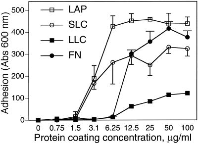
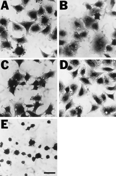
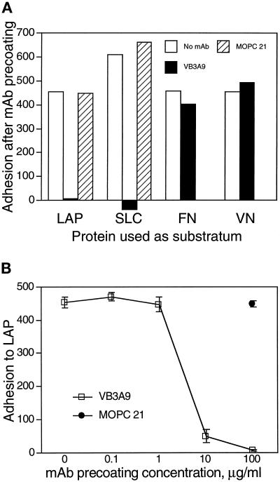
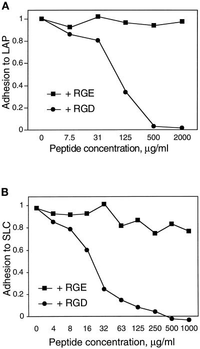
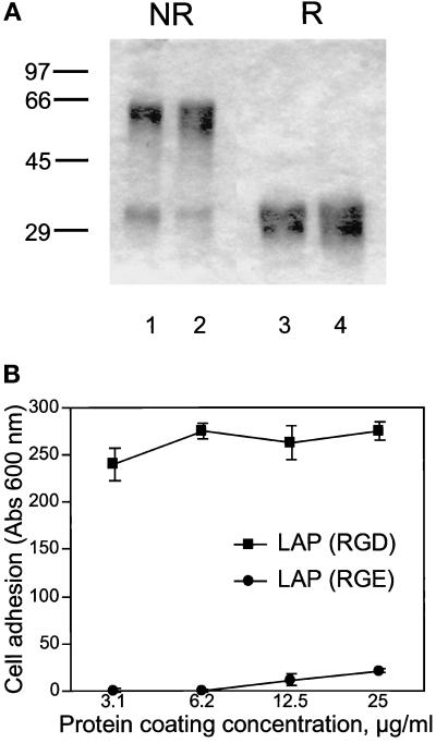
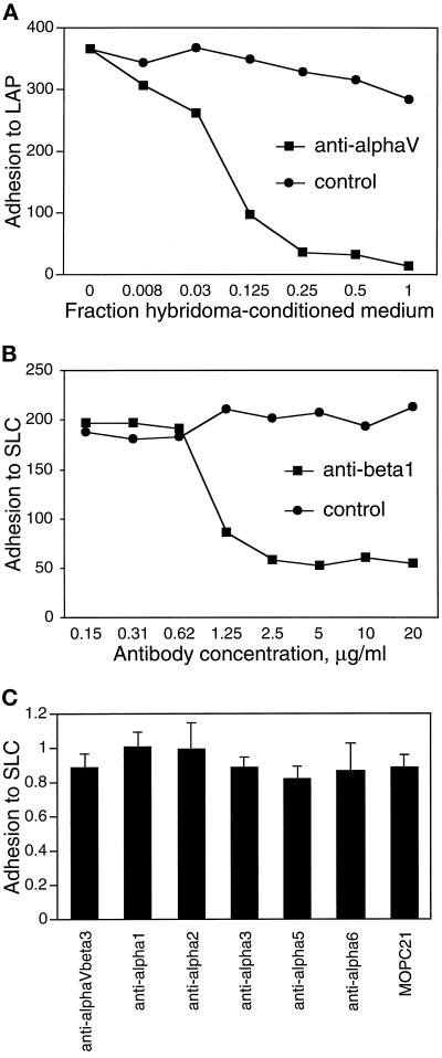
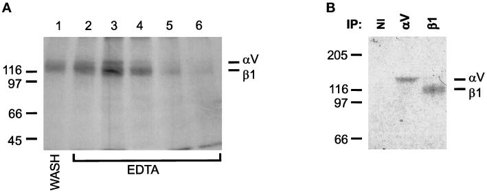
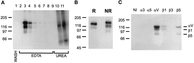
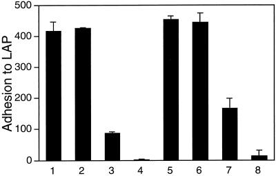
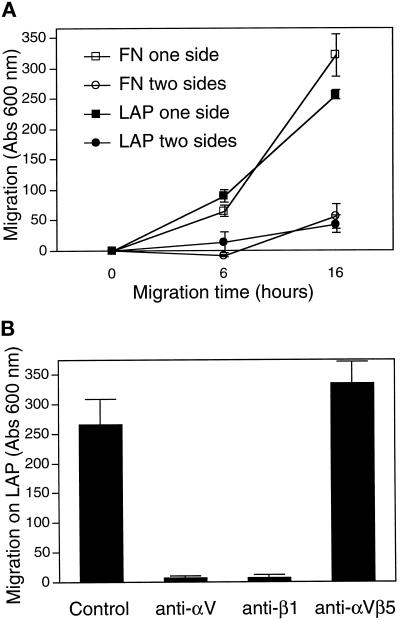
The multipotential cytokine transforming growth factor-beta (TGF-beta) is secreted in a latent form. Latency results from the noncovalent association of TGF-beta with its processed propeptide dimer, called the latency-associated peptide (LAP); the complex of the two proteins is termed the small latent complex. Disulfide bonding between LAP and latent TGF-beta-binding protein (LTBP) produces the most common form of latent TGF-beta, the large latent complex. The extracellular matrix (ECM) modulates the activity of TGF-beta. LTBP and the LAP propeptides of TGF-beta (isoforms 1 and 3), like many ECM proteins, contain the common integrin-binding sequence RGD. To increase our understanding of latent TGF-beta function in the ECM, we determined whether latent TGF-beta1 interacts with integrins. A549 cells adhered and spread on plastic coated with LAP, small latent complex, and large latent complex but not on LTBP-coated plastic. Adhesion was blocked by an RGD peptide, and cells were unable to attach to a mutant form of recombinant LAP lacking the RGD sequence. Adhesion was also blocked by mAbs to integrin subunits alphav and beta1. We purified LAP-binding integrins from extracts of A549 cells using LAP bound to Sepharose. alphavbeta1 eluted with EDTA. After purification in the presence of Mn2+, a small amount of alphavbeta5 was also detected. A549 cells migrated equally on fibronectin- and LAP-coated surfaces; migration on LAP was alphavbeta1 dependent. These results establish alphavbeta1 as a LAP-beta1 receptor. Interactions between latent TGF-beta and alphavbeta1 may localize latent TGF-beta to the surface of specific cells and may allow the TGF-beta1 gene product to initiate signals by both TGF-beta receptor and integrin pathways.


Figures

Figure 1
Figure 1
A549 cell adhesion to LAP, SLC, LLC, and FN. (A) Wells were coated with solutions of LAP, SLC, LLC, and FN, at various concentrations as shown, and blocked with BSA. A549 cells were allowed to adhere to the coated wells for 90 min, and nonadherent cells were washed off. Absorption at 600 nm indicates the amount of stain associated with adherent cells and is proportional to cells bound.
Figure 2
Figure 2
A549 cells adhere and spread on plastic coated with LAP (A), SLC (B), LLC (C), and FN (D) but not on plastic coated with poly-d-lysine (E). Bar, 100 μm.
Figure 3
Figure 3
A mAb to LAP blocks adhesion of A549 cells to LAP and SLC. Adhesion assays were done as described in Figure 1 using wells coated with 50-μg/ml solutions of LAP, SLC, FN, and VN. After being coated with a specific protein and blocked with BSA, wells were incubated with 0.1- to 100-μg/ml solutions of the mAbs VB3A9 (anti-LAP) and MOPC 21 (irrelevant control) and then washed with PBS and used for measuring cell adhesion. In A, mAbs were used at 100 μg/ml. (Binding to SLC was measured in a separate experiment; the greater binding to SLC than to LAP in the absence of mAb likely represents a difference in the number of cells added.) (B) The ability of VB3A9 preincubation to block adhesion to LAP is dose dependent.
Figure 4
Figure 4
An RGD-containing peptide blocks adhesion of A549 cells to LAP and SLC. Adhesion of A549 cells to wells coated with 33 μg/ml LAP (A) or SLC (B) was measured as described in Figure 1. Cells were incubated with the indicated concentrations of an RGD-containing peptide or control (RGE) peptide during the adhesion period. Adhesion is normalized to cells bound to poly-d-lysine in the absence of peptide.
Figure 5
Figure 5
A549 cells do not bind to a mutant form of recombinant LAP lacking the RGD site. (A) Recombinant LAP is homogeneous by SDS-PAGE. LAP was produced in insect cells and purified by affinity chromatography as described in MATERIALS AND METHODS. One form consists of the simian LAP sequence with a C33S mutation (lanes 2 and 4); the other form is identical except that the RGD sequence is changed to RGE (lanes 1 and 3). Three micrograms were loaded per lane. Samples were run reduced (R) or nonreduced (NR) and stained with Coomassie brilliant blue. (B) The two forms of recombinant LAP were coated on plastic microtiter wells and used for adhesion assays with A549 cells, as described in Figure 1.
Figure 6
Figure 6
mAbs against integrin subunits αv and β1 block adhesion of A549 cells to LAP. Adhesion of A549 cells to wells coated with 50 μg/ml LAP or SLC was determined as described in Figure 1, in the presence of the indicated concentrations of mAbs against integrins αv (A) and β1 (B). L230 (anti-αv) was used as dilutions of hybridoma-conditioned medium; medium conditioned by the irrelevant hybridoma 9E10 was used as a control. MOPC 21 served as a control for the anti-β1 mAb 4B4. (C) Blocking mAbs against other integrins or integrin subunits had no significant effect on A549 cell adhesion to SLC. Anti-αvβ3 was used at 20 μg/ml; all other mAbs were used at 25 μg/ml. Control attachment levels for the different conditions were similar. The mAbs used are listed in MATERIALS AND METHODS.
Figure 7
Figure 7
Affinity purification of LAP-binding integrins. Recombinant LAP was coupled to Sepharose 4B beads as described in MATERIALS AND METHODS. A549 cells were surface labeled with 125I and extracted with OSGP. Extracts were loaded on the LAP column and washed in buffer containing 1 mM Ca2+ and Mg2+. Retained proteins were eluted with 10 mM EDTA and collected in 1-ml fractions. (A) EDTA-eluted integrins. Aliquots of each fraction were separated by SDS-PAGE and autoradiographed. Lane 1, last wash fraction; lanes 2–6, elution with EDTA. (B) Immunoprecipitation with polyclonal Abs against cytoplasmic domains of αv and β1. Fractions 2–6 were pooled; equal volumes were immunoprecipitated with antisera to αv and β1 or with nonimmune serum (NI). Immunoprecipitates were separated by SDS-PAGE and autoradiographed. Gels were 8% and nonreducing. The positions of protein standards are shown to the side of each gel (masses in kilodaltons).
Figure 8
Figure 8
Affinity purification of LAP-binding integrins in the presence of Mn2+. LAP-Sepharose affinity chromatography was done as described in Figure 8, except that buffers contained 1 mM Ca2+, Mg2+, and Mn2+. (A) Autoradiograph of fractions from the LAP affinity column, run nonreduced on a 7% gel. Lane 1: last wash fraction; lanes 2–8, elution with EDTA; lanes 9–11, elution with 6 M urea. (B) The EDTA-eluted fractions shown in B were pooled. Aliquots were electrophoresed under reducing (R) and nonreducing (NR) conditions on 7% gels and autoradiographed. (C) The same sample was divided into equal aliquots and immunoprecipitated with polyclonal antisera to cytoplasmic domains of the indicated integrin subunits or with nonimmune rabbit serum (NI). Immunoprecipitates were separated nonreduced on 7% gels and autoradiographed.
Figure 9
Figure 9
Effect of αvβ5 blocking antibody on A549 cell adhesion to LAP. Adhesion of A549 cells to immobilized LAP was measured as described in Figure 1, with the addition of mAbs to β1 (4B4, 10 μg/ml), αvβ5 (P1F6, 1:1000 dilution of ascites fluid), or both during the adhesion period. Adhesion was tested with or without addition of 0.5 mM MnCl2 to the medium. Additions are indicated in the bottom panel.
Figure 10
Figure 10
A549 cells migrate equally on LAP and FN. (A) Cell motility assays were performed in Boyden chambers as described in MATERIALS AND METHODS. The results indicate the number of cells that migrated from the top to the bottom of a porous filter that was coated on the bottom or on both sides with a test protein. (B) Cells were allowed to migrate on LAP as in A for 16 h, in the presence or absence of mAbs to αv (50% L230 hybridoma medium), β1 (10 μg/ml 4B4), or αvβ5 (1:500 dilution of P1F6 ascites fluid).

References

    1. Akiyama SK, Yamada SS, Chen WT, Yamada KM. Analysis of fibronectin receptor function with monoclonal antibodies: roles in cell adhesion, migration, matrix assembly, and cytoskeletal organization. J Cell Biol. 1989;109:863–875. - PMC - PubMed
    1. Balzac F, Retta SF, Albini A, Melchiorri A, Koteliansky VE, Geuna M, Silengo L, Tarone G. Expression of beta1B integrin isoform in CHO cells results in a dominant negative effect on cell adhesion and motility. J Cell Biol. 1994;127:557–565. - PMC - PubMed
    1. Bodary SC, McLean JW. The integrin β1 subunit associates with the vitronectin receptor αv subunit to form a novel vitronectin receptor in a human embryonic kidney cell line. J Biol Chem. 1990;265:5938–5941. - PubMed
    1. Brooks PC, Stromblad S, Sanders LC, von Schalscha TL, Aimes RT, Stetler-Stevenson WG, Quigley JP, Cheresh DA. Localization of matrix metalloproteinase MMP-2 to the surface of invasive cells by interaction with integrin αvβ3. Cell. 1996;85:683–693. - PubMed
    1. Dallas SL, Miyazono K, Skerry TM, Mundy GR, Bonewald LF. Dual role for the latent transforming growth factor-β binding protein in storage of latent TGF-β in the extracellular matrix and as a structural protein. J Cell Biol. 1995;131:539–549. - PMC - PubMed

Publication types

LinkOut - more resources