. 1998 Sep;64(9):3134-9.
doi: 10.1128/AEM.64.9.3134-3139.1998.

Signal transduction in the protozoan host Hartmannella vermiformis upon attachment and invasion by Legionella micdadei

Affiliations

Signal transduction in the protozoan host Hartmannella vermiformis upon attachment and invasion by Legionella micdadei

Y Abu Kwaik et al. Appl Environ Microbiol. 1998 Sep.

Abstract

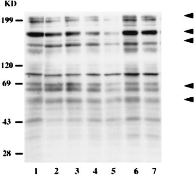
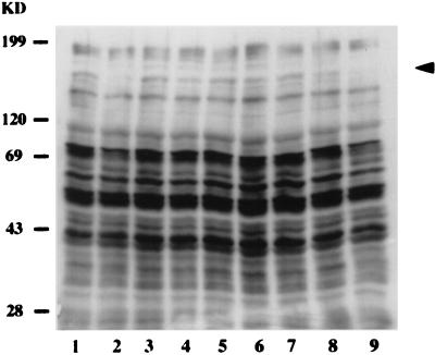
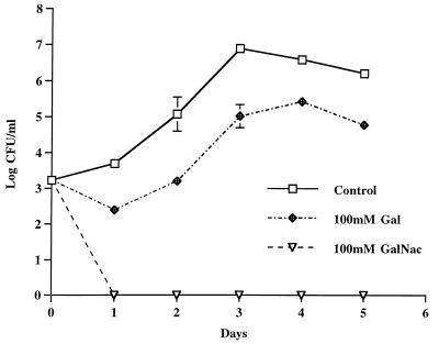
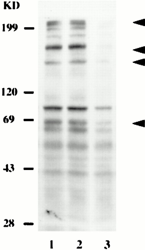
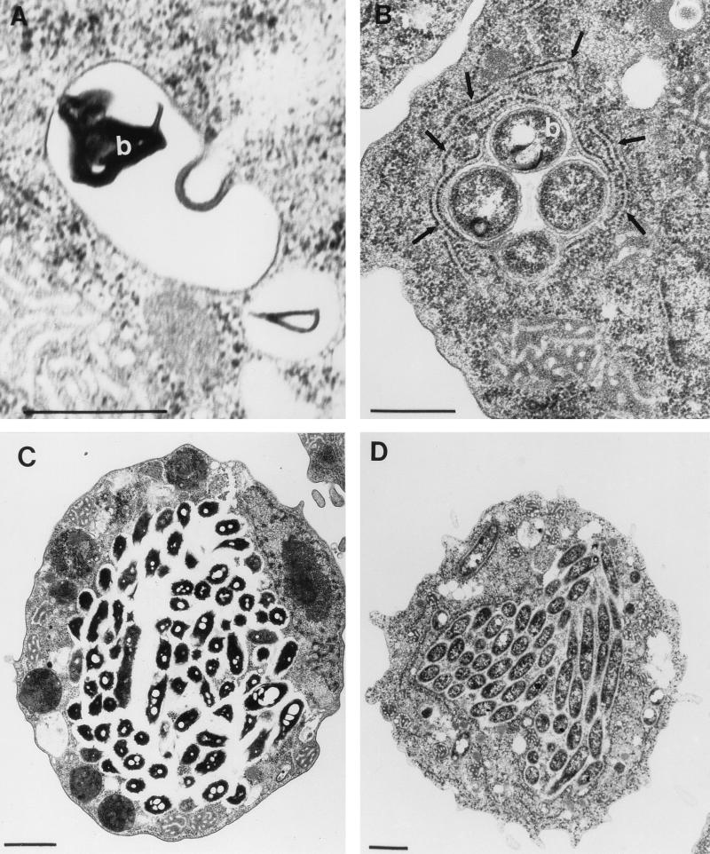
The intracellular pathogens Legionella micdadei and Legionella pneumophila are the two most common Legionella species that cause Legionnaires' disease. Intracellular replication within pulmonary cells is the hallmark of Legionnaires' disease. In the environment, legionellae are parasites of protozoans, and intracellular bacterial replication within protozoans plays a major role in the transmission of Legionnaires' disease. In this study, we characterized the initial host signal transduction mechanisms involved during attachment to and invasion of the protozoan host Hartmannella vermiformis by L. micdadei. Bacterial attachment prior to invasion of H. vermiformis by L. micdadei is associated with tyrosine dephosphorylation of multiple host cell proteins, including a 170-kDa protein. We have previously shown that this 170-kDa protein is the galactose N-acetylgalactosamine (Gal/GalNAc)-inhibitable lectin receptor that mediates attachment to and invasion of H. vermiformis by L. pneumophila. Subsequent bacterial entry targets L. micdadei into a phagosome that is not surrounded by the rough endoplasmic reticulum (RER). In contrast, uptake of L. pneumophila mediated by attachment to the Gal/GalNAc lectin is followed by targeting of the bacterium into an RER-surrounded phagosome. These results indicate that despite similarities in the L. micdadei and L. pneumophila attachment-mediated signal transduction mechanisms in H. vermiformis, the two bacterial species are targeted into morphologically distinct phagosomes in their natural protozoan host.


Figures

FIG. 1
FIG. 1
Attachment to and invasion of H. vermiformis by L. micdadei induces reversible tyrosine dephosphorylation of several host cell proteins. H. vermiformis cell extracts were prepared from uninfected cells (lane 1) or cells infected for 1, 5, 15, and 30 min (lanes 2 through 5), subjected to sodium dodecyl sulfate-polyacrylamide gel electrophoresis, and probed with anti-phosphotyrosine antibody. Lane 6 contained cell extracts from uninfected cells incubated at 37°C for 30 min. Lane 7 contained extracts from H. vermiformis infected for 30 min; then the extracellular bacteria were washed off and the amoebae were incubated for 15 min. Arrowheads indicate the positions of proteins that were markedly tyrosine dephosphorylated. The second arrowhead from the top indicates the position of the 170-kDa Gal/GalNAc lectin (34). The results are representative of the results of at least two independent experiments. KD, kilodaltons.
FIG. 2
FIG. 2
Tyrosine dephosphorylation of H. vermiformis proteins induced by L. micdadei is specific and requires attachment of the bacteria to the Gal/GalNAc lectin. H. vermiformis cell extracts were prepared from uninfected cells (lane 1) or cells infected for 30 min with live (lane 2) or dead (lane 3) bacteria. Amoebal extracts were also prepared after stimulation with latex beads (lane 4), bacterial culture supernatants (lane 5), or supernatant collected from an infection (lane 6) as additional controls. Lanes 7 through 9 contained host cell extracts prepared from infections in the presence of Gal, GalNAc, and mannose, respectively. The arrowhead indicates the position of the 170-kDa Gal/GalNAc lectin (34). The results are representative of the results of at least two independent experiments. KD, kilodaltons.
FIG. 3
FIG. 3
Inhibition of invasion of H. vermiformis by L. micdadei in the presence of different sugar monomers. The growth kinetics of L. pneumophila in cocultures with H. vermiformis were determined in the absence or presence of sugars at a concentration of 100 mM. At several times after infection, the numbers of bacteria in the cocultures were determined following growth on agar plates. The bacteria did not replicate extracellularly in the cocultures, and thus the increase in the number of bacteria was due to intracellular replication.
FIG. 4
FIG. 4
Tyrosine dephosphorylation of H. vermiformis proteins induced by L. micdadei does not require entry of the bacteria. H. vermiformis cell extracts were prepared from uninfected cells in the absence (lane 1) or presence (lane 2) of methylamine. Lane 3 contained cell lysates prepared after 30 min of infection of H. vermiformis with L. micdadei in the presence of methylamine. The arrowheads indicate the positions of the proteins that were tyrosine dephosphorylated. The second arrowhead from the top indicates the position of the 170-kDa Gal/GalNAc lectin (34). The results are representative of the results of at least two independent experiments. KD, kilodaltons.
FIG. 5
FIG. 5
L. micdadei and L. pneumophila are targeted into morphologically distinct phagosomes in H. vermiformis: transmission electron micrographs of H. vermiformis infected with L. micdadei (A and C) or L. pneumophila (B and D). The micrographs were obtained 4 h postinfection (A and B) or 18 h postinfection (C and D). The arrows in panel B indicate the RER-surrounded phagosome. b, bacteria. (A and B) Bars = 0.5 μm. (C and D) Bars = 1 μm.

References

    1. Abu Kwaik Y. The phagosome containing Legionella pneumophila within the protozoan Hartmanella vermiformis is surrounded by the rough endoplasmic reticulum. Appl Environ Microbiol. 1996;62:2022–2028. - PMC - PubMed
    1. Abu Kwaik Y. Induced expression of the Legionella pneumophila gene encoding a 20-kilodalton protein during intracellular infection. Infect Immun. 1998;66:203–212. - PMC - PubMed
    1. Abu Kwaik Y, Eisenstein B I, Engleberg N C. Phenotypic modulation by Legionella pneumophila upon infection of macrophages. Infect Immun. 1993;61:1320–1329. - PMC - PubMed
    1. Abu Kwaik Y, Engleberg N C. Cloning and molecular characterization of a Legionella pneumophila gene induced by intracellular infection and by various in vitro stress stimuli. Mol Microbiol. 1994;13:243–251. - PubMed
    1. Abu Kwaik Y, Fields B S, Engleberg N C. Protein expression by the protozoan Hartmannella vermiformis upon contact with its bacterial parasite, Legionella pneumophila. Infect Immun. 1994;62:1860–1866. - PMC - PubMed

Publication types

MeSH terms

LinkOut - more resources