. 1998 Aug;9(2):247-56.
doi: 10.1016/s1074-7613(00)80607-x.

CD30 is a CD40-inducible molecule that negatively regulates CD40-mediated immunoglobulin class switching in non-antigen-selected human B cells

Affiliations

CD30 is a CD40-inducible molecule that negatively regulates CD40-mediated immunoglobulin class switching in non-antigen-selected human B cells

A Cerutti et al. Immunity. 1998 Aug.

Abstract

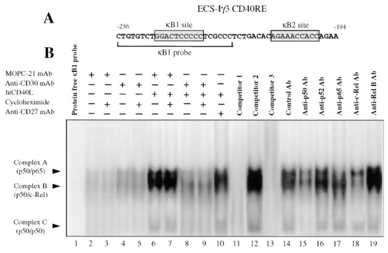
We used our monoclonal model of germinal center maturation, CL-01 B cells, to investigate the role of CD30 in human B cell differentiation. CL-01 cells are IgM+ IgD+ CD30+ and switch to IgG, IgA, and IgE when exposed to CD40L and IL-4. Switching is hampered by CD30 coengagement, possibly through interference with the CD40-mediated NF-kappaB-dependent transcriptional activation of downstream C(H) genes. The physiological relevance of this phenomenon is emphasized by similar CD30-mediated effects in naive B cells. Expression of CD30 by these cells is induced by CD40L but is inhibited by B cell receptor coengagement and/or exposure to IL-6 and IL-12. Our data suggest that CD30 critically regulates the CD40-mediated differentiation of non-antigen-selected human B cells.


Figures

Figure 1
Figure 1. sIgM+sIgD+ CL-01 B Cells Express Surface CD30 Whose Engagement Negatively Modulates CD40L and IL-4-Induced Switching to IgG, IgA, and IgE but Not Surface CD40 Expression
(A and B) Upon incubation for 2 days in medium only or in medium containing 0.5 μg/ ml of htCD40L, 5 μg/ml of control MOPC-21 MAb, or 5 μg/ml of MAb to human CD30, CL-01 cells were analyzed for expression of surface CD30 or CD40. (C and D) CL-01 cells were cultured for 5 days in medium only or in medium containing IL-4 alone (100 U/ml), htCD40L alone (0.5 μg/ml), or htCD40L and IL-4 in the presence of either MOPC-21 or anti-CD30 MAbs (5 μg/ml). Equal amounts of template cDNA were used to PCR amplify IH-CH (C), VHDJH-CH (D), and β-actin transcripts (C and D). Amplified products were then fractionated into 2% ethidium bromide–stained agarose gel. These findings were derived from one of three experiments yielding comparable results.
Figure 2
Figure 2. Normal sIgM+sIgD+ B Cells Express CD30 mRNA, Surface CD30, and GC Markers upon CD40 Engagement by CD40L; BCR Coengagement Down-Regulates Surface CD30 Expression
Naive B cells were cultured for 3 days in medium only (A) or in medium containing 0.5 μg/ml of htCD40L (B) or htCD40L and 2 μg/ml of anti-κ and anti-λ chain (anti-BCR) MAbs (C) and then analyzed for expression of surface CD19, CD30, CD71, CD80, CD86, and CD95. β-actin and CD30 cDNAs from the same cells were PCR amplified and fractioned into a 2% ethidium bromide–stained agarose gel. These findings were derived from one of five experiments yielding comparable results.
Figure 3
Figure 3. Stimuli Other than CD40L Do Not Induce CD30 Expression in Normal sIgM+sIgD+ B Cells
Naive B cells were cultured for 3 days in medium only or in medium containing IL-1β (10 ng/ml), IL-2 (200 U/ml), IL-4 (200 U/ml), IL-7 (100 U/ml), IL-10 (200 ng/ml), IL-13 (5 ng/ml), IFNγ (200 U/ml), IFNα (200 U/ml), TNFα (2.5 ng/ml), LTβ (2.5 ng/ml), TGFβ (0.5 ng/ml), IL-6 (200 ng/ml), IL-12 (1 ng/ml), immobilized anti-κ and anti-λ (anti-BCR) MAbs (2 μg/ml each), and a combination of IL-6, IL-12, and anti-BCR Ab in the presence (full histograms) or in the absence (open histograms) of htCD40L (0.5 μg/ml). Cells were then reacted with FITC-conjugated anti-CD30 MAb and PE-conjugated anti-CD19 MAbs and analyzed by flow cytometric analysis. The percentage of CD19+CD30+ B cells corresponds to the proportion of cells reacted with the above MAbs that display a fluorescence intensity higher than that displayed by similar cells reacted with control FITC- and PE-conjugated isotype matched MAbs. Values are mean ± SD of three determinations from three independent experiments.
Figure 4
Figure 4. CD30 Engagement in the Absence of BCR Coengagement Inhibits Ig Class Switching but Not Proliferation in Normal sIgM+sIgD+ B Cells Exposed to CD40L and Appropriate Cytokines
Naive B cells were preincubated for 2 days with medium only, htCD40L (0.5 μg/ml), or htCD40L and immobilized anti-κ and anti-λ chain (anti-BCR) MAbs (2 μg/ml each). Cells were then harvested, washed, and seeded in culture plates coated with either MOPC-21 (open histograms) or anti-CD30 (full histograms) MAbs (5 μg/ml) in the presence of medium only or medium containing IL-4 (100 U/ml) and IL-10 (100 ng/ml); htCD40L; or htCD40L, IL-4, and IL-10. In selected experiments, TGFβ1 (0.5 ng/ml) was added to htCD40L, IL-4, and IL-10 to optimize IgA secretion. After 8 days, the concentration of IgM, IgG, IgA, and IgE in the culture fluids was measured by ELISA. To measure DNA synthesis, B cells (1 × 10⁵/well) were cultured as above for 5 days and then pulsed for 18 hr with [3H]TdR. B cells were harvested at day 3 and counted (200 cells) by trypan blue dye exclusion to measure cell viability. Values are mean ± SD of four determinations from three independent experiments.
Figure 5
Figure 5
CD30 Engagement Down-Regulates the CD40-Mediated NF-κB-Dependent Activation of the Human Cγ3 Germline Gene Promoter CL-01 cells were transiently cotransfected with a pRL-CMV control luciferase plasmid and either a wild-type (wt) ECS-Iγ3-pGL3 (LUC) plasmid (A) or an ECS-Iγ3 CD40RE-SV40-pGL3 (LUC) minimal promoter plasmid (B). The top graphics depict the organization of these constructs. The initiation of transcription within the wt ECS-Iγ3 promoter is indicated by an arrow, and numbering is such that +1 corresponds to the first residue after this initiation site. After transfection, B cells were seeded in culture plates coated with either MOPC-21 (open histograms) or agonistic anti-CD30 MAbs (full histograms) in medium only or in medium containing IL-4 (500 U/ml); htCD40L (0.5 μg/ml); htCD40L and IL-4; or htCD40L, IL-4, and anti-IFNγ Abs (1:500). Luciferase activity was measured after 24 hr. Values are mean ± SD of three independent experiments.
Figure 6
Figure 6. CD30 Engagement Down-Regulates the CD40-Mediated Binding of NF-κB to the CD40RE of the Human Cγ3 Germline Gene Promoter
(A) Sequence of the ECS-Iγ3 CD40RE encompassed by the κB1 probe. The two κB sites within the CD40RE are boxed. (B) CL-01 cells were cultured with MOPC-21 MAb (5 μg/ml), anti-CD30 MAb (5 μg/ml), MOPC-21 MAb and htCD40L (0.5 μg/ml), anti-CD30 MAb and htCD40L, or anti-CD27 MAb (5 μg/ml) and htCD40L in the presence or in the absence of 100 μg/ml of cycloheximide (CHX). Nuclear proteins were extracted after 6 hr of culture, incubated with a radiolabeled κB1 oligonucleotide for 30 min, and then fractionated through a 6% polyacrylamide gel (lanes 2–10; lane 1 corresponds to the protein-free probe). The specificity of shifted complexes A, B, and C, and their identity to NF-κB complexes were established by pre-incubating nuclear proteins from CD40L-induced cells with competitors 1 (unlabeled κB1 probe), 2 (unlabeled κB1 mutant), and 3 (palindromic NF-κB binding sequence from the Ig κ promoter) (lanes 11–13). The composition of complexes A, B, and C was determined by preincubating nuclear proteins with rabbit Abs to human thyreoglobulin (control), p50, p52, p65, c-Rel, or Rel B proteins (lanes 14–19).
Figure 7
Figure 7. Role of CD30 in the Regulation of Human CD40-Dependent B Cell Differentiation
In the upper limb of the scheme, a naive CD30 B cell differentiates into a GC B cell and undergoes switching to IgG, IgA, or IgE upon CD40 engagement by CD40L expressed by an activated CD4+ T cell and exposure to T cell–derived cytokines. BCR coengagement by Ag as well as IL-6 and IL-12 secreted by an activated dendritic cell inhibit the expression of CD30 by this GC B cell. In the lower limb, a naive B cell is activated by a CD4+CD40L+ T cell in the absence of BCR coengagement by Ag complexes on the surface of a dendritic cell and, therefore, expresses CD30. In this non-Ag-selected B cell, CD40L-induced switching to IgG, IgA, or IgE is negatively modulated through CD30 engagement by CD30L expressed on either a CD4+ or a CD8+ T cell.

Similar articles

Cited by

References

    1. Ansieau S, Scheffrahn I, Mosialos G, Brand H, Duyster J, Kaye K, Harada J, Dougall B, Hubinger G, Kieff E, et al. Tumor necrosis factor receptor associated (TRAF)-1, TRAF-2, and TRAF-3 interact in vivo with the CD30 cytoplasmic domain; TRAF-2 mediates CD30-induced nuclear factor κ B activation. Proc Natl Acad Sci USA. 1996;93:14053–14058. - PMC - PubMed
    1. Armitage RJ, Sato AT, Macduff BM, Clifford KN, Alpert AR, Smith A, Fanslow WC. Identification of a source of biologically active CD40 ligand. Eur J Immunol. 1992;22:2071–2076. - PubMed
    1. Aruffo A, Farrington M, Hollenbaugh D, Li X, Milatovich A, Nonoyama S, Bajorath J, Grosmaire LS, Stenkamp R, Nebauer M, Roberts RL, et al. The CD40 ligand, gp39, is defective in activated T cells from patients with X-linked hyper IgM syndrome. Cell. 1993;72:291–300. - PubMed
    1. Cerutti A, Zan H, Schaffer A, Bergsagel L, Harindranath N, Max EE, Casali P. CD40 ligand and appropriate cytokines induce switching to IgG, IgA, and IgE and coordinated germinal center and plasmacytoid phenotypic differentiation in a human monoclonal IgM+ IgD+ B cell line. J Immunol. 1998;160:2145–2157. - PMC - PubMed
    1. Choe J, Kim HS, Zhang X, Armitage RJ, Choi YS. Cellular and molecular factors that regulate the differentiation and apoptosis of germinal center B cells. Anti-Ig down-regulates Fas expression on CD40 ligand-stimulated germinal center B cells and inhibits Fas-mediated apoptosis. J Immunol. 1996;157:1006–1016. - PubMed

Publication types

MeSH terms