. 1998 Sep;5(5):645-53.
doi: 10.1128/CDLI.5.5.645-653.1998.

Human immunodeficiency virus type 1-like DNA sequences and immunoreactive viral particles with unique association with breast cancer

Affiliations

Human immunodeficiency virus type 1-like DNA sequences and immunoreactive viral particles with unique association with breast cancer

E M Rakowicz-Szulczynska et al. Clin Diagn Lab Immunol. 1998 Sep.

Abstract

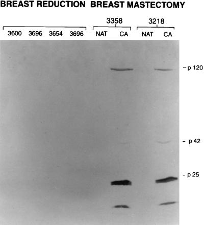
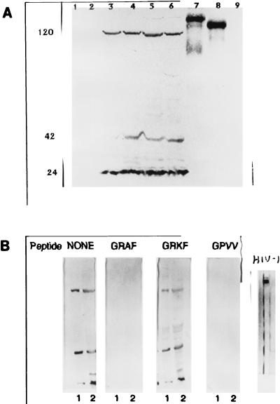
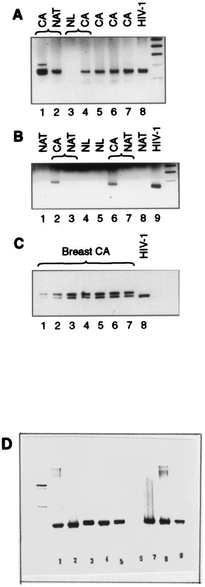
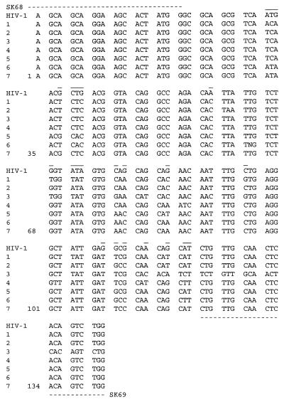
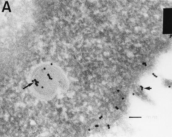
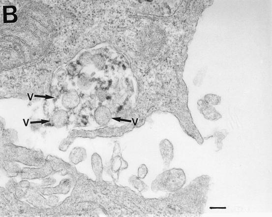
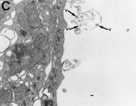
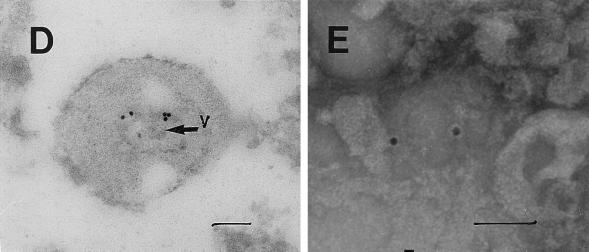
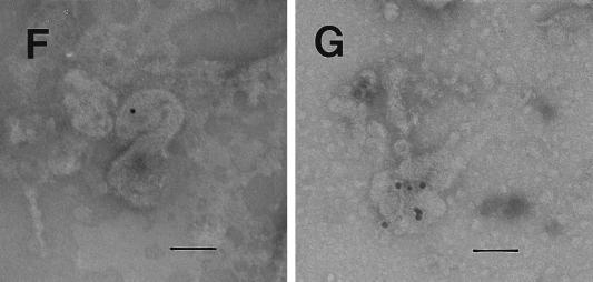
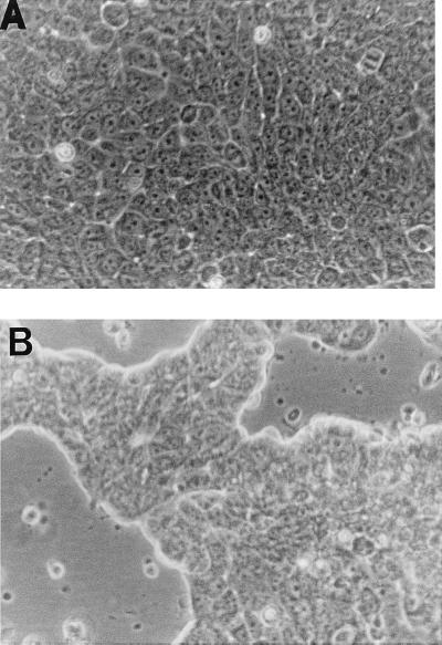
RAK antigens p120, p42, and p25 exhibit molecular and immunological similarity to the proteins encoded by human immunodeficiency virus type 1 (HIV-1) and are expressed by 95% of breast and gynecological cancer cases in women and prostate cancer cases in men. The binding of an epitope-specific anti-HIV-1 gp120 monoclonal antibody (MAb) (amino acids 308 to 322) to cancer RAK antigens has been found to be inhibited by a peptide derived from variable loop V3 of HIV-1. Breast cancer DNAs of 40 patients were PCR amplified with HIV-1 gp41-derived primers, and all of the samples were found to be positive. The DNA fragments amplified in seven blindly selected breast cancer samples were sequenced. The breast cancer DNA sequences showed at least 90% homology to the HIV-1 gene for gp41. Antisense oligonucleotides complementary to the HIV-1-like sequences inhibited reverse transcriptase activity and inhibited the growth of breast cancer cells in vitro. Viral particles detected in breast cancer cell lines were strongly immunogold labeled with the anti-HIV-1 gp120 MAb. The results obtained strongly suggest that the long-postulated breast cancer virus may, in fact, be related to HIV-1.


Figures

FIG. 1
FIG. 1
Electrophoretic analysis (10% polyacrylamide gel) and Western blotting of cytoplasmic proteins with MAb RAK-BrI. In a blind experiment, both breast cancers (CA) (3358 and 3218) tested RAK antigen positive and NAT samples tested RAK antigen negative. No RAK antigens were detected in the four breast tissue samples obtained during breast reduction (3600, 3696, 3654, and 3696).
FIG. 2
FIG. 2
Reactivity of an anti-HIV-1 gp120 MAb with breast and gynecological cancer cytoplasmic proteins and HIV-1 proteins. (A) Lanes: 1 and 2, normal breast and normal uterine proteins, respectively; 3 to 5, breast cancer tissues from three different patients; 6, uterine cancer; 7 and 8, HIV-1 gp160 and gp120, respectively; 9, HIV-1 p24 (negative control). The anti-HIV-1 MAb reacted with three cancer proteins (RAK p120, p42, and p25) but also with HIV-1 gp120 and its precursor gp160. The positions of p120, p42, and p24 are shown on the left. (B) Reactivity of the anti-HIV-1 gp120 MAb with breast cancer proteins obtained from two different patients (lanes 1 and 2) before and after preincubation with the indicated peptides. The peptides containing the consensus sequence GRAF or GRVV inhibited the binding of the anti-HIV-1 MAb to all three cancer antigens. Positively charged lysine in the peptide GRKF did not allow MAb binding, and the peptide did not affect interaction with cancer antigens.
FIG. 3
FIG. 3
Electrophoretic pattern of PCR products amplified by HIV-1 (gp41 Env)-derived primers SK68 and SK69 (A to C) or globin primers (D), separated in a 1.5% (A and B) or a 4% (C) agarose gel and stained with ethidium bromide. CA, cancer; NL, normal breast tissue. (A) Lanes: 1 and 2, CA and NAT samples from one patient that both tested positive (but the PCR with NAT was weaker); 3 and 4, NL sample that tested negative and CA sample from the same patient that tested positive; 5, 6, and 7, CA samples from different patients that tested positive; 8, HIV-1-positive control. (B) Lanes 1 and 8, NAT samples from two different patients that tested negative; 2, 3, 6, and 7, CA samples that tested positive and NAT samples that tested negative; 4 and 5, NL samples from different patients that tested negative; 9, HIV-1-positive control. (C) PCR amplification patterns of breast cancer DNAs selected for sequencing. The lower band (142 bp) corresponded in size to the HIV-1 band. (D) PCR amplification patterns obtained with globin primers. Lanes: 1 to 3, normal breast DNA; 4, breast milk DNA; 5, NAT sample DNA; 7 to 9, breast cancer DNA. Sample 6 tested globin negative and was discarded. Globin-positive samples 1 to 5 were all negative with primers SK68 and SK69.
FIG. 4
FIG. 4
DNA sequences amplified in breast cancer samples from seven different patients with HIV-1 gp41-derived primers SK68 and SK69. Broken lines indicate primer locations. Lines over the sequences indicate variable sequences that are different in at least two patients from that of HIV-1.
FIG. 5
FIG. 5
Transmission electron micrographs of cellular and extracellular vacuoles carrying viral particles in SiHa (A, C, and D) and MCF 7 (B) cells. Viral particles were obtained by ultracentrifugation (100,000 × g, 1 h) of cell culture media and negatively stained with uranyl acetate (E to G). In A and D to G, samples were also immunogold labeled with an anti-HIV-1 gp120 MAb. The sizes of the immunogold particles (arrows in A) were 15 (A and D) and 10 (E) nm. V, virus-like particles. Bars, 100 nm. The original magnification was 30,000 (C) or 75,000 (A, B, and D to G).
FIG. 5
FIG. 5
Transmission electron micrographs of cellular and extracellular vacuoles carrying viral particles in SiHa (A, C, and D) and MCF 7 (B) cells. Viral particles were obtained by ultracentrifugation (100,000 × g, 1 h) of cell culture media and negatively stained with uranyl acetate (E to G). In A and D to G, samples were also immunogold labeled with an anti-HIV-1 gp120 MAb. The sizes of the immunogold particles (arrows in A) were 15 (A and D) and 10 (E) nm. V, virus-like particles. Bars, 100 nm. The original magnification was 30,000 (C) or 75,000 (A, B, and D to G).
FIG. 6
FIG. 6
Effect of antisense oligonucleotide RAK-I on growth of breast cancer cell line MCF 7. Breast cancer MCF 7 cells were grown for 4 days in the absence (A) or presence (B) of antisense oligonucleotide RAK-I (5′-CCAGACTGTGAGTTGCAACAG-3′), which was added daily to concentrations of 100 μg/ml (day 1) and 50 μg/ml (days 2 and 3). The oligonucleotide (5′-TGTGACATCAGGCTCAAATC-3′) used in control experiments did not affect cell growth (data not shown).

Similar articles

Cited by

References

    1. Andersson M L, Medstrand P, Yin H, Blomberg J. Differential expression of human endogenous retroviral sequences similar to mouse mammary tumor virus in normal peripheral blood mononuclear cells. AIDS Res Hum Retroviruses. 1996;12:833–840. - PubMed
    1. Biesecker B B, Boehnke M, Calzone K, Markel D S, Garber J E, Collins F S, Weber B L. Genetic counseling for families with inherited susceptibility to breast and ovarian cancer. JAMA. 1993;269:1970–1974. - PubMed
    1. Bouton A H, Parsons J T. Retroviruses and cancer: models for cancer in animals and humans. Cancer Invest. 1993;11:70–79. - PubMed
    1. Chang Y, Cesarman E, Pessin M S, Lee F, Culpepper J, Knowles D M, Moore P S. Identification of herpes virus-like DNA sequences in AIDS-associated Kaposi’s sarcoma. Science. 1994;266:1865–1869. - PubMed
    1. Chopra H C, Feller W F. Virus-like particles in human breast cancer. Tex Rep Biol Med. 1969;27:945–954. - PubMed

Publication types

MeSH terms