Review
. 1998 Sep;62(3):597-635.
doi: 10.1128/MMBR.62.3.597-635.1998.

Molecular and biotechnological aspects of microbial proteases

Affiliations
Free PMC article
Review

Molecular and biotechnological aspects of microbial proteases

M B Rao et al. Microbiol Mol Biol Rev. 1998 Sep.
Free PMC article

Abstract

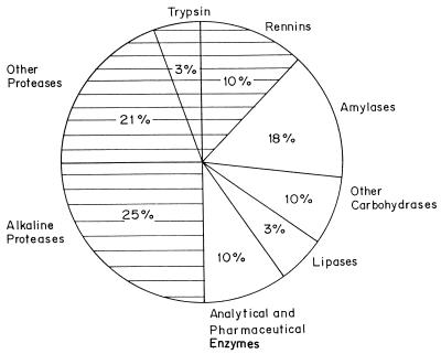
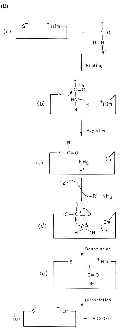
Proteases represent the class of enzymes which occupy a pivotal position with respect to their physiological roles as well as their commercial applications. They perform both degradative and synthetic functions. Since they are physiologically necessary for living organisms, proteases occur ubiquitously in a wide diversity of sources such as plants, animals, and microorganisms. Microbes are an attractive source of proteases owing to the limited space required for their cultivation and their ready susceptibility to genetic manipulation. Proteases are divided into exo- and endopeptidases based on their action at or away from the termini, respectively. They are also classified as serine proteases, aspartic proteases, cysteine proteases, and metalloproteases depending on the nature of the functional group at the active site. Proteases play a critical role in many physiological and pathophysiological processes. Based on their classification, four different types of catalytic mechanisms are operative. Proteases find extensive applications in the food and dairy industries. Alkaline proteases hold a great potential for application in the detergent and leather industries due to the increasing trend to develop environmentally friendly technologies. There is a renaissance of interest in using proteolytic enzymes as targets for developing therapeutic agents. Protease genes from several bacteria, fungi, and viruses have been cloned and sequenced with the prime aims of (i) overproduction of the enzyme by gene amplification, (ii) delineation of the role of the enzyme in pathogenecity, and (iii) alteration in enzyme properties to suit its commercial application. Protein engineering techniques have been exploited to obtain proteases which show unique specificity and/or enhanced stability at high temperature or pH or in the presence of detergents and to understand the structure-function relationships of the enzyme. Protein sequences of acidic, alkaline, and neutral proteases from diverse origins have been analyzed with the aim of studying their evolutionary relationships. Despite the extensive research on several aspects of proteases, there is a paucity of knowledge about the roles that govern the diverse specificity of these enzymes. Deciphering these secrets would enable us to exploit proteases for their applications in biotechnology.


Figures

FIG. 1
FIG. 1
Distribution of enzyme sales. The contribution of different enzymes to the total sale of enzymes is indicated. The shaded portion indicates the total sale of proteases.
FIG. 2
FIG. 2
Active sites of proteases. The catalytic site of proteases is indicated by ∗ and the scissile bond is indicated by  formula image 
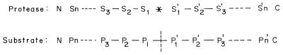
; S1 through Sn and S1′ through Sn′ are the specificity subsites on the enzyme, while P1 through Pn and P1′ through Pn′ are the residues on the substrate accommodated by the subsites on the enzyme.
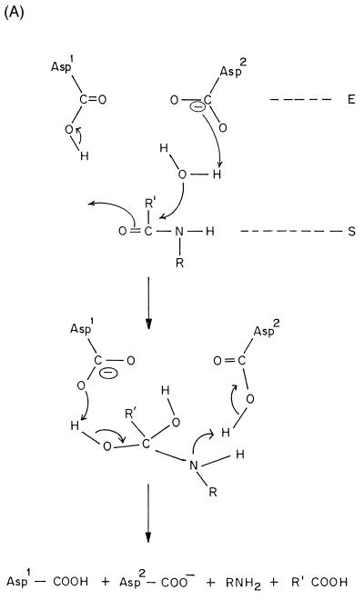
FIG. 3
FIG. 3
Mechanism of action of proteases. (A) Aspartic proteases. (B) Cysteine proteases. Im and +HIm refer to the imidazole and protonated imidazole, respectively.
FIG. 3
FIG. 3
Mechanism of action of proteases. (A) Aspartic proteases. (B) Cysteine proteases. Im and +HIm refer to the imidazole and protonated imidazole, respectively.
FIG. 4
FIG. 4
Homology alignment of the protease sequences. The protease sequences have been selected from the SWISS-PROT and PIR entries, and some have been deduced from the nucleotide sequences obtained from the EMBL database. These are aligned by using CLUSTAL W software for multiple alignment (291). (A) Acidic proteases. (B) Neutral proteases. (C) Alkaline proteases. The key to the sequences is given in Table 5. Numbering of the amino acid residues is based on the first sequence in the list. Identical (○) and conserved (•) residues are boxed; those involved in the active site are indicated by ∗.
FIG. 4
FIG. 4
Homology alignment of the protease sequences. The protease sequences have been selected from the SWISS-PROT and PIR entries, and some have been deduced from the nucleotide sequences obtained from the EMBL database. These are aligned by using CLUSTAL W software for multiple alignment (291). (A) Acidic proteases. (B) Neutral proteases. (C) Alkaline proteases. The key to the sequences is given in Table 5. Numbering of the amino acid residues is based on the first sequence in the list. Identical (○) and conserved (•) residues are boxed; those involved in the active site are indicated by ∗.
FIG. 5
FIG. 5
Dendrogram showing the relationships among the proteases, created by the TreeView package (213). The proteases are grouped as acidic proteases (a), neutral proteases (b), and alkaline proteases (c). Abbreviations of the species described are those used in Table 6. The differences between the sequences are proportional to the length along the horizontal axis.

References

    1. Akimkina T V, Nosovskaya E A, Kostrov S V. Cloning and expression of the neutral proteinase gene of Bacillus cereus in Bacillus subtiliscells. Mol Biol. 1992;26:418–423. - PubMed
    1. Allen C, Stromer V K, Smith F D, Lacy G H, Mount M S. Complementation of an Erwinia carotovora subsp. carotovoraprotease mutant with a protease encoding cosmid. Mol Gen Genet. 1986;202:276–279.
    1. Amerik A, Chistyakova L G, Ostroumova N I, Gurevich A I, Antonov V K. Cloning, expression and structure of the truncated functionally active lon gene of Escherichia coli. Bioorg Khim. 1988;14:408–411. - PubMed
    1. Ammerer G, Hunter C P, Rothman J H, Saari G C, Valls L A, Stevens T M. PEP4 gene of Saccharomyces cerevisiaeencodes proteinase A, a vacuolar enzyme required for processing of vacuolar precursors. Mol Cell Biol. 1986;6:2490–2497. - PMC - PubMed
    1. Argos P. A sensitive procedure to compare amino acid sequences. J Mol Biol. 1987;193:385–396. - PubMed

Publication types

MeSH terms

LinkOut - more resources