

Multidrug resistance protein 1 protects the oropharyngeal mucosal layer and the testicular tubules against drug-induced damage
- PMID: 9730882
- PMCID: PMC2213389
- DOI: 10.1084/jem.188.5.797
Multidrug resistance protein 1 protects the oropharyngeal mucosal layer and the testicular tubules against drug-induced damage
Abstract
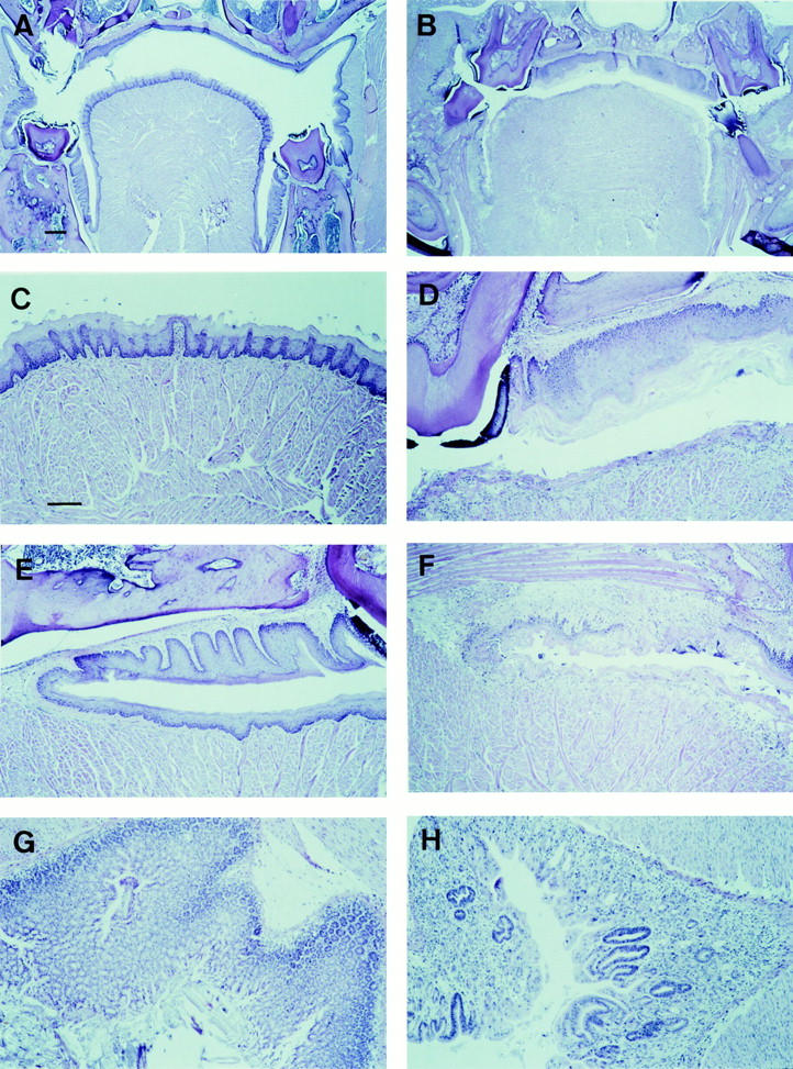
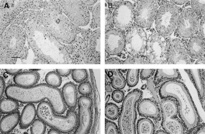
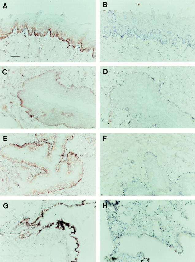
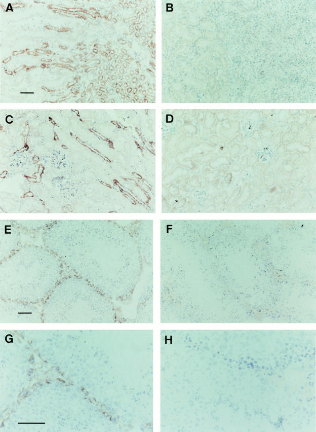
The multidrug resistance protein 1 (MRP1) gene encodes a transporter protein that helps to protect cells against xenobiotics. Elevated levels of MRP1 in tumor cells can result in active extrusion of a wide range of (anticancer) drugs with different cellular targets, a phenomenon called multidrug resistance (MDR). To explore the protective function of the mouse mrp1 protein during drug treatment, we investigated the toxicity caused by the anticancer drug etoposide-phosphate (ETOPOPHOS) in mice lacking the mrp1 gene (mrp1(-/-) mice). We show here that the lack of mrp1 protein results in increased etoposide-induced damage to the mucosa of the oropharyngeal cavity and to the seminiferous tubules of the testis. The high concentrations of mrp1 that we find in the basal layers of the oropharyngeal mucosa and in the basal membrane of the Sertoli cells in the testis apparently protect wild-type mice against this tissue damage. We also find drug-induced polyuria in mrp1(-/-) mice, which correlates with the presence of mrp1 protein in the urinary collecting tubules, the major site of kidney water reabsorption. Our results indicate that specific inhibitors of MRP1 used to reverse MDR, in combination with carcinostatic drugs transported by MRP1, might lead to drug-induced mucositis, (temporary) infertility, and diabetes insipidus.
Figures
References
-
- Gottesman MM, Hrycyna CA, Schoenlein PV, Germann UA, Pastan I. Genetic analysis of the multidrug transporter. Annu Rev Genet. 1995;29:607–649. - PubMed
-
- Zaman, G.J.R., and P. Borst. 1996. MRP, mode of action and role in MDR. In Multidrug Resistance in Cancer Cells. S. Gupta and T. Tsuruo, editors. John Wiley & Sons Ltd., Chichester, UK. 95–107.
-
- Loe, D.W., R.G. Deeley, and S.P.C. Cole. 1996. Biology of the multidrug resistance-associated protein, MRP. Eur. J. Cancer. 32A:945–957. - PubMed
-
- Cole SPC, Bhardwaj G, Gerlach JH, Mackie JE, Grant CE, Almquist KC, Stewart AJ, Kurz EU, Duncan AMV, Deeley RG. Overexpression of a transporter gene in a multidrug-resistant human lung cancer cell line. Science. 1992;258:1650–1654. - PubMed
Publication types
MeSH terms
Substances
LinkOut - more resources
Full Text Sources
Molecular Biology Databases
Research Materials
