. 1998 Sep 7;188(5):855-66.
doi: 10.1084/jem.188.5.855.

Chemokine sequestration by viral chemoreceptors as a novel viral escape strategy: withdrawal of chemokines from the environment of cytomegalovirus-infected cells

Affiliations

Chemokine sequestration by viral chemoreceptors as a novel viral escape strategy: withdrawal of chemokines from the environment of cytomegalovirus-infected cells

B Bodaghi et al. J Exp Med. .

Abstract

Human cytomegalovirus (HCMV), a betaherpesvirus, has developed several ways to evade the immune system, notably downregulation of cell surface expression of major histocompatibility complex class I heavy chains. Here we report that HCMV has devised another means to compromise immune surveillance mechanisms. Extracellular accumulation of both constitutively produced monocyte chemoattractant protein (MCP)-1 and tumor necrosis factor-superinduced RANTES (regulated on activation, normal T cell expressed and secreted) was downregulated in HCMV-infected fibroblasts in the absence of transcriptional repression or the expression of polyadenylated RNA for the cellular chemokine receptors CCR-1, CCR-3, and CCR-5. Competitive binding experiments demonstrated that HCMV-infected cells bind RANTES, MCP-1, macrophage inflammatory protein (MIP)-1beta, and MCP-3, but not MCP-2, to the same receptor as does MIP-1alpha, which is not expressed in uninfected cells. HCMV encodes three proteins with homology to CC chemokine receptors: US27, US28, and UL33. Cells infected with HCMV mutants deleted of US28, or both US27 and US28 genes, failed to downregulate extracellular accumulation of either RANTES or MCP-1. In contrast, cells infected with a mutant deleted of US27 continues to bind and downregulate those chemokines. Depletion of chemokines from the culture medium was at least partially due to continuous internalization of extracellular chemokine, since exogenously added, biotinylated RANTES accumulated in HCMV-infected cells. Thus, HCMV can modify the chemokine environment of infected cells through intense sequestering of CC chemokines, mediated principally by expression of the US28-encoded chemokine receptor.


Figures

Figure 1
Figure 1
Uninfected and infected cells were incubated with TNF (5 ng/ml) from time 0 (relative to the start of infection) for varying periods of time: A, no treatment with TNF; B, treated with TNF from 0 to 24 h; C, treated with TNF from 0 to 48 h; D, treated with TNF from 0 to 72 h. RANTES production was measured in culture supernatants collected from infected and uninfected cells at the intervals indicated in the x axis (0–24, 24– 48, and 48–72 h relative to the start of infection or the beginning of treatment). At each time, cells were washed and re-fed medium with or without TNF as indicated. The experiment was run in triplicate. Extracellular RANTES (pg/ml) was quantified by ELISA.
Figure 2
Figure 2
Cells were infected with wild-type HCMV for the times indicated (PI) or were uninfected (NI). Poly A RNA was extracted, reverse transcribed, and amplified by PCR using primers specific for RANTES, MCP-1, or actin. PC, positive control of the corresponding cDNA cloned in pCDNA; NC, the negative control consisting of nonretrotranscribed poly A RNA. Molecular weight markers were the 100-bp ladder.
Figure 3
Figure 3
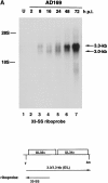
Kinetics of HCMV UL33 (A) and US27/US28 RNA (B) expression. Total cytoplasmic RNA from uninfected (U) or HCMV strain AD169-infected HFF cells (multiplicity of infection 3) were isolated at the indicated hour pi and analyzed by blot hybridization. Riboprobes used for each blot, as well as the expected transcripts (40) are indicated in the schematic drawing. T, transcription initiation site; An, polyadenylation site; E, early kinetic class transcript; L, late kinetic class transcript.
Figure 3
Figure 3
Kinetics of HCMV UL33 (A) and US27/US28 RNA (B) expression. Total cytoplasmic RNA from uninfected (U) or HCMV strain AD169-infected HFF cells (multiplicity of infection 3) were isolated at the indicated hour pi and analyzed by blot hybridization. Riboprobes used for each blot, as well as the expected transcripts (40) are indicated in the schematic drawing. T, transcription initiation site; An, polyadenylation site; E, early kinetic class transcript; L, late kinetic class transcript.
Figure 4
Figure 4
Time course and specificity of MIP-1α receptor expression. (A) 125I-labeled MIP-1α binding to uninfected (UNINF) or HCMV strain AD169-infected HFF cells was examined at the indicated time pi, in either the absence or presence of excess unlabeled MIP-1α (h+MIP-1α) at a final concentration of 100 nM. cpm of bound radiolabeled MIP-1α are given above each bar. (B) Percentage of displaceable binding of 125I-labeled MIP-1α using the indicated unlabeled chemokine (200 nM final concentration) in infected HFF cells at 72 h pi.
Figure 4
Figure 4
Time course and specificity of MIP-1α receptor expression. (A) 125I-labeled MIP-1α binding to uninfected (UNINF) or HCMV strain AD169-infected HFF cells was examined at the indicated time pi, in either the absence or presence of excess unlabeled MIP-1α (h+MIP-1α) at a final concentration of 100 nM. cpm of bound radiolabeled MIP-1α are given above each bar. (B) Percentage of displaceable binding of 125I-labeled MIP-1α using the indicated unlabeled chemokine (200 nM final concentration) in infected HFF cells at 72 h pi.
Figure 5
Figure 5
Genome organization and growth of HCMV mutants. The organization of the HCMV genome (A) and the BamHI-S fragment (B), which contains the US27 and US28 genes, are shown in the schematic drawing. In B, the US27 and US28 transcripts are shown, as well as the sites of the β-gluc expression cassette insertions in RV91, RV92, or RV101. The arrowhead is the site of a point insertion (RV91) and dark rectangles indicate deletions (RV92 and RV101). (C) Schematic drawing of the 2.85-kb β-gluc expression cassette used in the construction of HCMV mutants. (D) Single cycle growth analysis of HCMV mutants.
Figure 6
Figure 6
MIP-1α binding by cells infected with HCMV mutants. Uninfected or HCMV-infected HFF cells (multiplicity of infection 2.5) were analyzed for their ability to bind 125I-labeled MIP-1α at 72 h pi. RV92-2 and RV92-15 are independent isolates containing the identical US28 deletion. In some experiments, excess unlabeled MIP-1α (200 nM final concentration) was used (AD169+MIP-1α). cpm of bound radiolabeled MIP-1α are given above each bar.
Figure 6
Figure 6
MIP-1α binding by cells infected with HCMV mutants. Uninfected or HCMV-infected HFF cells (multiplicity of infection 2.5) were analyzed for their ability to bind 125I-labeled MIP-1α at 72 h pi. RV92-2 and RV92-15 are independent isolates containing the identical US28 deletion. In some experiments, excess unlabeled MIP-1α (200 nM final concentration) was used (AD169+MIP-1α). cpm of bound radiolabeled MIP-1α are given above each bar.
Figure 7
Figure 7
(A) RANTES expressed in picogram/milliliter or (B) MCP-1 expressed in nanogram/milliliter were measured by ELISA in supernatants collected at 0–24 h (black bars), 24–48 (white bars), and 48–72 h (gray bars) after infection from either uninfected cells (NF) or cells infected with wild-type or mutant HCMVs: AD (wild-type), RV91 (ΔUS27), RV92 (ΔUS28), RV101 (ΔUS27/ΔUS28). At each interval, cells were washed and re-fed growth medium.
Figure 8
Figure 8
Fibroblasts infected for 48 h with wild-type (AD) or mutant HCMVs (RV101, RV92, RV91) were washed with PBS, incubated for 3 min at room temperature with low pH glycine buffer to strip membranes of chemokine, and then incubated with biotinylated RANTES (100 nM) for 3 h at 37°C. Cells were then washed again with PBS, treated with low-pH glycine buffer, trypsinized, and extracted in a high salt buffer. Extracts were separated in 15% SDS-PAGE and transferred to nitrocellulose. After saturation with 3% gelatin/1% BSA in PBS/0.1% Tween 20, blots were incubated with peroxidase-labeled streptavidin (1:400) and developed using a chemiluminescence technique for 5 s. RANTES-B (Rantes-B, 125 ng) was run in parallel. AD = infected with wild-type HCMV; RV91 = infected with ΔUS27 mutant HCMV; RV92 = infected with ΔUS28 mutant HCMV; RV101 = infected with ΔUS27/ ΔUS28 mutant HCMV; NF = uninfected cell extract. Representative of two experiments.

References

    1. Loetscher P, Seitz M, Baggiolini M, Moser B. Interleukin 2 regulates CC chemokine receptor expression and chemotactic responsiveness in T lymphocytes. J Exp Med. 1996;184:569–577. - PMC - PubMed
    1. Davis-Poynter NJ, Farrell HE. Masters of deception: a review of herpesvirus immune evasion strategies. Immunol Cell Biol. 1996;74:513–522. - PubMed
    1. Moore PS, Boshoff C, Weiss RA, Chang Y. Molecular mimicry of human cytokine and cytokine response pathway genes by KSHV. Science. 1996;274:1739–1744. - PubMed
    1. Murphy PM. Molecular piracy of chemokine receptors by herpesviruses. Infect Agents Dis. 1994;3:137–154. - PubMed
    1. Smith GL. Virus proteins that bind cytokines, chemokines or interferons. Curr Opin Immunol. 1996;8:467–471. - PubMed

Publication types

MeSH terms