. 1998 Sep 7;188(5):973-8.
doi: 10.1084/jem.188.5.973.

Qa-1b binds conserved class I leader peptides derived from several mammalian species

Affiliations

Qa-1b binds conserved class I leader peptides derived from several mammalian species

Z Kurepa et al. J Exp Med. .

Abstract

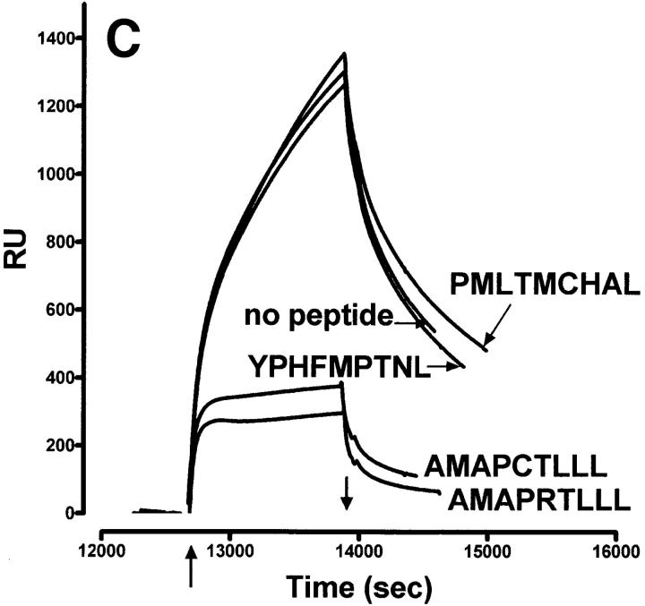
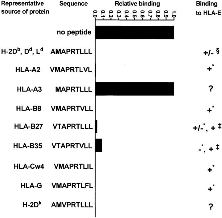
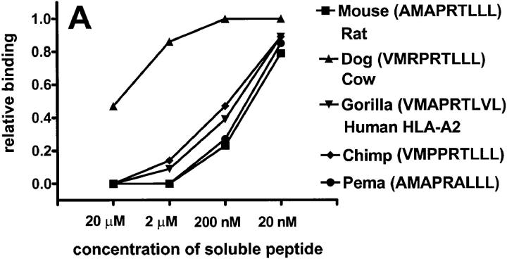
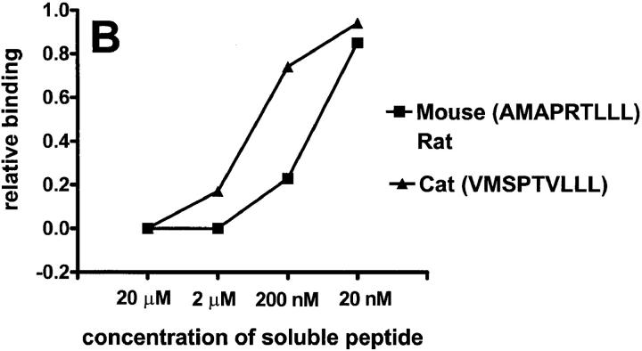
Qa-1b binds a peptide (AMAPRTLLL), referred to as Qdm (for Qa-1 determinant modifier), derived from the signal sequence of murine class Ia molecules. This peptide binds with high affinity and accounts for almost all of the peptides associated with this molecule. Human histocompatibility leukocyte antigen (HLA)-E, a homologue of Qa-1b, binds similar peptides derived from human class Ia molecules and interacts with CD94/NKG2 receptors on natural killer cells. We used surface plasmon resonance to determine the ability of Qa-1b to bind related ligands representing peptides derived from the leaders of class I molecules from several mammalian species. All of the peptides reported to bind HLA-E bound readily to Qa-1b. In addition, peptides derived from leader segments of different mammals also bound to Qa-1b, indicating a conservation of this "Qdm-like" epitope throughout mammalian evolution. We have attempted to define a minimal peptide on a polyglycine backbone that binds Qa-1b. Our previous findings showed that P2 and P9 are important but not sufficient for binding to Qa-1b. Although a minimum peptide (GMGGGGLLL) bound Qa-1(b), its interaction was relatively weak, as were peptides sharing five or six residues with Qdm, indicating that multiple native residues are required for a strong interaction. This finding is consistent with the observation that this molecule preferentially binds this single ligand.


Figures

Figure 1
Figure 1
Characteristics of sQa-1b2m secreted from transfected D. melanogaster cells and its specific binding to immobilized QdmC5 peptide. (A) After concentration and purification, the supernatant from Qa-1b2m-transfected Drosophila cells was resolved on a 15% SDS-PAGE, and stained using Coomassie brilliant blue. Arrows, Qa-1b heavy chain (Hc) and β2m. (B–D) SPR demonstrating binding of sQa-1b2m to immobilized QdmC5. Sensorgrams were obtained using injection volumes of 20 μl at a rate of 1 μl/min. Mass increase due to macromolecular binding is measured in resonance units (RU). Arrowheads, Start (↑ ) and end (↓ ) of the injection. (B) Injection of 0.5 μM sQa-1b or sM3. (C) 0.5 μM sQa-1b was run over the chip alone, or in the presence of 20 μM Qdm (AMAPRTLLL), QdmC5 (AMAPCTLLL), or two control peptides, PMLTMCHAL and YPHFMPTNL. (D) 0.5 μM sQa-1b was run at 1 μl/min for 20 min over immobilized QdmC5 in the absence or presence of competitor peptides. Results are presented as relative binding of sQa-1b, where 0 represents binding in the presence of 20 μM Qdm (25 RU), and 1 is the binding in the absence of peptide (263 RU). *, +, and O represent binding in the presence of 20 μM of the entire 24-mer Dd leader, MGAMAPRTL and MAPRTLLLL, respectively.
Figure 1
Figure 1
Characteristics of sQa-1b2m secreted from transfected D. melanogaster cells and its specific binding to immobilized QdmC5 peptide. (A) After concentration and purification, the supernatant from Qa-1b2m-transfected Drosophila cells was resolved on a 15% SDS-PAGE, and stained using Coomassie brilliant blue. Arrows, Qa-1b heavy chain (Hc) and β2m. (B–D) SPR demonstrating binding of sQa-1b2m to immobilized QdmC5. Sensorgrams were obtained using injection volumes of 20 μl at a rate of 1 μl/min. Mass increase due to macromolecular binding is measured in resonance units (RU). Arrowheads, Start (↑ ) and end (↓ ) of the injection. (B) Injection of 0.5 μM sQa-1b or sM3. (C) 0.5 μM sQa-1b was run over the chip alone, or in the presence of 20 μM Qdm (AMAPRTLLL), QdmC5 (AMAPCTLLL), or two control peptides, PMLTMCHAL and YPHFMPTNL. (D) 0.5 μM sQa-1b was run at 1 μl/min for 20 min over immobilized QdmC5 in the absence or presence of competitor peptides. Results are presented as relative binding of sQa-1b, where 0 represents binding in the presence of 20 μM Qdm (25 RU), and 1 is the binding in the absence of peptide (263 RU). *, +, and O represent binding in the presence of 20 μM of the entire 24-mer Dd leader, MGAMAPRTL and MAPRTLLLL, respectively.
Figure 2
Figure 2
Blocking of the binding of sQa-1b to immobilized QdmC5 by peptides derived from leader sequences of human class I molecules. Results are presented as relative binding, where 0 is the binding of sQa-1b in the presence of 20 μM Qdm (32 RU), and 1 is the binding in the absence of peptides (345 RU). 0.5 μM sQa-1b was run over immobilized QdmC5 for 20 min at the rate of 1 μl/min in the presence of 20 μM competitor peptides. For HLA-E binding, + indicates strong binding, − indicates no binding, and +/− indicates weak binding. Data taken from * Table 1 in reference ,  reference , and § reference .
Figure 3
Figure 3
Putative peptides from leaders of various mammalian class I molecules bind to Qa-1b. Results are presented at relative binding, where 0 is the binding of sQa-1b in the presence of 20 μM Qdm (25 RU in A, 26 RU in B), and 1 is the binding in the absence of blockers (276 RU in A, 230 RU in B). Running buffer was Hepes-buffered saline (HBS) in A and 2% DMSO in HBS in B.
Figure 3
Figure 3
Putative peptides from leaders of various mammalian class I molecules bind to Qa-1b. Results are presented at relative binding, where 0 is the binding of sQa-1b in the presence of 20 μM Qdm (25 RU in A, 26 RU in B), and 1 is the binding in the absence of blockers (276 RU in A, 230 RU in B). Running buffer was Hepes-buffered saline (HBS) in A and 2% DMSO in HBS in B.

References

    1. Hunt DF, Henderson RA, Shabanowitz J, Sakaguchi K, Michel H, Sevilir N, Cox AL, Appella E, Engelhard VH. Characterization of peptides bound to the class I MHC molecule HLA-A2.1 by mass spectrometry. Science. 1992;255:1261–1263. - PubMed
    1. Aldrich CJ, DeCloux A, Woods AS, Cotter RJ, Soloski MJ, Forman J. Identification of a TAP-dependent leader peptide recognized by alloreactive T cells specific for a class Ib antigen. Cell. 1994;79:649–658. - PubMed
    1. DeCloux A, Woods AS, Cotter RJ, Soloski MJ, Forman J. Dominance of a single peptide bound to the class Ib molecule, Qa-1b . J Immunol. 1997;158:2183–2191. - PubMed
    1. Braud V, Jones EY, McMichael A. The human major histocompatibility complex class Ib molecule HLA-E binds signal sequence-derived peptides with primary anchor residues at position 2 and 9. Eur J Immunol. 1997;27:1164–1169. - PubMed
    1. O'Callaghan CA, Tormo J, Willcox BE, Braud VM, Jakobsen BK, Stuart DI, McMichael AJ, Bell JI, Jones EY. Structural features impose tight peptide binding specificity in the nonclassical MHC molecule HLA-E. Mol Cell. 1998;1:531–541. - PubMed

Publication types

MeSH terms