. 1998 Sep 7;188(5):991-5.
doi: 10.1084/jem.188.5.991.

Paired immunoglobulin-like receptor (PIR)-A is involved in activating mast cells through its association with Fc receptor gamma chain

Affiliations

Paired immunoglobulin-like receptor (PIR)-A is involved in activating mast cells through its association with Fc receptor gamma chain

A Maeda et al. J Exp Med. .

Abstract

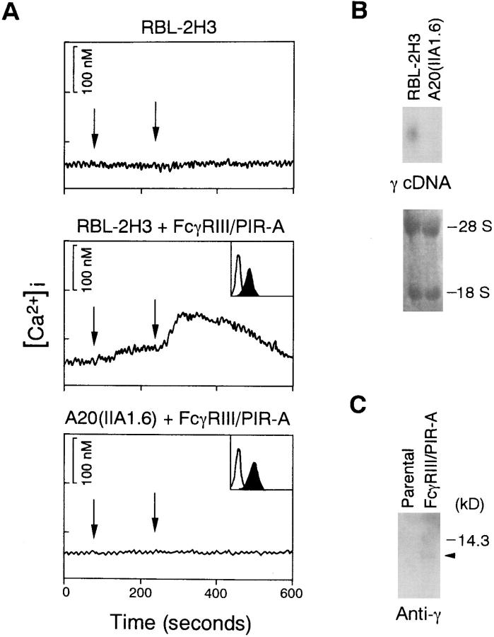
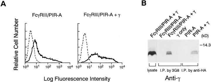
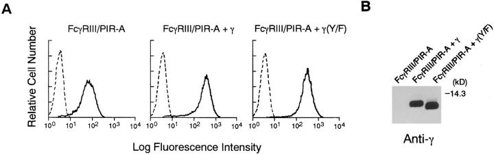
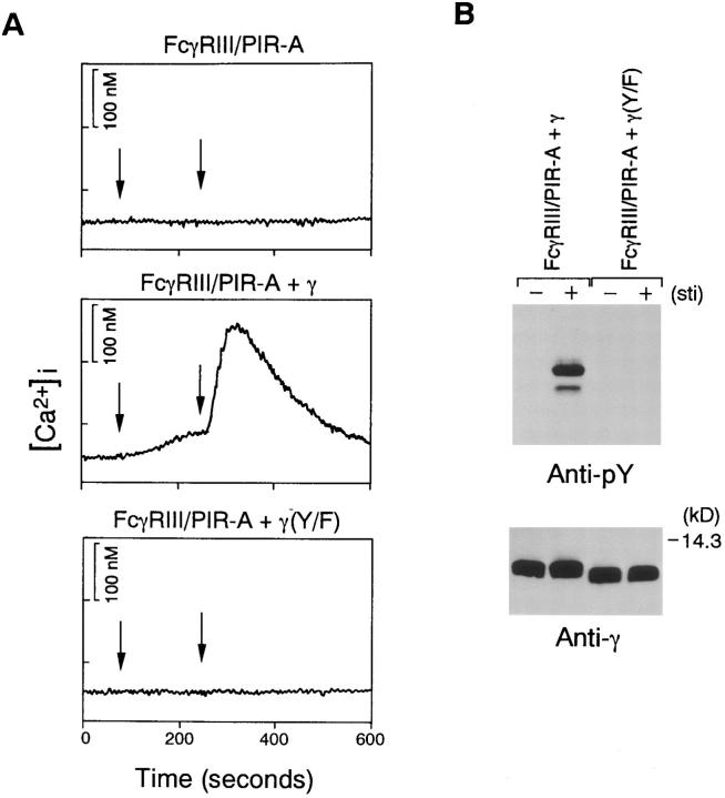
Paired immunoglobulin-like receptor (PIR)-A and PIR-B possess similar ectodomains with six immunoglobulin-like loops, but have distinct transmembrane and cytoplasmic domains. PIR-B bears immunoreceptor tyrosine-based inhibitory motif (ITIM) sequences in its cytoplasmic domain that recruit Src homology (SH)2 domain-containing tyrosine phosphatases SHP-1 and SHP-2, leading to inhibition of B and mast cell activation. In contrast, the PIR-A protein has a charged Arg residue in its transmembrane region and a short cytoplasmic domain that lacks ITIM sequences. Here we show that Fc receptor gamma chain, containing an immunoreceptor tyrosine-based activation motif (ITAM), associates with PIR-A. Cross-linking of this PIR-A complex results in mast cell activation such as calcium mobilization in an ITAM-dependent manner. Thus, our data provide evidence for the existence of two opposite signaling pathways upon PIR aggregation. PIR-A induces the stimulatory signal by using ITAM in the associated gamma chain, whereas PIR-B mediates the inhibitory signal through its ITIMs.


Figures

Figure 1
Figure 1
FcγRIII–PIR-A induces calcium mobilization and associates with FcRγ chain in RBL-2H3 cells. (A) Calcium mobilization by cross-linking of FcγRIII–PIR-A. Left arrows indicate application of 3G8 or biotin-tagged 3G8, and right arrows indicate application of F(ab′)2 anti– mouse IgG or streptavidin in RBL-2H3 or A20 IIA1.6 cells, respectively. Surface expression levels of FcγRIII–PIR-A are indicated in inset boxes. The x and y axes for the histograms indicate fluorescence intensity (log scales) and relative cell number, respectively. (B) Northern blot analysis using mouse γ cDNA as a probe. Bottom panel shows 28S and 18S ribosomal RNA pattern. (C) Association of FcγRIII–PIR-A with γ in RBL-2H3 cells. Immunoprecipitates with 3G8 were separated by 15% SDS-PAGE gel and detected by anti-γ Ab.
Figure 2
Figure 2
Association of FcγRIII–PIR-A with γ chain in 293 T cells. (A) FACS® (Becton Dickinson, San Jose, CA) analysis of 293 T cells transiently expressing FcγRIII– PIR-A in the presence or absence of γ. Cells were labeled with FITC-conjugated 3G8. Nontransfected 293 T cells were used as a negative control. (B) Association was detected as described in legend to Fig. 1. In the case of association of HA-tagged PIR-A with γ, anti-HA mAb was used for immunoprecipitation instead of 3G8.
Figure 3
Figure 3
Expression of FcγRIII–PIR-A and γ in DT40 transformants. (A) FACS® analysis of DT40 cells by using FITC-conjugated 3G8. Parental DT40 cells were used as a negative control. (B) Expression of γ by Western blot analysis using anti-γ Ab. Transformant cells were directly dissolved in SDS-sample buffer.
Figure 4
Figure 4
The FcγRIII–PIR-A complex transmits activation signals in the presence of γ. (A) Calcium mobilization by cross-linking of FcγRIII– PIR-A. Transformants shown in Fig. 3 were stimulated by 3G8 (left arrows) and F(ab′)2 anti–mouse IgG (right arrows). (B) Tyrosine phosphorylation of γ by cross-linking of FcγRIII–PIR-A. Transformant cells were stimulated with 3G8 (3 min), and then F(ab′)2 anti–mouse IgG (3 min). Cells were dissolved by NP-40 buffer, immunoprecipitated by anti-γ Ab, separated by 15% SDS-PAGE gel, and probed with antiphosphotyrosine mAb 4G10. Membrane was reprobed with anti-γ Ab (bottom).

References

    1. Weiss A, Littman DR. Signal transduction by lymphocyte antigen receptors. Cell. 1994;76:263–274. - PubMed
    1. Kurosaki T. Molecular mechanisms in B cell antigen receptor signaling. Curr Opin Immunol. 1997;9:309–318. - PubMed
    1. DeFranco AL. The complexity of signaling pathways activated by the BCR. Curr Opin Immunol. 1997;9:296–308. - PubMed
    1. Cambier JC. Inhibitory receptors abound? . Proc Natl Acad Sci USA. 1997;94:5993–5995. - PMC - PubMed
    1. Yokoyama WM. What goes up must come down: the emerging spectrum of inhibitory receptors. J Exp Med. 1997;186:1803–1808. - PMC - PubMed

Publication types