. 1998 Sep 1;12(17):2770-81.
doi: 10.1101/gad.12.17.2770.

Ribonuclease E organizes the protein interactions in the Escherichia coli RNA degradosome

Affiliations

Ribonuclease E organizes the protein interactions in the Escherichia coli RNA degradosome

N F Vanzo et al. Genes Dev. .

Abstract

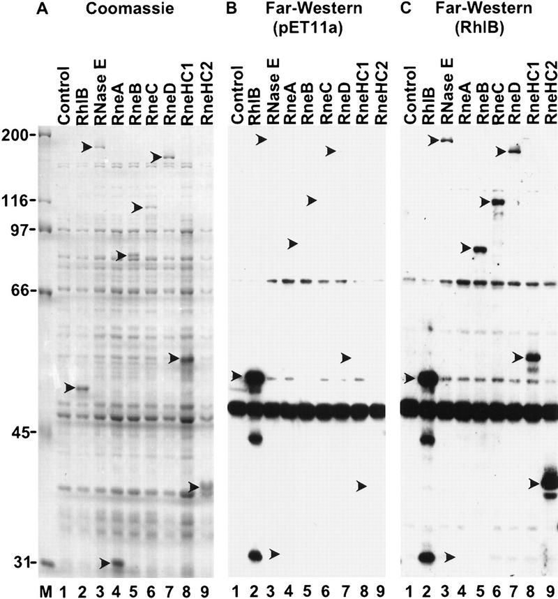
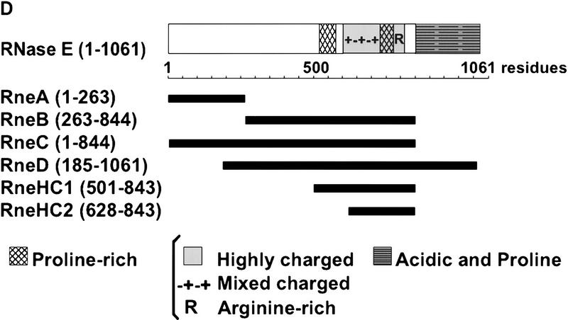
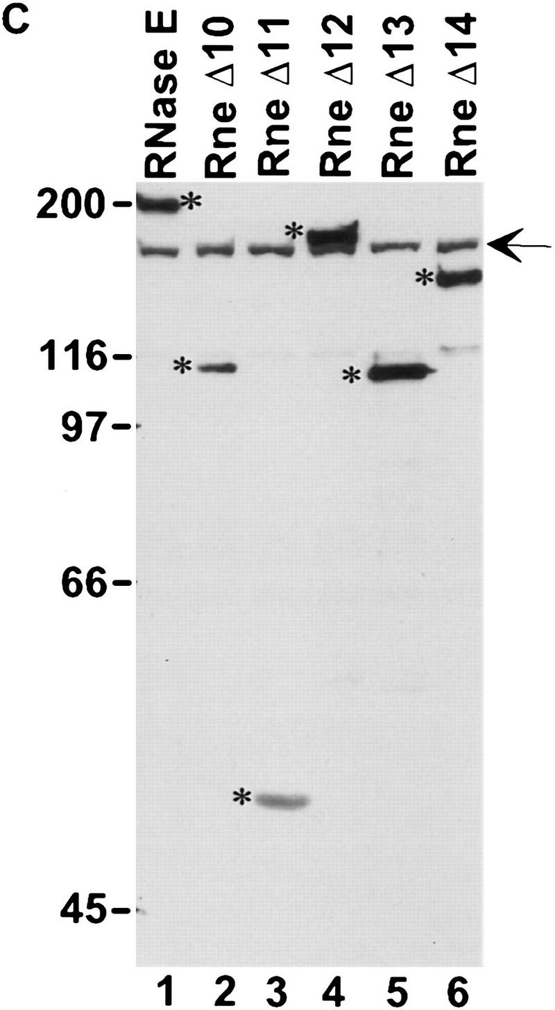
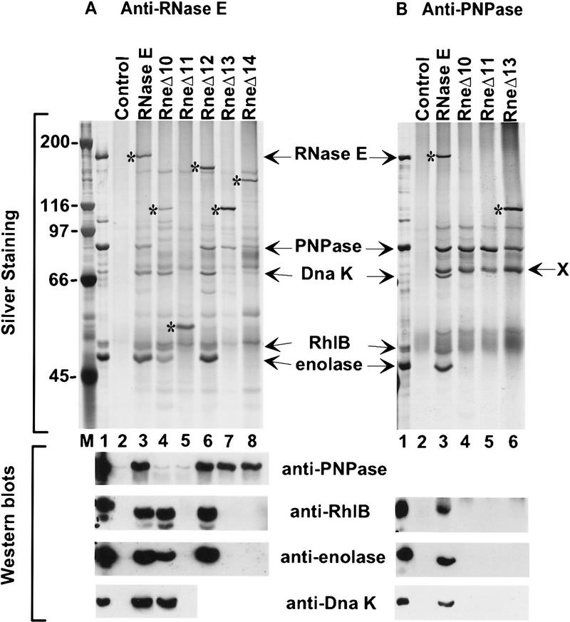
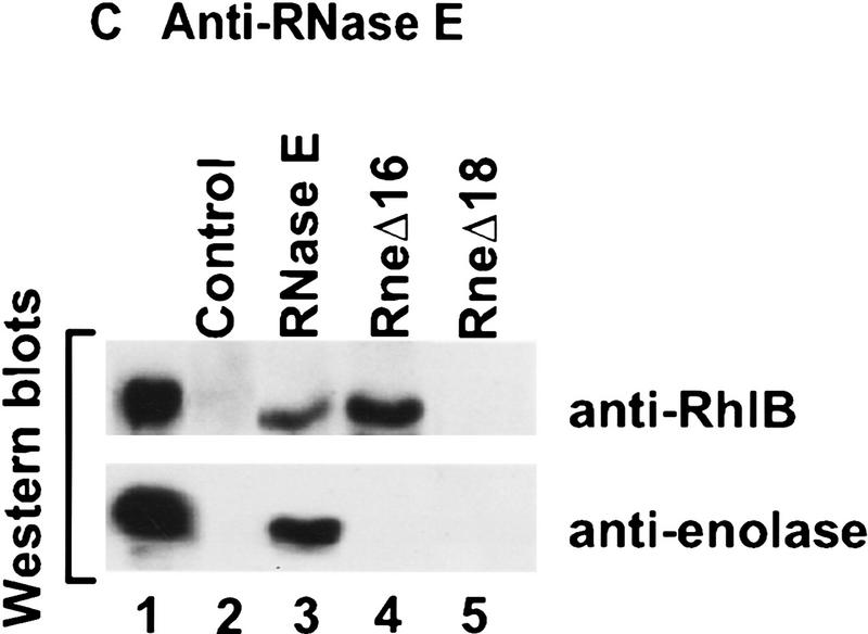
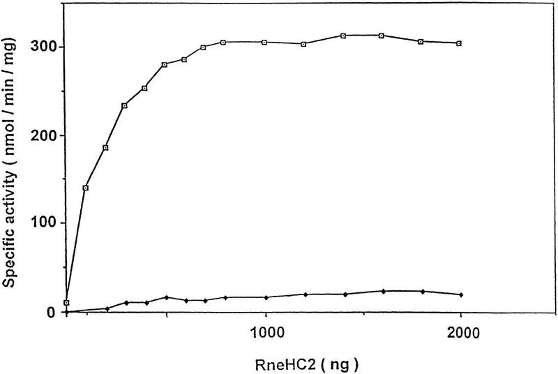
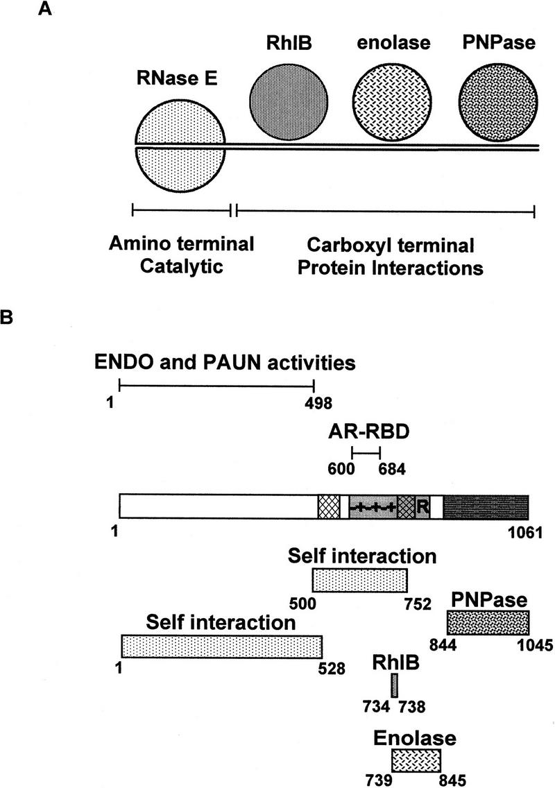
The Escherichia coli RNA degradosome is the prototype of a recently discovered family of multiprotein machines involved in the processing and degradation of RNA. The interactions between the various protein components of the RNA degradosome were investigated by Far Western blotting, the yeast two-hybrid assay, and coimmunopurification experiments. Our results demonstrate that the carboxy-terminal half (CTH) of ribonuclease E (RNase E) contains the binding sites for the three other major degradosomal components, the DEAD-box RNA helicase RhlB, enolase, and polynucleotide phosphorylase (PNPase). The CTH of RNase E acts as the scaffold of the complex upon which the other degradosomal components are assembled. Regions for oligomerization were detected in the amino-terminal and central regions of RNase E. Furthermore, polypeptides derived from the highly charged region of RNase E, containing the RhlB binding site, stimulate RhlB activity at least 15-fold, saturating at one polypeptide per RhlB molecule. A model for the regulation of the RhlB RNA helicase activity is presented. The description of RNase E now emerging is that of a remarkably complex multidomain protein containing an amino-terminal catalytic domain, a central RNA-binding domain, and carboxy-terminal binding sites for the other major components of the RNA degradosome.


Figures

Figure 1
Figure 1
RhlB interacts with the highly charged region of RNase E. (A) Crude extracts of BL21(DE3) containing the control vector pET11a (lane 1) or plasmids overexpressing RhlB, RNase E, RneA, RneB, RneC, RneD, RneHC1, and RneHC2 (lanes 2–9) were analyzed on a 9% SDS-polyacrylamide gel and stained with Coomassie blue. (C) For the Far Western analysis, the proteins were blotted to a membrane, incubated with RhlB in solution, and probed with an anti-RhlB antibody. The RhlB in solution was partially purified from E. coli with a pET derivative expressing RhlB. (B) A control in which the membrane was incubated with a mock preparation from E. coli containing the pET11a vector. In A, protein molecular masses (kD) are marked at left. The position of the recombinant proteins was determined with Ponceau red and is indicated by arrowheads. (D) A schematic representation of the RNase E polypeptides overexpressed by the pET system. The carboxy-terminal half can be roughly divided, by amino acid content, into a proline-rich segment (residues 524–568, 33% proline), a highly charged (HC) domain (residues 600–820, 45% charged), and an acidic terminal part rich in proline residues (845–1061, 20% acidic, 11% proline). The highly charged domain consists of three successive regions: a mixed charged region alternating arginine and acidic residues (600–738), a second proline-rich segment (739–778), and a short region mainly charged in arginine (789–820).
Figure 1
Figure 1
RhlB interacts with the highly charged region of RNase E. (A) Crude extracts of BL21(DE3) containing the control vector pET11a (lane 1) or plasmids overexpressing RhlB, RNase E, RneA, RneB, RneC, RneD, RneHC1, and RneHC2 (lanes 2–9) were analyzed on a 9% SDS-polyacrylamide gel and stained with Coomassie blue. (C) For the Far Western analysis, the proteins were blotted to a membrane, incubated with RhlB in solution, and probed with an anti-RhlB antibody. The RhlB in solution was partially purified from E. coli with a pET derivative expressing RhlB. (B) A control in which the membrane was incubated with a mock preparation from E. coli containing the pET11a vector. In A, protein molecular masses (kD) are marked at left. The position of the recombinant proteins was determined with Ponceau red and is indicated by arrowheads. (D) A schematic representation of the RNase E polypeptides overexpressed by the pET system. The carboxy-terminal half can be roughly divided, by amino acid content, into a proline-rich segment (residues 524–568, 33% proline), a highly charged (HC) domain (residues 600–820, 45% charged), and an acidic terminal part rich in proline residues (845–1061, 20% acidic, 11% proline). The highly charged domain consists of three successive regions: a mixed charged region alternating arginine and acidic residues (600–738), a second proline-rich segment (739–778), and a short region mainly charged in arginine (789–820).
Figure 2
Figure 2
Construction of E. coli strains expressing various deletions in RNase E. (A) A schematic representation of the experimental system. The PBRN1 strain carries the rne gene with an amber mutation in its first tyrosine codon and the temperature-sensitive tRNA suppressor supFts that inserts tyrosine. At 30°C, wild-type RNase E protein is produced because of the suppression by supFts. At 42°C, a polypeptide containing only the first 25 amino acids of RNase E is produced and growth is supported by a plasmid encoding the wild-type RNase E (pAM–rne). (B) The structure of the mutant proteins with the boundaries of the deletions noted in parentheses. The messages encoding these proteins, expressed from the rne promoter (Prne), contain the natural 5′ and 3′ UTR encoded by the rne gene. PBRN1 containing pAM–rne and its derivatives was grown at 42°C to the stationary phase. Total cell extracts were analyzed by Western blotting with an anti-RNase E antibody. (C) Expression of RNase E and mutant peptides at 42°C. The RNase E polypeptides are marked by asterisks. The signal indicated by the arrow is the result of a cross-reaction of the antiserum with another protein present in all the extracts.
Figure 2
Figure 2
Construction of E. coli strains expressing various deletions in RNase E. (A) A schematic representation of the experimental system. The PBRN1 strain carries the rne gene with an amber mutation in its first tyrosine codon and the temperature-sensitive tRNA suppressor supFts that inserts tyrosine. At 30°C, wild-type RNase E protein is produced because of the suppression by supFts. At 42°C, a polypeptide containing only the first 25 amino acids of RNase E is produced and growth is supported by a plasmid encoding the wild-type RNase E (pAM–rne). (B) The structure of the mutant proteins with the boundaries of the deletions noted in parentheses. The messages encoding these proteins, expressed from the rne promoter (Prne), contain the natural 5′ and 3′ UTR encoded by the rne gene. PBRN1 containing pAM–rne and its derivatives was grown at 42°C to the stationary phase. Total cell extracts were analyzed by Western blotting with an anti-RNase E antibody. (C) Expression of RNase E and mutant peptides at 42°C. The RNase E polypeptides are marked by asterisks. The signal indicated by the arrow is the result of a cross-reaction of the antiserum with another protein present in all the extracts.
Figure 3
Figure 3
Analysis of the composition of the RNA degradosome. The PBRN1 strain, containing pAM–rne or its derivatives and pAPT–rhlB was grown at 42°C to stationary phase.Plasmid pAPT–rhlB is a p15A-derived plasmid expressing RhlB under the control of the Plac expression signals. Protein extracts were prepared as described in the Materials and Methods. Anti-RNase E antibody (A,C) or anti-PNPase antibody (B), cross-linked to protein A–Sepharose beads, was used to immunopurify the protein complexes. The composition of the immunopurified protein complexes was analyzed by SDS-PAGE and silver staining (top, A, B) or by Western blotting (bottom, A–C). (Lane 1) The RNA degradosome preparation from Py et al. (1996). (Lane 2) A control immunopurification using the preimmune serum for each antibody. The RNase E protein and its mutants are marked with asterisks. In B, X indicates an unrelated protein present in all the cell extracts that react with the anti-PNPase antibody. The molecular masses of markers (lane M) are indicated at left (in kD).
Figure 3
Figure 3
Analysis of the composition of the RNA degradosome. The PBRN1 strain, containing pAM–rne or its derivatives and pAPT–rhlB was grown at 42°C to stationary phase.Plasmid pAPT–rhlB is a p15A-derived plasmid expressing RhlB under the control of the Plac expression signals. Protein extracts were prepared as described in the Materials and Methods. Anti-RNase E antibody (A,C) or anti-PNPase antibody (B), cross-linked to protein A–Sepharose beads, was used to immunopurify the protein complexes. The composition of the immunopurified protein complexes was analyzed by SDS-PAGE and silver staining (top, A, B) or by Western blotting (bottom, A–C). (Lane 1) The RNA degradosome preparation from Py et al. (1996). (Lane 2) A control immunopurification using the preimmune serum for each antibody. The RNase E protein and its mutants are marked with asterisks. In B, X indicates an unrelated protein present in all the cell extracts that react with the anti-PNPase antibody. The molecular masses of markers (lane M) are indicated at left (in kD).
Figure 4
Figure 4
The RneHC2 polypeptide stimulates RhlB ATPase activity. The specific ATPase activity of wild-type (□) and mutant (♦) RhlB protein was determined in the presence of increasing concentrations of the RneHC2 polypeptide. The values are the average of duplicate assays. The reactions, containing 1 μg of RhlB and 0–2 μg of the RneHC2 polypeptide, were as described (Py et al. 1996), except that the yeast RNA was increased to 200 μg/ml. Less than 15% of the ATP was converted to ADP and Pi under the conditions of maximal activity. RhlB* carries the E166K point mutation in the DEAD motif (DEAD→DKAD). RhlB, RhlB*, and RneHC2 were partially purified from cells overexpressing these proteins. Each protein was judged to be at least 70% pure by SDS-PAGE. Protein concentrations were determined by Lowry assay using a BSA standard.
Figure 5
Figure 5
Organization of the protein interactions within the RNA degradosome. (A) Cartoon of the E. coli RNA degradosome illustrating the central role of the CTH of RNase E in the protein interactions. The actual stoichiometry and structure of the components within the degradosome are not certain. It seems reasonable to suppose that RNase E is dimeric although a trimeric or higher order structure is possible (see Discussion). (B) Summary diagram illustrating the known activities and binding regions in RNase E.

References

    1. Alberts B. The cell as a collection of protein machines: Preparing the next generation of molecular biologists. Cell. 1998;92:291–294. - PubMed
    1. Anderson JSJ, Parker R. The 3′ to 5′ degradation of yeast mRNA is a general mechanism for mRNA turnover that requires the SKI2 DEVH box protein and 3′ to 5′ exonucleases of the exosome complex. EMBO J. 1998;17:1497–1506. - PMC - PubMed
    1. Belasco JG, Higgins CF. Mechanisms of mRNA decay in bacteria: A perspective. Gene. 1988;72:15–23. - PubMed
    1. Blum E, Py B, Carpousis AJ, Higgins CF. Polyphosphate kinase is a component of the Escherichia coli RNA degradosome. Mol Microbiol. 1997;26:387–398. - PubMed
    1. Braun F, Hajnsdorf E, Régnier P. Polynucleotide phosphorylase is required for the rapid degradation of the RNase E-processed rpsO mRNA of Escherichia coli devoid of its 3′ hairpin. Mol Microbiol. 1996;19:997–1005. - PubMed

Publication types

MeSH terms

LinkOut - more resources