. 1998 Sep 7;142(5):1223-33.
doi: 10.1083/jcb.142.5.1223.

Degradation of misfolded endoplasmic reticulum glycoproteins in Saccharomyces cerevisiae is determined by a specific oligosaccharide structure

Affiliations

Degradation of misfolded endoplasmic reticulum glycoproteins in Saccharomyces cerevisiae is determined by a specific oligosaccharide structure

C A Jakob et al. J Cell Biol. .

Abstract

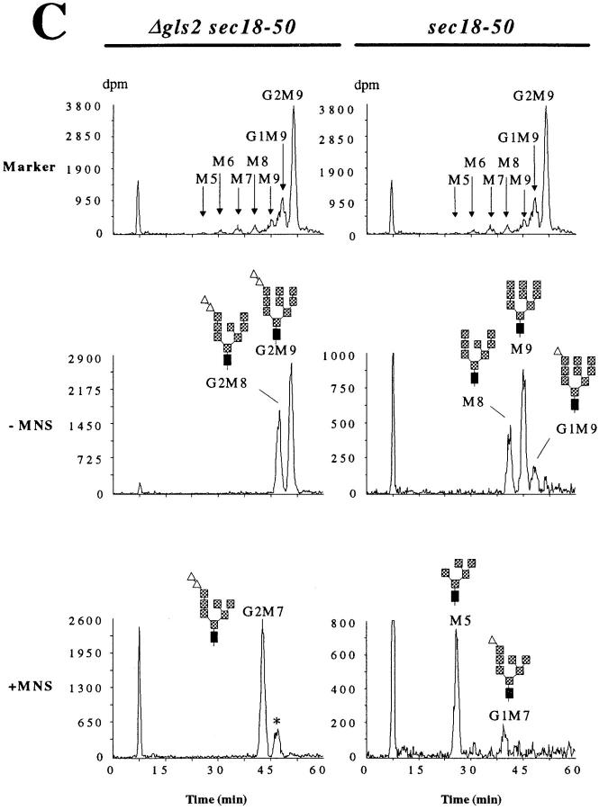
In Saccharomyces cerevisiae, transfer of N-linked oligosaccharides is immediately followed by trimming of ER-localized glycosidases. We analyzed the influence of specific oligosaccharide structures for degradation of misfolded carboxypeptidase Y (CPY). By studying the trimming reactions in vivo, we found that removal of the terminal alpha1,2 glucose and the first alpha1,3 glucose by glucosidase I and glucosidase II respectively, occurred rapidly, whereas mannose cleavage by mannosidase I was slow. Transport and maturation of correctly folded CPY was not dependent on oligosaccharide structure. However, degradation of misfolded CPY was dependent on specific trimming steps. Degradation of misfolded CPY with N-linked oligosaccharides containing glucose residues was less efficient compared with misfolded CPY bearing the correctly trimmed Man8GlcNAc2 oligosaccharide. Reduced rate of degradation was mainly observed for misfolded CPY bearing Man6GlcNAc2, Man7GlcNAc2 and Man9GlcNAc2 oligosaccharides, whereas Man8GlcNAc2 and, to a lesser extent, Man5GlcNAc2 oligosaccharides supported degradation. These results suggest a role for the Man8GlcNAc2 oligosaccharide in the degradation process. They may indicate the presence of a Man8GlcNAc2-binding lectin involved in targeting of misfolded glycoproteins to degradation in S. cerevisiae.


Figures

Figure 1
Figure 1
Biosynthesis and trimming of oligosaccharides in the ER lumen of Saccharomyces cerevisiae. The lipid-linked oligosaccharide precursors are synthesized at the ER membrane. Some of the involved mannosyltransferases (encoded by the ALG3, ALG9, and ALG12 loci) and their products as well as the glucosyltransferases (encoded by the ALG6, ALG8 and ALG10 loci) and their respective products are depicted. The fully assembled Glc3Man9GlcNAc2 oligosaccharide precursor is transferred to asparagine residues of the N-X-S/T sequence of polypeptides. This is followed by trimming involving glucosidase I (GLS1), glucosidase II (GLS2) and mannosidase I (MNS1) to yield the protein-bound Man8GlcNAc2 oligosaccharide.
Figure 2
Figure 2
In vivo kinetics of N-linked oligosaccharide trimming. Two different yeast strains were used. Strain YG557 (Δgls2 sec18-50; left) carries a deletion of the glucosidase II–encoding locus and a mutation in the SEC18 locus resulting in a temperature-sensitive protein transport from the ER to the Golgi compartment. Strain YG556 (sec18-50; right) is fully competent in oligosaccharide trimming in the ER. Cells were labeled with a 1-min pulse of 3H-mannose, followed by a chase with an excess of unlabeled mannose. (A) Protein-bound oligosaccharides were isolated at the time indicated after initiation of the chase and analyzed by HPLC. The elution of radioactivity was monitored. The proposed oligosaccharide structure is given for the individual peaks. For explanation of the symbols, see Fig. 1. (B) Kinetics of maturation of N-linked oligosaccharides. The HPLC profiles shown in A were quantified using the FLO-ONE software (version 3.6; Packard Instrument Co., Meriden, CT). The amount of radioactivity present in each individual peak was determined, expressed as a percentage of total radioactivity present and plotted against time. The value for the G2M8 and the M8 oligosaccharide, respectively, was corrected for the absence of one mannose residue. (C) Verification of NLO species by exo-α1,2-mannosidase digestion. The NLO obtained from 5-min (sec18-50) and 10-min (Δgls2 sec18-50) chase periods (A) were treated with exo-α1,2-mannosidase from A. saitoi and separated by HPLC. Endo H–treated LLO, obtained from a Δalg10 Δgls2 (G2) strain, was used as marker for the oligosaccharides. The proposed oligosaccharide structure is given for the individual peaks. For explanation of the symbols, see Fig. 1. −MNS, NLO without exo-α1,2-mannosidase treatment; +MNS, NLO after A. saitoi exo-α1,2-mannosidase cleavage.
Figure 2
Figure 2
In vivo kinetics of N-linked oligosaccharide trimming. Two different yeast strains were used. Strain YG557 (Δgls2 sec18-50; left) carries a deletion of the glucosidase II–encoding locus and a mutation in the SEC18 locus resulting in a temperature-sensitive protein transport from the ER to the Golgi compartment. Strain YG556 (sec18-50; right) is fully competent in oligosaccharide trimming in the ER. Cells were labeled with a 1-min pulse of 3H-mannose, followed by a chase with an excess of unlabeled mannose. (A) Protein-bound oligosaccharides were isolated at the time indicated after initiation of the chase and analyzed by HPLC. The elution of radioactivity was monitored. The proposed oligosaccharide structure is given for the individual peaks. For explanation of the symbols, see Fig. 1. (B) Kinetics of maturation of N-linked oligosaccharides. The HPLC profiles shown in A were quantified using the FLO-ONE software (version 3.6; Packard Instrument Co., Meriden, CT). The amount of radioactivity present in each individual peak was determined, expressed as a percentage of total radioactivity present and plotted against time. The value for the G2M8 and the M8 oligosaccharide, respectively, was corrected for the absence of one mannose residue. (C) Verification of NLO species by exo-α1,2-mannosidase digestion. The NLO obtained from 5-min (sec18-50) and 10-min (Δgls2 sec18-50) chase periods (A) were treated with exo-α1,2-mannosidase from A. saitoi and separated by HPLC. Endo H–treated LLO, obtained from a Δalg10 Δgls2 (G2) strain, was used as marker for the oligosaccharides. The proposed oligosaccharide structure is given for the individual peaks. For explanation of the symbols, see Fig. 1. −MNS, NLO without exo-α1,2-mannosidase treatment; +MNS, NLO after A. saitoi exo-α1,2-mannosidase cleavage.
Figure 3
Figure 3
Maturation of CPY in wild-type and mannosidase-deficient mutant cells. Logarithmically growing cells were pulsed with 35S-methionine for 5 min at 26°C and then chased for 30 min. The cells were broken, CPY immunoprecipitated, separated by SDS-PAGE and visualized by autoradiography. The processing of CPY in wild-type cells (wt, strain SS328) is shown in the top panel, that of mannosidase-deficient cells (mns1, strain YG746) in the bottom panel. The positions of the ER-modified form of proCPY (p1CPY); of the Golgi-modified form of proCPY (p2CPY) and of the vacuolar mature CPY (mCPY) are indicated.
Figure 4
Figure 4
Degradation of misfolded CPY with glucosylated oligosaccharides. (A) Cells were labeled with a short pulse with 35S-labeled methionine, chased with an excess of unlabeled methionine, lyzed at the time indicated after the chase and CPY* was precipitated using CPY-specific antiserum. Precipitated CPY* was resolved by SDS-PAGE, autoradiography was performed using a PhosphoImager System and the level of CPY* was determined. The CPY* level at initiation of the chase was taken as 100%. Degradation rates of misfolded CPY were calculated from two independent experiments. (B) Autoradiography of immunoprecipitated misfolded CPY resolved by SDS-PAGE. The time of the chase is indicated above the lanes. The following strains indicated on the left side of A and above the different autoradiographs (B) were used in the analysis: wt: prc1-1, YG618; G0: Δalg6 Δgls2 prc1-1, YG623; G1: Δalg8 Δgls2 prc1-1, YG624; G2: Δalg10 Δgls2 prc1-1, YG625.
Figure 5
Figure 5
N-linked oligosaccharide structure affects degradation of misfolded CPY. Yeast cells were grown into stationary phase (A and C, cells grown in YPD; D, growth in minimal medium), equal cell numbers harvested and their proteins were extracted, separated by SDS-PAGE, transferred to nitrocellulose and probed with antiserum for CPY. The membranes were stripped and reprobed with antiserum directed against hexokinase (HXK) which served as internal standard. All strains analyzed in this figure contained the prc1-1 mutation. The relevant genotype of the strains analyzed is given above each lane or below each column, respectively. Strains: YG618 (wt, wild-type, lanes A1, C1, and D1); YG797 (Δalg3, lanes A2 and D3); YG796 (Δalg9, lanes A3 and D5); YG807 (Δalg12, lanes A4 and D7); YG777 (Δmns1, lanes A5 and C3); YG620 (Δalg6, lane A6); YG619 (Δgls2, lane C2); YG821 (wild-type + YEp352, lane D2); YG822 (Δalg3 + pALG3, lane D4); YG823 (Δalg9 + pALG9, lane D6); YG824 (Δalg12 + pALG12, lane D8); Abbreviations: wt, wild-type; CPY*, misfolded CPY; HXK, hexokinase. (A) Degradation of misfolded CPY is dependent on core mannose residues. The position of malfolded CPY* is indicated. The mobility of CPY* in SDS-PAGE varied due to the different oligosaccharide structures. (B) Quantification of degradation of misfolded CPY. Protein amounts (mean ± SD) of three independent Western blot experiments as shown in A were quantified by using a CCD camera and the Wincam V2.1 software (Cybertech, Berlin, Germany) and normalized to the hexokinase levels. The amount of misfolded CPY of the wild-type strain was set as 1.0. (C) Degradation of misfolded CPY is reduced in Δgls2 and Δmns1 cells but not in wild-type cells. The mobility of CPY* in SDS-PAGE varied due to the different oligosaccharide structures. (D) Degradation of CPY* is dependent on the altered oligosaccharide structures. Wild-type and mutant strains with altered oligosaccharide biosynthesis (indicated above the lanes) with (+) or without (−) the corresponding complementing plasmid were analyzed for degradation of CPY* by Western blot analysis. The position of CPY* is indicated. Hexokinase served as a control protein.
Figure 6
Figure 6
Processing of wild-type CPY occurs independent of NLO structure. Yeast cells were grown into stationary phase, equal cell numbers harvested and their proteins extracted. Extracts were analyzed before (−) or after endo H (+) treatment. The proteins were separated by SDS-PAGE, transferred to nitrocellulose and probed with antiserum to detect CPY. After exposure, membranes were stripped and reprobed with antiserum directed against hexokinase (HXK). Δalg3 strains (lanes 1, 2, 7, and 8) and Δalg9 strains (lanes 3, 4, 9, and 10) carrying a mutant prc1-1 locus expressing CPY* (lanes 1–4) or a wild-type PRC1 locus (lanes 5–10) were analyzed. The positions of the ER form p1CPY* and deglycosylated proCPY* (dpCPY*) are shown at the left (lanes 1–4). Mature, wild-type CPY (mCPY), lacking one (−1) or two (−2) oligosaccharides and deglycosylated, mature CPY (dCPY) are indicated on the right side (lanes 5–10). The following strains were used: YG797 (Δalg3 prc1-1), YG796 (Δalg9 prc1-1), SS328 (PRC1), YG228 (Δalg3 PRC1), and YG414 (Δalg9 PRC1).
Figure 7
Figure 7
A role of N-linked oligosaccharides in the degradation of glycoproteins in yeast. A secreted glycoprotein folds in the lumen of the ER with the help of chaperone(s). The N-linked oligosaccharide of the glycoprotein is trimmed by glycosidases (indicated by the three arrows) to the Man8GlcNAc2 structure. The correctly folded protein is exported to the Golgi compartment. If folding of the glycoprotein is not completed within the time required for complete oligosaccharide trimming, the misfolded glycoprotein, bearing oligosaccharides of the Man8GlcNAc2 structure and associated with chaperone(s), is targeted for export to the cytosol, where degradation by the proteasome occurs. A lectin, recognizing specifically the Man8GlcNAc2 structure, is involved in the targeting of the malfolded protein to the degradation pathway.

References

    1. Aebi M, Gassenhuber J, Domdey H, te Heesen S. Cloning and characterization of the ALG3 gene of Saccharomyces cerevisiae. . Glycobiology. 1996;6:439–444. - PubMed
    1. Alonso, J.M., A. Santa-Cecilia, and P. Calvo. Effect of bromoconduritol on glucosidase II from rat liver. Eur. J. Biochem. 215:37–42. - PubMed
    1. Biederer T, Volkwein C, Sommer T. Degradation of subunits of the Sec61p complex, an integral component of the ER membrane, by the ubiquitin-proteasome pathway. EMBO (Eur Mol Biol Organ) J. 1996;15:2069–2076. - PMC - PubMed
    1. Bischoff J, Kornfeld R. Evidence for an α-mannosidase in endoplasmic reticulum of rat liver. J Biol Chem. 1983;258:7907–7910. - PubMed
    1. Bonifacino JS. Reversal of fortune for nascent proteins. Nature. 1996;384:405–406. - PubMed

Publication types

Associated data