. 1998 Sep 7;142(5):1257-67.
doi: 10.1083/jcb.142.5.1257.

Development of approaches to improve cell survival in myoblast transfer therapy

Affiliations

Development of approaches to improve cell survival in myoblast transfer therapy

Z Qu et al. J Cell Biol. .

Abstract

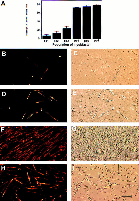
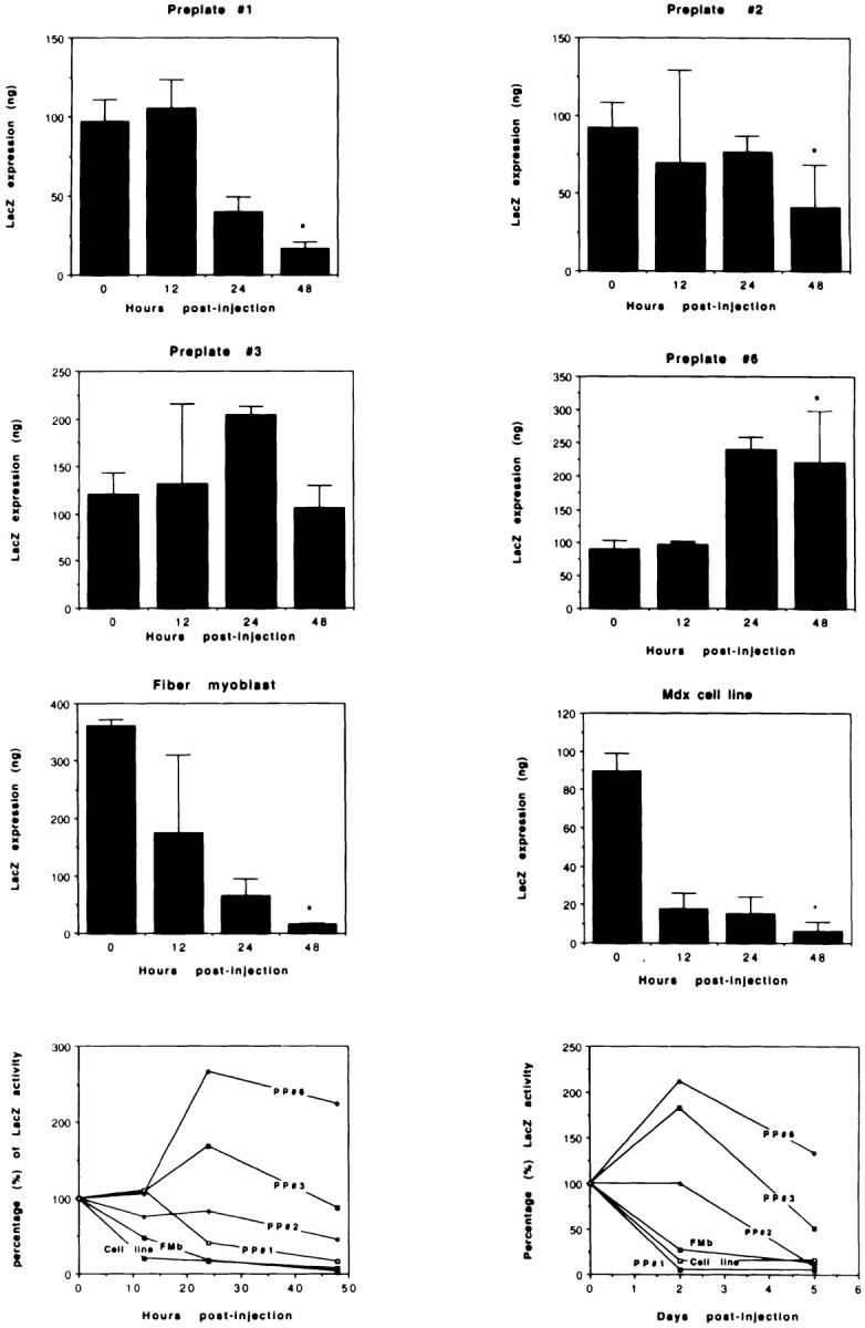
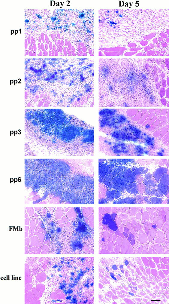
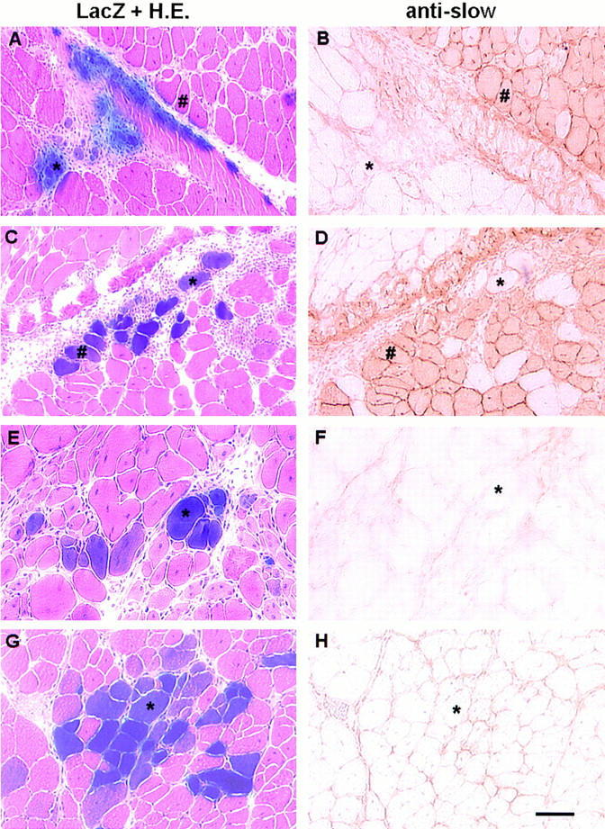
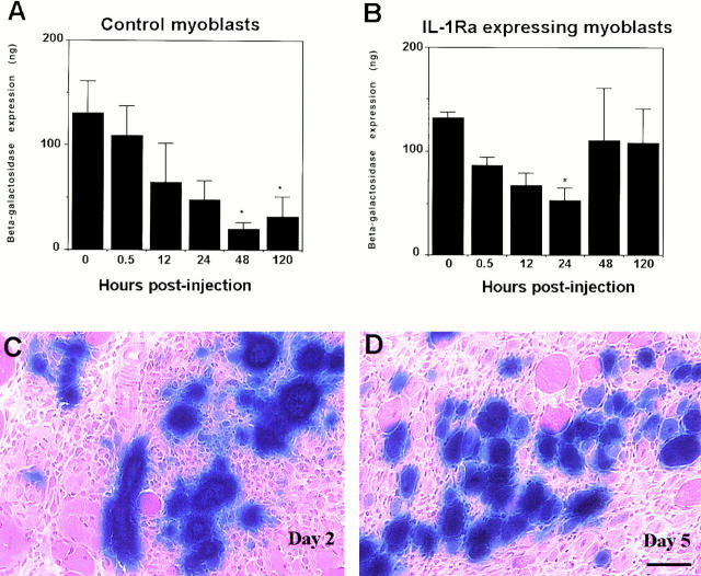
Myoblast transplantation has been extensively studied as a gene complementation approach for genetic diseases such as Duchenne Muscular Dystrophy. This approach has been found capable of delivering dystrophin, the product missing in Duchenne Muscular Dystrophy muscle, and leading to an increase of strength in the dystrophic muscle. This approach, however, has been hindered by numerous limitations, including immunological problems, and low spread and poor survival of the injected myoblasts. We have investigated whether antiinflammatory treatment and use of different populations of skeletal muscle-derived cells may circumvent the poor survival of the injected myoblasts after implantation. We have observed that different populations of muscle-derived cells can be isolated from skeletal muscle based on their desmin immunoreactivity and differentiation capacity. Moreover, these cells acted differently when injected into muscle: 95% of the injected cells in some populations died within 48 h, while others richer in desmin-positive cells survived entirely. Since pure myoblasts obtained from isolated myofibers and myoblast cell lines also displayed a poor survival rate of the injected cells, we have concluded that the differential survival of the populations of muscle-derived cells is not only attributable to their content in desmin-positive cells. We have observed that the origin of the myogenic cells may influence their survival in the injected muscle. Finally, we have observed that myoblasts genetically engineered to express an inhibitor of the inflammatory cytokine, IL-1, can improve the survival rate of the injected myoblasts. Our results suggest that selection of specific muscle-derived cell populations or the control of inflammation can be used as an approach to improve cell survival after both myoblast transplantation and the myoblast-mediated ex vivo gene transfer approach.


Figures

Figure 1
Figure 1
Characterization of the different populations of muscle-derived cells in vitro. The populations of muscle-derived cells after preplate (pp) displayed different desmin immunoreactivities ranging between 7 and 78% (A). The first preplate contained only 7% of desmin-positive cells, while the sequential preplates were enriched in myoblast content: pp2 = 14%, pp3 = 25%, pp4 = 72%, pp5 = 75%, and pp6 = 78% (A). Moreover, the ability of the muscle-derived cells to fuse into myotubes was found higher in pp5 (F and G) and pp6 (H and I) in comparison with PP1 (B and C) and PP3 (D and E). The desmin immunofluorescence is shown in B, D, F, and H, and the corresponding phase contrast field is shown in C, E, G, and I, respectively. Bar, 120 μm.
Figure 2
Figure 2
Characterization of the survival of different populations of muscle-derived cells after transplantation. Injection of the muscle-derived cells obtained after preplate 1 was rapidly lost 48 h after injection: only 17% of the transgene expression present on the injected myoblasts before injection was measured in the injected muscle. However, the cells isolated at preplate 2 led to 55% of the myoblast loss; preplate 3 led to a 12% loss, and preplate 6 gained 124% of the level of transgene expression present in the cells before transplantation. Surprisingly, a 96% loss of the pure population of myoblasts isolated from single myofibers was still observed 48 h after transplantation (fiber myoblast, FMb). Similarly, the immortalized myoblast cell line was rapidly lost after transplantation: 93% of the level of transgene expression present in the cell culture after implantation had disappeared 2 d after injection (Mdx cell line). Even though PP3 and PP6 displayed a better cell survival 2 d after injection, a decrease was still observed in the amount of LacZ reporter gene in the injected muscle 5 d after injection. However, the cells that displayed a better survival (PPs 3 and 6) remained with a higher level of gene transfer 5 d after injection. *P < 0.05 when compared with transduced noninjected myoblasts (0 h).
Figure 3
Figure 3
Determination of the ability of the different populations of muscle-derived cells to achieve gene transfer in muscle 2 and 5 d after injection. The population of purified primary myoblasts pp6 and, to a lesser extent, pp3, offered a better gene transfer than the population of muscle-derived cells isolated in pp1 and pp2. The myoblast cell line (cell line) and the highly pure myoblast culture isolated from fast single myofibers (FMb) also displayed a reduction in gene transfer when compared with the muscle-derived cells isolated in pp6. Bar, 50 μm.
Figure 4
Figure 4
Expression of slow MyHC in LacZ-positive cells in soleus (slow) and gastrocnemius (fast) muscles 5 d after FMb (A, B, E, F ) and PP6 (C, D, G, H) implantation. Serial cryostat sections were used to reveal the colocalization of LacZ-expressing myofibers with the presence of myofibers expressing slow MyHC. The transduced myoblasts isolated from single myofibers (FMb) either fused together or with host myoblasts and formed immature myofibers in which no slow myosin heavy chain was detected (*, A and B), or fused with host myofibers that similarily did not express slow MyHC (*, E and F). This result suggested that the myoblasts from fast single fibers did not fuse with myofibers expressing slow MyHC (#, A and B) and may preferentially have fused with host muscle fibers of the same phenotype. In contrast, preplate 6 fused with host muscle fibers expressing (#, C and D) or not expressing slow myosin heavy chain (*, C, D, G, and H), suggesting that the muscle derived cells at PP6 had the ability to fuse with both fast- and slow-twitch muscle fibers. Bar, 50 μm.
Figure 5
Figure 5
Characterization of the ability of engineered myoblast expressing antiinflammatory substance to reduce the poor survival of the injected cells. The survival of the myoblasts engineered to express IL-1Ra was compared with the nonengineered control cells. The nonengineered cells were rapidly lost 48 h after injection (Control myoblast). In contrast, the cells engineered to express IL-1Ra significantly reduced the early loss of the injected cells (IL-1Ra– expressing myoblast): only 20% of the injected cells seemed to be lost 48 h after injection. However, a significant reduction in the amount of β-galactosidase expression was observed 24 h after injection in comparison to the noninjected myoblasts. We observed a high number of transduced myofibers that persisted between day 2 and day 5 after injection of the IL-1Ra expressing myoblasts (C and D). The absence of significant difference for both populations of cells at 0 and 0.5 h after injection suggested that the loss of myoblasts was minimal during injection. *P < 0.05 when compared with transduced noninjected myoblasts (0 h). Bar, 50 μm.

References

    1. Acsadi G, Dickson G, Love DR, Jani A, Walsh FS, Gurusinghe A, Wolff JA, Davies KE. Human dystrophin expression in mdx mice after intramuscular injection of DNA constructs. Nature. 1991;352:815–818. - PubMed
    1. Acsadi G, Jani A, Massie B, Simoneau M, Holland P, Blaschuk K, Karpati G. A differential efficiency of adenovirus-mediated in vivo gene transfer into skeletal muscle cells at different maturity. Hum Mol Genet. 1994a;3:579–584. - PubMed
    1. Acsadi G, Jani A, Huard J, Blaschuk K, Massie B, Holland P, Lochmuller H, Karpati G. Cultured human myoblasts and myotubes show markedly different transducibility by replication-defective adenovirus recombinant. Gene Ther. 1994b;1:338–340. - PubMed
    1. Acsadi G, Lochmuller H, Jani A, Huard J, Massie B, Prescott S, Simoneau M, Petrof BJ, Karpati G. Dystrophin expression in muscles of mdx mice after adenovirus-mediated in vivo gene transfer. Hum Gene Ther. 1996;7:129–140. - PubMed
    1. Alamedine HS, Dehaupas M, Fardeau M. Regeneration of skeletal muscle fibers from autologous satellite cells multiplied in-vitro. Muscle Nerve. 1989;12:544–555. - PubMed

Publication types