. 1998 Sep 7;142(5):1289-99.
doi: 10.1083/jcb.142.5.1289.

The yeast V159N actin mutant reveals roles for actin dynamics in vivo

Affiliations

The yeast V159N actin mutant reveals roles for actin dynamics in vivo

L D Belmont et al. J Cell Biol. .

Abstract

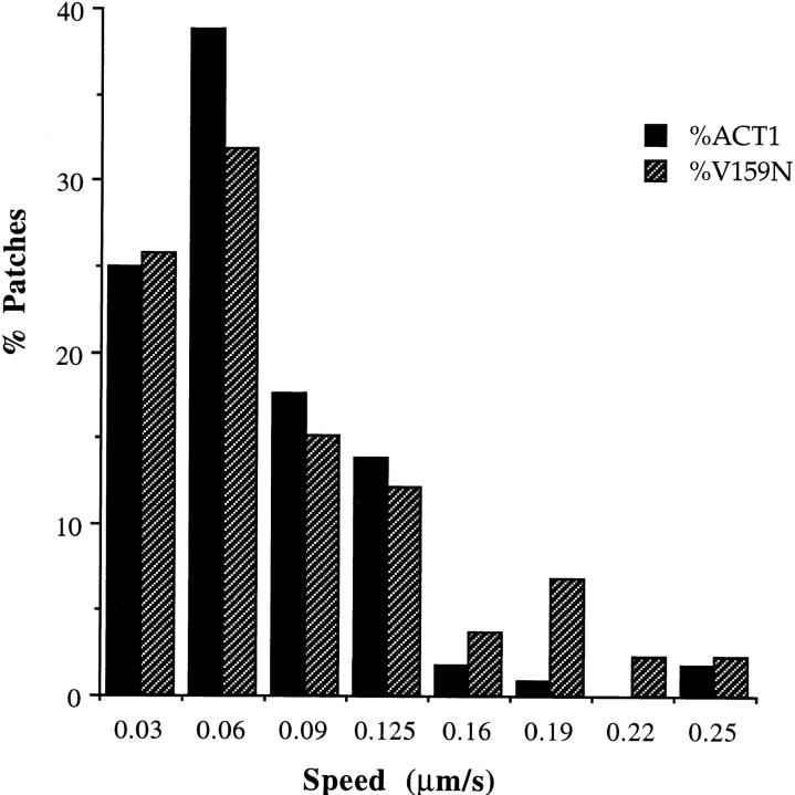
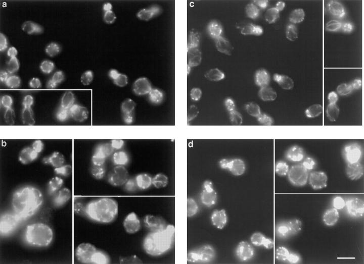
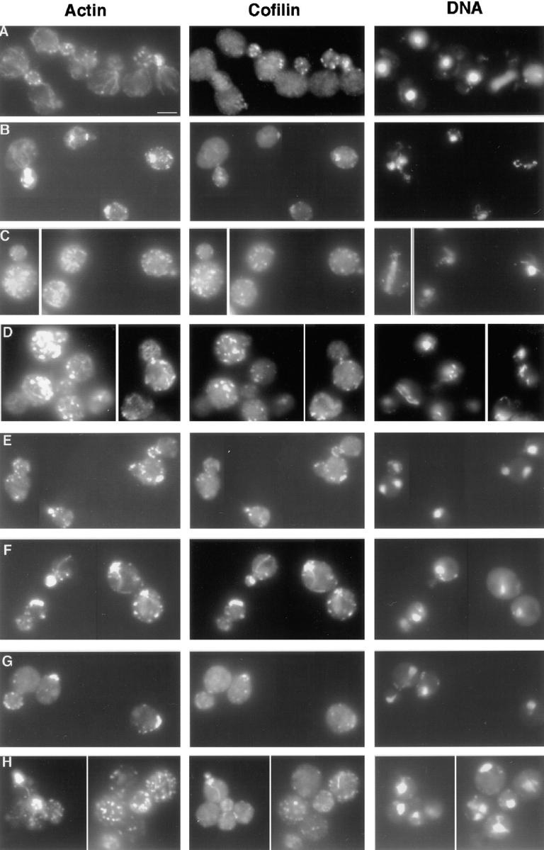
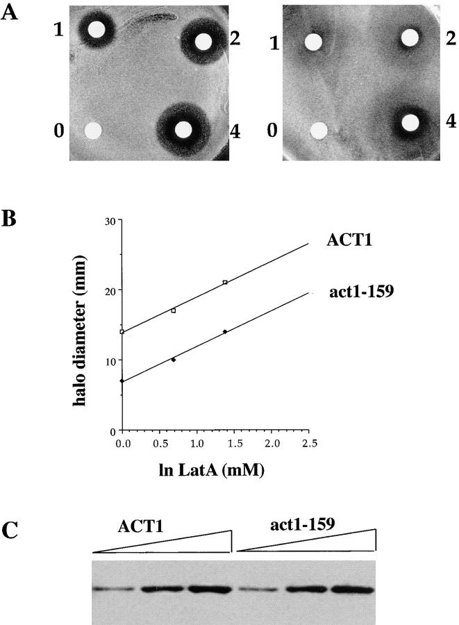
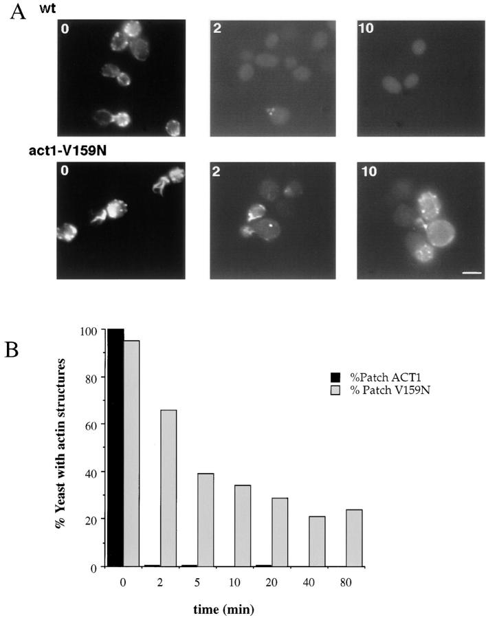
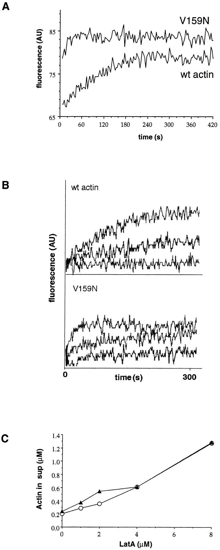
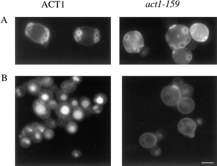
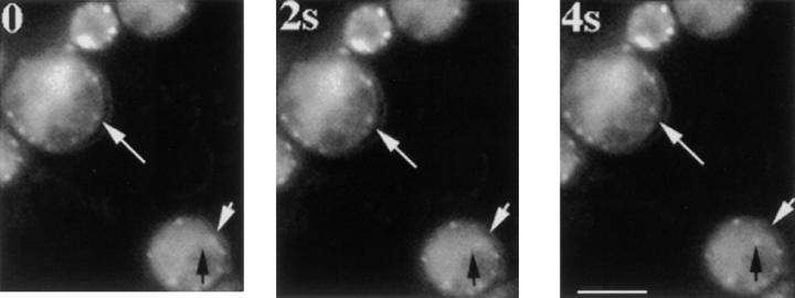
Actin with a Val 159 to Asn mutation (V159N) forms actin filaments that depolymerize slowly because of a failure to undergo a conformational change after inorganic phosphate release. Here we demonstrate that expression of this actin results in reduced actin dynamics in vivo, and we make use of this property to study the roles of rapid actin filament turnover. Yeast strains expressing the V159N mutant (act1-159) as their only source of actin have larger cortical actin patches and more actin cables than wild-type yeast. Rapid actin dynamics are not essential for cortical actin patch motility or establishment of cell polarity. However, fluid phase endocytosis is defective in act1-159 strains. act1-159 is synthetically lethal with cofilin and profilin mutants, supporting the conclusion that mutations in all of these genes impair the polymerization/ depolymerization cycle. In contrast, act1-159 partially suppresses the temperature sensitivity of a tropomyosin mutant, and the loss of cytoplasmic cables seen in fimbrin, Mdm20p, and tropomyosin null mutants, suggesting filament stabilizing functions for these actin-binding proteins. Analysis of the cables in these double-mutant cells supports a role for fimbrin in organizing cytoplasmic cables and for Mdm20p and tropomyosin in excluding cofilin from the cables.


Figures

Figure 6
Figure 6
Rates of actin patch motility are unaffected by the act1-159 mutation. Actin cortical patches were visualized using GFP-ABP1, and images were collected at 4-s intervals. The speed of each moving patch was measured at every interval. n = 114 (wild-type actin); n = 132 (V159N actin). There was no significant difference in the speed of patch movement.
Figure 1
Figure 1
Rhodamine-phalloidin staining of yeast expressing V159N actin. Yeast cells were grown in rich media at 25°C. The F-actin was stained with rhodamine-phalloidin. (a) Wild-type haploids (DDY1495), (b) act1-159 haploids (DDY1493), (c) wild-type diploids (DDY479), and (d) act1-159/ACT1 heterozygotes (DDY1491). Images for haploids (a and b) and diploids (c and d) were captured using identical exposure times and gain settings to allow comparison of the intensity of rhodamine-phalloidin staining. Bar, 5 μm.
Figure 8
Figure 8
Immunofluorescence of double mutants. Log phase cells grown at 25°C were fixed with formaldehyde and stained with anti-actin and anti-cofilin antibodies. (A) Wild-type yeast (DDY1495), (B) act1-159 (DDY1493), (C) sac6Δ (haploid progeny of DDY216 × DDY1493), (D) sac6Δ act1-159 (from DDY216 × DDY1493), (E) tpm1Δ (from DDY487 × DDY1493), (F) tpm1Δ act1-159 (from DDY487 × DDY1493), (G) mdm20Δ (from DDY1415 × DDY1493), (H) act1-159 mdm20Δ (from DDY1415 × DDY1493). Cofilin is localized exclusively to cortical actin patches in wild-type, act1-159, and act1-159 sac6Δ cells, but in the tpm1Δ act1-159 and act1-159 mdm20Δ double mutants, cofilin is seen on structures that look like cables. Bar, 5 μm.
Figure 2
Figure 2
LAT-A halo assay and Western blot of actin. (A) LAT-A was placed on sterile disks at concentrations of 0, 1 mM, 2 mM, and 4 mM. These disks were placed on nascent lawns of wild-type (left) or act1-159 (right) yeast and allowed to grow at 25°C for 3 d. (B) The concentration of LAT-A required to produce a halo of complete growth inhibition of a given size is fourfold higher in the act1-159 mutant compared with wild-type yeast. (C) Western blot of an SDS polyacrylamide gel loaded with 3, 6, and 9 μl of yeast extracts from wild-type and act1-159 strains. Actin bands from three separate blots were measured by densitometry. Tubulin was measured as a loading control for normalization (not shown). The expression levels of wild-type and V159N actin were the same within an experimental error of 15%.
Figure 3
Figure 3
Actin patch persistence in 400 μM LAT-A. LAT-A, a drug that sequesters actin monomers, was added to the growth medium. Cells were fixed in formaldehyde at various time points, and stained with rhodamine phalloidin. (A) Wild-type (DDY1495) and act1-159 mutant (DDY1493) cells are shown at 0, 2, and 10 min after drug addition. (B) The percentage of cells with detectable actin structures at various time points. More than 200 cells were counted for each condition. Bar, 5 μm.
Figure 4
Figure 4
LAT-A binding and ATP exchange. (A) ATP exchange was measured by monitoring the increase in fluorescence of ε-ATP upon binding to monomeric actin. Each measurement was repeated three times, and the t 1/2 of V159N actin was 15 s (15 s, 15 s, 14 s), and the t 1/2 of wild-type actin was ∼100 s (100 s, 102 s, 100 s). Typical plots of fluorescence are shown. The beginning and ending fluorescence levels for wild-type actin are 68 and 80. The beginning and ending fluorescence levels for the V159N actin are 80 and 85. There is a dead time of ∼4 s before the first measurement can be recorded, so it is possible that the starting point for the V159N actin is similar to that of the wild-type actin. This would make the value of 15 s an overestimate of the t 1/2 of ATP exchange, because most of the exchange would have occurred in the dead time. (B) LAT-A was added to the ATP exchange reactions to a final concentration of 0, 2.5, and 5 μM. The t 1/2 of ATP exchange in 2.5 μM LAT-A is approximately doubled for both V159N and wild-type actin, suggesting similar affinities for LAT-A. (C) Increasing concentrations of LAT-A were added to 4 μM polymerized actin. After 1 h, the polymerized actin was pelleted and the amount of actin left in the supernatant was quantified (○, wild-type actin; ▴, V159N actin).
Figure 5
Figure 5
act1-159 mutants exhibit random budding and a defect in endocytosis. (A) Calcofluor staining showing bud scars in wild-type and act1-159 diploids. (B) Endocytosis was evaluated by observing the uptake of Lucifer yellow into the vacuole using fluorescence microscopy. The weak fluorescence from the act1-159 mutant strain indicates a defect in fluid phase endocytosis. Bar, 5 μm.
Figure 7
Figure 7
Cortical actin patches in act1-159 continue to move in the presence of LAT-A. LAT-A was added to 400 μM, and samples were taken during a 2-h time period and images were recorded to assess patch motility using GFP-ABP1. The cells pictured here had been growing in LAT-A–containing media for 86 min. The top white arrow points to a patch that moves away from the arrow towards the center of the cell. In the cell on the bottom right, the white arrow marks the starting position of an actin patch, and the black arrow points to its final destination. The low signal to noise ratio is due to decreased actin patch fluorescence intensity resulting from sequestration of actin monomers by LAT-A. Frames are 2-s apart. Bar, 5 μm.

References

    1. Adams AE, Botstein D, Drubin DG. Requirement of yeast fimbrin for actin organization and morphogenesis in vivo. Nature. 1991;354:404–408. - PubMed
    1. Ayscough KR, Stryker J, Pokala N, Sanders M, Crews P, Drubin DG. High rates of actin filament turnover in budding yeast and roles for actin in establishment and maintenance of cell polarity revealed using the actin inhibitor latrunculin-A. J Cell Biol. 1997;137:399–416. - PMC - PubMed
    1. Berstein BW, Bamburg JR. Tropomyosin binding to F-actin protects the F-actin from disassembly by brain actin-depolymerizing-factor (ADF) Cell Motil. 1982;2:1–8. - PubMed
    1. Carlier MF. Actin polymerization and ATP hydrolysis. Adv Biophys. 1990;26:51–73. - PubMed
    1. Carlier MF, Pantaloni D. Direct evidence for ADP-Pi-F-actin as the major intermediate in ATP-actin polymerization. Rate of dissociation of Pi from actin filaments. Biochemistry. 1986;25:7789–7792. - PubMed

Publication types

MeSH terms