. 1998 Sep 7;142(5):1301-12.
doi: 10.1083/jcb.142.5.1301.

Involvement of an actomyosin contractile ring in Saccharomyces cerevisiae cytokinesis

Affiliations

Involvement of an actomyosin contractile ring in Saccharomyces cerevisiae cytokinesis

E Bi et al. J Cell Biol. .

Abstract

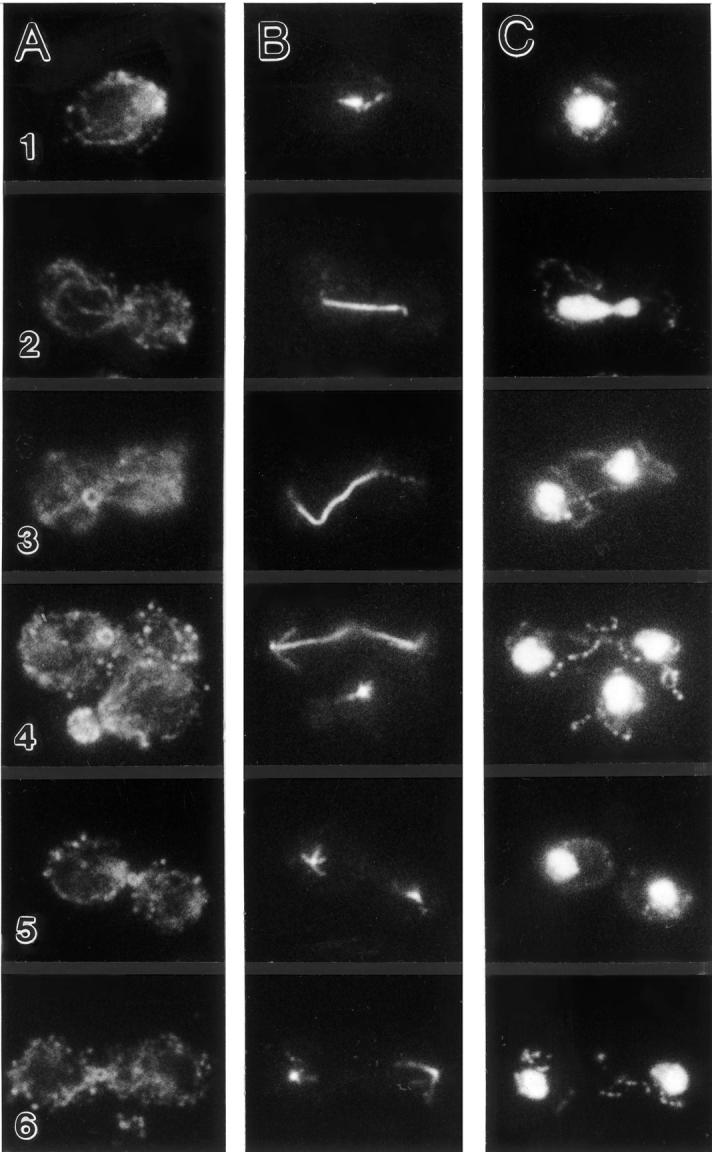
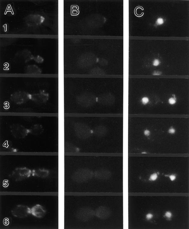
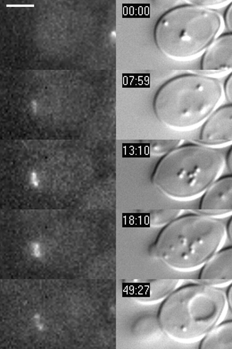
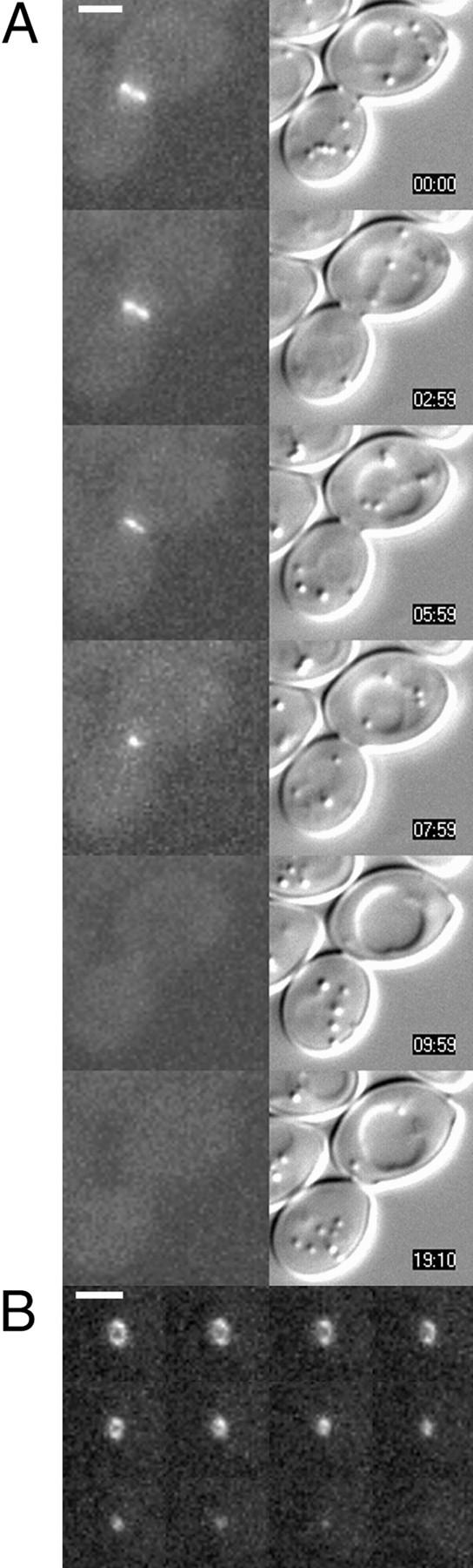
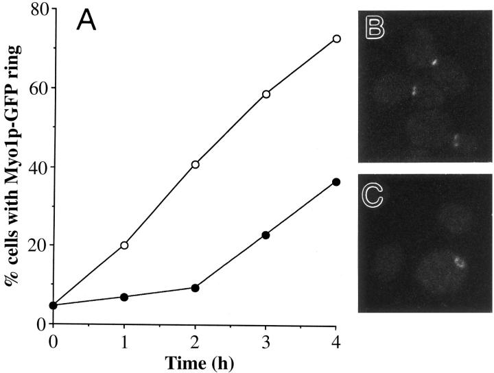
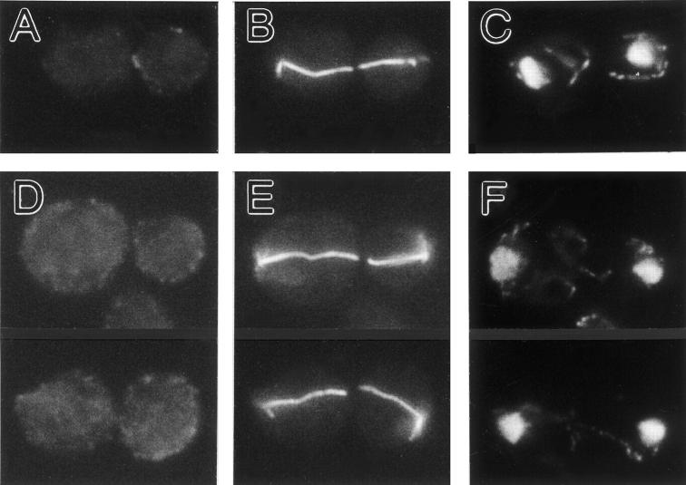
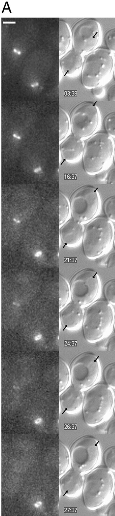
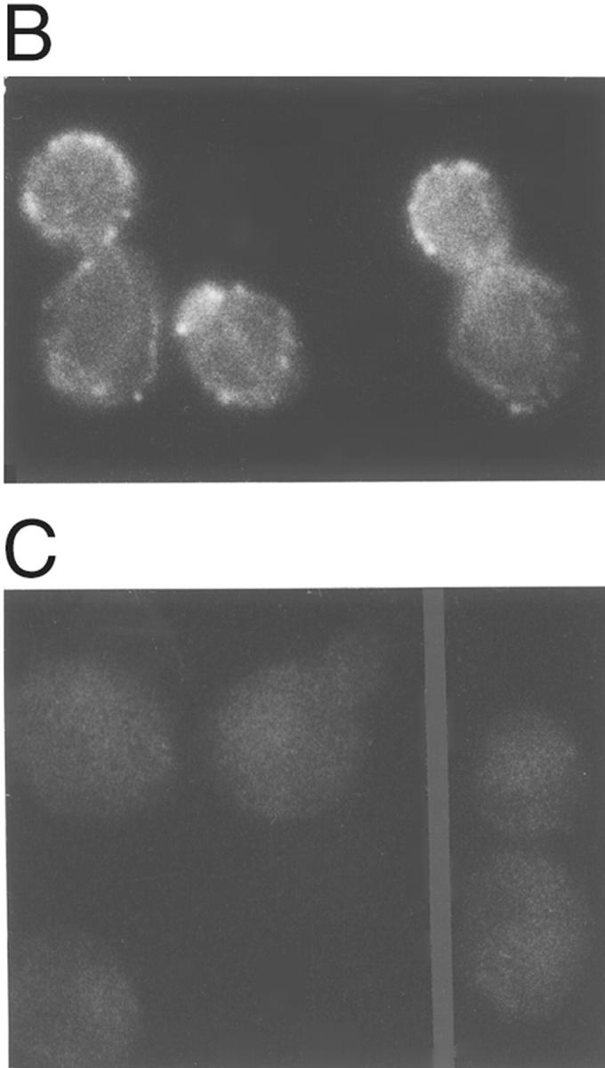
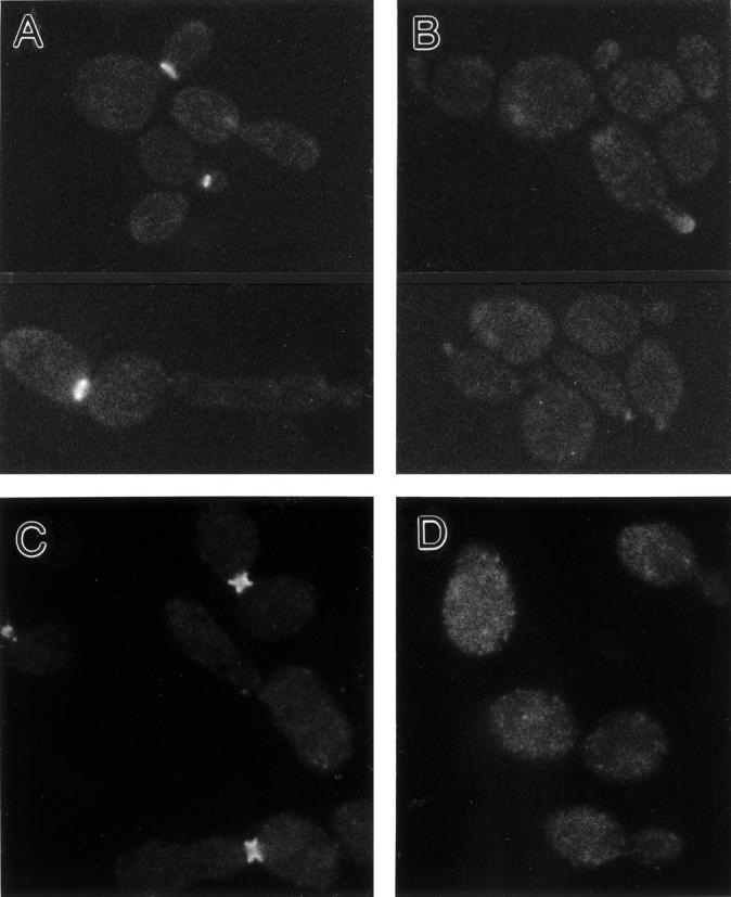
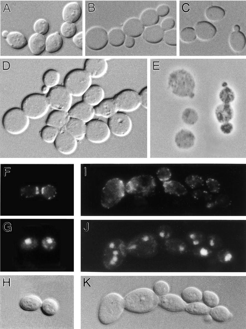
In Saccharomyces cerevisiae, the mother cell and bud are connected by a narrow neck. The mechanism by which this neck is closed during cytokinesis has been unclear. Here we report on the role of a contractile actomyosin ring in this process. Myo1p (the only type II myosin in S. cerevisiae) forms a ring at the presumptive bud site shortly before bud emergence. Myo1p ring formation depends on the septins but not on F-actin, and preexisting Myo1p rings are stable when F-actin is depolymerized. The Myo1p ring remains in the mother-bud neck until the end of anaphase, when a ring of F-actin forms in association with it. The actomyosin ring then contracts to a point and disappears. In the absence of F-actin, the Myo1p ring does not contract. After ring contraction, cortical actin patches congregate at the mother-bud neck, and septum formation and cell separation rapidly ensue. Strains deleted for MYO1 are viable; they fail to form the actin ring but show apparently normal congregation of actin patches at the neck. Some myo1Delta strains divide nearly as efficiently as wild type; other myo1Delta strains divide less efficiently, but it is unclear whether the primary defect is in cytokinesis, septum formation, or cell separation. Even cells lacking F-actin can divide, although in this case division is considerably delayed. Thus, the contractile actomyosin ring is not essential for cytokinesis in S. cerevisiae. In its absence, cytokinesis can still be completed by a process (possibly localized cell-wall synthesis leading to septum formation) that appears to require septin function and to be facilitated by F-actin.


Figures

Figure 1
Figure 1
Formation and contraction of an actin ring during the cell cycle. Cells of strain YEF473 growing exponentially in SC medium were triple-stained for F-actin (A), tubulin (B), and DNA (C) as described in Materials and Methods. Cells shown are in G1 or S phase (1), S or G2 phase (4, bottom cell), early anaphase (2), late anaphase (3 and 4, top cell), or after anaphase (5 and 6). All cells are shown at the same magnification. The cells shown were chosen for presentation because they had been distorted enough during preparation (note also the bend in the spindle of cell 3) to allow the actin rings to be seen more or less en face. More typical side views of the actin ring can be seen in Fig. 4.
Figure 4
Figure 4
Coincidence of the actin and Myo1p rings. Cells of strain YEF1698 growing exponentially in YM-P medium were fixed with ice-cold 70% ethanol (refer to Materials and Methods) and then stained for F-actin (A) and DNA (C). The GFP signal was also recorded (B). Cells 1–6 show successive stages in the cell cycle. All cells are shown at the same magnification.
Figure 2
Figure 2
Formation of the Myo1p–GFP ring relative to bud emergence. Cells of strain YEF1698 growing exponentially in SC medium were spotted onto SC medium containing 25% gelatin and observed by time-lapse photomicroscopy as described in Materials and Methods. Pairs of GFP (left) and DIC (right) images were recorded at the indicated times. Similar series of images were obtained for five different cells undergoing bud emergence. Formation of the Myo1p ring before bud emergence was also supported by observations on fixed cells (Fig. 4, cell 1; Fig. 7; and data not shown). Bar, 2 μm.
Figure 3
Figure 3
Contraction of the Myo1p–GFP ring late in the cell cycle. Time-lapse analysis of strain YEF1698 was performed as in Fig. 2. (A) Pairs of GFP fluorescence and DIC images were recorded at the indicated times from a cell positioned such that the Myo1p–GFP ring was seen from the side. Very similar series of images were obtained from four different cells. (B) GFP fluorescence images were recorded at 1-min intervals from a cell positioned such that its Myo1p–GFP ring was seen en face. Very similar series of images were obtained for two additional cells. Bars, 2 μm.
Figure 7
Figure 7
Formation of Myo1p–GFP rings in the absence of F-actin. Unbudded cells of strain YEF1698 were isolated as described in Materials and Methods and reinoculated into fresh YM-P medium in the absence or presence of 200 μM LAT-A. (A) Time-course of Myo1p–GFP ring formation in the absence (open circles) or presence (closed circles) of LAT-A. (B and C) Representative cells from the cultures without (B) and with (C) LAT-A, photographed 4 h after reinoculation.
Figure 5
Figure 5
Dependence of actin ring formation on Myo1p. Cells of wild-type strain YEF473 (A–C) and myo1Δ/myo1Δ strain YEF1820 (D–F) growing exponentially in SC medium were triple-stained for F-actin (A and D), tubulin (B and E), and DNA (C and F).
Figure 6
Figure 6
Dependence of Myo1p ring contraction, but not of Myo1p ring maintenance, on F-actin. (A) Cells of strain YEF1698 growing exponentially in SC medium were treated with 200 μM LAT-A for 10 min and then spotted onto solid SC/25% gelatin medium containing 200 μM LAT-A and observed by time-lapse video microscopy. Pairs of GFP fluorescence (left) and DIC (right) images were recorded at the times indicated. Arrows in DIC images, ends of the late-anaphase nucleus. Recording was continued for an additional 35 min beyond the images shown without obvious change in the appearance of the Myo1p–GFP ring in the right-hand cell. (B and C) Rhodamine-phalloidin staining of cells before (B) or 10 min after (C) the beginning of LAT-A treatment. Bar, 2 μm.
Figure 6
Figure 6
Dependence of Myo1p ring contraction, but not of Myo1p ring maintenance, on F-actin. (A) Cells of strain YEF1698 growing exponentially in SC medium were treated with 200 μM LAT-A for 10 min and then spotted onto solid SC/25% gelatin medium containing 200 μM LAT-A and observed by time-lapse video microscopy. Pairs of GFP fluorescence (left) and DIC (right) images were recorded at the times indicated. Arrows in DIC images, ends of the late-anaphase nucleus. Recording was continued for an additional 35 min beyond the images shown without obvious change in the appearance of the Myo1p–GFP ring in the right-hand cell. (B and C) Rhodamine-phalloidin staining of cells before (B) or 10 min after (C) the beginning of LAT-A treatment. Bar, 2 μm.
Figure 8
Figure 8
Dependence of Myo1p–GFP ring formation and maintenance on the septins. Cells of strain YEF1798 were fixed with 70% ethanol (refer to Materials and Methods) and processed for the observation of Myo1p-GFP (A and B) or fixed with formaldehyde and processed for the visualization of Cdc11p (C and D) during exponential growth in YM-P medium at 25°C (A and C) or 1 h after a shift to 37°C (B and D).
Figure 9
Figure 9
Phenotypic effects of MYO1 deletions. (A–E) Cells of wild-type strain YEF473 (A) and of myo1Δ/myo1Δ strains JMY1318 (B and C) and YEF1820 (D and E) were fixed during exponential growth in SC medium and examined by DIC (A–D) or phase-contrast (E) microscopy. Cells were examined without additional treatments (A, B, and D), after light sonication (C), or after treatment with lytic enzyme to remove cell walls (refer to Materials and Methods) (E). (F–K) Cells of strains YEF473 (F–H) and YEF1820 (I–K) were fixed during exponential growth in YM-P medium, double stained for F-actin (F and I) and DNA (G and J), and observed also by DIC (H and K). All cells are shown at the same magnification.

References

    1. Adams, A.E.M. 1984. Cellular morphogenesis in the yeast Saccharomyces cerevisiae. Ph.D. thesis. University of Michigan, Ann Arbor, MI. 179 pp.
    1. Adams AEM, Pringle JR. Relationship of actin and tubulin distribution to bud growth in wild-type and morphogenetic-mutant Saccharomyces cerevisiae. . J Cell Biol. 1984;98:934–945. - PMC - PubMed
    1. Alfa CE, Hyams JS. Distribution of tubulin and actin through the cell division cycle of the fission yeast Schizosaccharomyces japonicus var. versatilis: a comparison with Schizosaccharomyces pombe. . J Cell Sci. 1990;96:71–77. - PubMed
    1. Ayscough KR, Stryker J, Pokala N, Sanders M, Crews P, Drubin DG. High rates of actin filament turnover in budding yeast and roles for actin in establishment and maintenance of cell polarity revealed using the actin inhibitor latrunculin-A. J Cell Biol. 1997;137:399–416. - PMC - PubMed
    1. Baudin A, Ozier-Kalogeropoulos O, Denouel A, Lacroute F, Cullin C. A simple and efficient method for direct gene deletion in Saccharomyces cerevisiae. . Nucleic Acids Res. 1993;21:3329–3330. - PMC - PubMed

Publication types

MeSH terms

Associated data