. 1998 Sep;180(18):4804-13.
doi: 10.1128/JB.180.18.4804-4813.1998.

Transcription analysis of two disparate rRNA operons in the halophilic archaeon Haloarcula marismortui

Affiliations

Transcription analysis of two disparate rRNA operons in the halophilic archaeon Haloarcula marismortui

P P Dennis et al. J Bacteriol. 1998 Sep.

Abstract

The genome of the halophilic archaeon Haloarcula marismortui contains two rRNA operons designated rrnA and rrnB. Genomic clones of the two operons and their flanking regions have been sequenced, and primary transcripts and processing intermediates derived from each operon have been characterized. The 16S, 23S, and 5S genes from the two operons were found to differ at 74 of 1,472 positions, 39 of 2,922 positions, and 2 of 122 positions, respectively. This degree of sequence divergence for multicopy (paralogous) rRNA genes was 10- to 50-fold or more higher than anticipated. The two operons exhibit other profound differences that include (i) the presence in rrnA and the absence in rrnB of tRNAAla and tRNACys genes in the intergenic and distal regions, respectively, (ii) divergent 5' flanking sequences, and (iii) distinct pathways for processing and maturation of 16S rRNA. Processing and maturation of 16S and 23S rRNA from rrnA operon transcripts and of 23S rRNA from rrnB operon transcripts follow the canonical halophilic pathway, whereas maturation of 16S rRNA from rrnB operon transcripts follows an unusual and different pathway that is apparently devoid of any 5' processing intermediate.


Figures

FIG. 1
FIG. 1
Structure of rrnA and rrnB operons of H. marismortui. The chromosomal structures of the rrnA and rrnB operons are indicated. The 16S, 23S, 5S, tRNAAla, and tRNACys genes are represented by solid boxes, the processing inverted repeats are indicated by hatched boxes, and the other flanking and spacer sequences are indicated by open boxes. Flanking and spacer regions longer than 50 nt and greater than 90% identical in sequences between rrnA and rrnB are indicated by brackets. All gene sequences are greater than 95% identical (see Table 1). The complete nucleotide sequences of rrnA and rrnB have been deposited in GenBank (rrnA, AF034619; rrnB, AF034620).
FIG. 2
FIG. 2
Important sequence motifs located in the flanking and spacer regions of rrnA and rrnB. (A) Small portions of the helical structures of the processing inverted repeats surrounding the 16S and 23S genes in rrnA and rrnB are indicated. The BHB motif is noticeably absent from the rrnB 16S helix. The sites of cleavage detected within the bulges on the 5′ side (left) and 3′ side (right) of each helix are indicated by scissors, and the position of the nucleotide 3′ to the scissile phosphate is indicated. Where negative, numbers correspond to 5′ flanking sequences, with the first nucleotide of 16S rRNA being 1 for the rrnA and rrnB regions, respectively. i, 16S-23S intergenic spacer positions with the first nucleotide after 16S being iA1 or iB1; 23Sd, 23S-5S intergenic spacer with the first nucleotide after 23S being 23Sd1. The 23S-5S intergenic spacer is identical in rrnA and rrnB. (B) The point of sequence convergence at the junction between the 5′ flanking sequences and the beginning of the 16S genes in the rrnA and rrnB operons is indicated. The dots indicate identical nucleotides. (C) A 5′ flanking sequence element conserved between rrnA, rrnB, and the rrn operons of other genera of halophilic archaea are aligned. The numbers in parentheses are the first nucleotide positions of the sequences shown. Dots indicate conserved nucleotides in pairwise comparisons. The 3-nt bulge located in the 5′ portion of the 16S processing helix in rrnA and the single rrn operon of H. cutirubrum is boxed. A conserved sequence proposed to play a role in folding of a conserved pseudoknot within 16S rRNA is indicated (9). (D) 16S-23S intergenic sequence around the tRNAAla in rrnA and the corresponding region of rrnB. Endonucleolytic cleavages occur at or near the indicated positions at the 3′ end of the tRNAAla and tRNAAla-like sequences. Regions of secondary structure within tRNAAla are indicated by brackets; the potential for similar secondary structures in rrnB is also indicated. Dots represent identical nucleotides. The 3′ transcript end detected in the anticodon loop of the tRNAAla structure (position iA139) is indicated; the anticodon sequence is overlined. The position of a corresponding 3′ end in rrnB (position iB137) is also indicated. Finally, a 5′ end resulting from cleavage by RNase P at the 5′ end of tRNAAla is indicated (position iA105). No corresponding end was observed in rrnB (position iB121). (E) The sequence at the point of convergence within the 16S-23S intergenic space between rrnA and rrnB is shown. The UUAA sequence (overlined or underlined) immediately 3′ to the point of perfect convergence represents the archaeal TATA box for the putative intergenic promoters in rrnA and rrnB. The dots represent identical nucleotides. (F) The sequence at the point of divergence in the 5S 3′ flanking region is shown. 5Sd represents the 5S 3′ flanking sequence with the first nucleotide beyond 5S being 5Sd1. The dots represent identical nucleotides.
FIG. 3
FIG. 3
Alignment of promoter-like sequences from the 5′ flanking and intergenic spacer regions of rrnA and rrnB. The nucleotide sequences of the 5′ flanking promoter-like motifs from rrnA and rrnB (A) and the intergenic promoter-like motifs from rrnA and rrnB (B) are indicated. The sequences are aligned at the archaeal TATA box element and at the dinucleotide transcription initiation site (dot; usually G). The promoter designations and the nucleotide positions of the putative transcription start sites are indicated on the left.
FIG. 4
FIG. 4
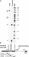
Analysis of 5′ transcript ends located in the 5′ flanking regions of rrnA and rrnB. Two respective AflIII restriction fragments specific for rrnA and rrnB were 5′ end labeled on the (−) strand at position 98 within the 16S sequence and used as probes against total cellular RNA in S1 nuclease protection assays. (A) The protection products were separated on denaturing polyacrylamide gels. The sizes (in nucleotides) of protection products and molecular length markers (MLM) are indicated beside the autoradiograms. (B) A diagram of rrnA and rrnB is shown. Each protection product is represented by an arrow with the length in nucleotides given on the right and the position of the 5′ transcript end given above the leftward pointing arrowheads. The positions of promoter-like sequence motifs and precursor processing sites are indicated. The 16S gene is a solid box, the 5′ portion of the processing repeat is a hatched box, and the remaining 5′ flanking region is unfilled. The dashed arrow in rrnB represents full-length protection of the probe by transcripts that were presumed to have been initiated at the upstream P1B promoter. At the bottom of panel B, the results of nuclease protection assays with 3′-end-labeled rrnB probes are illustrated (autoradiograms not shown). As above, the dashed arrows represent full-length protection of the probe, and the solid arrows represent partial-length protection of the probe. The length of each product is indicated on the left end and the position of the 3′ transcript end is indicated on the right end of each arrow (see text for details).
FIG. 5
FIG. 5
Analysis of 5′ and 3′ transcript ends located in the 16S-23S intergenic spaces of rrnA and rrnB. Two respective AvaI restriction fragments specific for rrnA and rrnB were 3′ end labeled on the (−) strand at position 1365 within the 16S gene or 5′ end labeled on the (−) strand at position 157 within the 23S gene and used as probes against total cellular RNA in S1 nuclease protection assays. The protection products were separated on denaturing polyacrylamide gels. The size in nucleotides of protection products and molecular length markers (MLM) are indicated beside each autoradiogram. (A) Autoradiograms with the rrnA probe either 3′ labeled (left) or 5′ labeled (right). The lanes marked A and A+G are Maxam Gilbert sequence reactions on the 3′-labeled probe. (B) Diagram illustrating the rrnA 383-nt 16S-23S intergenic space and a summary of the protection products obtained with the 3′-labeled probe (3′ transcript end sites are represented by rightward pointing arrows) and the 5′-labeled probe (5′ transcript end sites are represented by leftward pointing arrows). The estimated length of each product is indicated at the start of the arrow, and the positions of the respective 3′ or 5′ transcript ends within the intergenic spaces are indicated above the arrowhead. Opposing arrowheads represent sites of endonuclease cleavage. The gray arrow represents a product with a 3′ end at position iA105 that was not observed. The overlapping arrowheads at positions iA193 to iA205 represent multiple protection products. The dashed double-headed arrow represents full-length protection of both the 3′- and 5′-labeled probes with unprocessed transcripts from rrnA. The 16S, tRNAAla, and 23S genes are represented as solid boxes, the inverted processing repeats are indicated as hatched boxes, and other intergenic sequences are shown as open boxes. M16, maturation site at the 3′ end of 16S rRNA; BHB, BHB cleavage site; 5′TP, 5′ tRNA processing site (presumably RNase P); PPS, uncharacterized precursor processing site; 3′TP, 3′ tRNA processing site (apparently by an endonuclease; see the text); M23, 5′-end maturation of 23S rRNA (apparently an endonuclease; see the text). (C) Autoradiograms with the rrnB probe either 3′ labeled (left) or 5′ labeled (right). (D) Diagram of the rrnB 405-nt 16S-23S intergenic space. Other details are as described for panel B.
FIG. 6
FIG. 6
Analysis of 3′ transcript ends located in 23S-5S intergenic and flanking regions of rrnA and rrnB. Transcripts from both rrnA and rrnB were analyzed by using an AvaI fragment 3′ end labeled on the (−) strand at position 2859 within the 23S gene as a probe in S1 nuclease protection assays. (A) Autoradiogram illustrating protection products with the sizes in nucleotides of protection products (left) and of molecular length markers (MLM; right) indicated. (B) Diagram of the 23S-5S intergenic and 5S 3′ flanking regions. The 23S, 5S, and tRNACys genes are represented by solid boxes, the 23S inverted repeat is indicated by a hatched box, and other intergenic and flanking regions are indicated by open boxes. The dashed region, including the tRNACys gene, is unique to the rrnA operon. Other designations are as described in the legend to Fig. 5.

Similar articles

Cited by

References

    1. Branlant C A, Krol M A, Machott A, Pouyet J, Ebel J-P, Edwards K, Kossel H. E. coli 23S rRNA heterogeneity. Nucleic Acids Res. 1981;9:4303. - PMC - PubMed
    1. Brombach M, Specht T, Erdmann V A, Ulbrich N. Complete nucleotide sequence of a 23S ribosomal RNA gene from Halobacterium marismortui. Nucleic Acids Res. 1989;17:3293. - PMC - PubMed
    1. Chant J, Dennis P P. Archaebacteria: transcription and processing of ribosomal RNA sequences in Halobacterium cutirubrum. EMBO J. 1986;5:1091–1097. - PMC - PubMed
    1. Dennis, P. P. Expression of ribosomal RNA operons in halophilic archaea, In A. Oren (ed.), Microbiology and biogeochemistry of hypersaline environments, in press. CRC Press, Inc., Boca Raton, Fla.
    1. Dennis P P. Multiple promoters for the transcription of the ribosomal RNA gene cluster in Halobacterium cutirubrum. J Mol Biol. 1985;186:456–461. - PubMed

Publication types

LinkOut - more resources