. 1998 Sep;180(18):4856-64.
doi: 10.1128/JB.180.18.4856-4864.1998.

Oxidative stress response and characterization of the oxyR-ahpC and furA-katG loci in Mycobacterium marinum

Affiliations

Oxidative stress response and characterization of the oxyR-ahpC and furA-katG loci in Mycobacterium marinum

E Pagán-Ramos et al. J Bacteriol. 1998 Sep.

Abstract

Oxidative stress response in pathogenic mycobacteria is believed to be of significance for host-pathogen interactions at various stages of infection. It also plays a role in determining the intrinsic susceptibility to isoniazid in mycobacterial species. In this work, we characterized the oxyR-ahpC and furA-katG loci in the nontuberculous pathogen Mycobacterium marinum. In contrast to Mycobacterium smegmatis and like Mycobacterium tuberculosis and Mycobacterium leprae, M. marinum was shown to possess a closely linked and divergently oriented equivalents of the regulator of peroxide stress response oxyR and its subordinate gene ahpC, encoding a homolog of alkyl hydroperoxide reductase. Purified mycobacterial OxyR was found to bind to the oxyR-ahpC promoter region from M. marinum and additional mycobacterial species. Mobility shift DNA binding analyses using OxyR binding sites from several mycobacteria and a panel of in vitro-generated mutants validated the proposed consensus mycobacterial recognition sequence. M. marinum AhpC levels detected by immunoblotting, were increased upon treatment with H2O2, in keeping with the presence of a functional OxyR and its binding site within the promoter region of ahpC. In contrast, OxyR did not bind to the sequences upstream of the katG structural gene, and katG expression did not follow the pattern seen with ahpC. Instead, a new open reading frame encoding a homolog of the ferric uptake regulator Fur was identified immediately upstream of katG in M. marinum. The furA-katG linkage and arrangement are ubiquitous in mycobacteria, suggesting the presence of additional regulators of oxidative stress response and potentially explaining the observed differences in ahpC and katG expression. Collectively, these findings broaden our understanding of oxidative stress response in mycobacteria. They also suggest that M. marinum will be useful as a model system for studying the role of oxidative stress response in mycobacterial physiology, intracellular survival, and other host-pathogen interactions associated with mycobacterial diseases.


Figures

FIG. 1
FIG. 1
Genetic organization of the oxyR-ahpC and furA-katG loci in mycobacteria. (A) The genes oxyR (open boxes) and ahpC (shaded boxes) are tightly linked and divergently transcribed (arrows) in the majority of mycobacterial species with the exception of M. smegmatis (line indicates that the corresponding region upstream of ahpC has been sequenced and characterized but that no oxyR has been identified in this organism). In M. tuberculosis, oxyR has been inactivated via multiple, naturally occurring mutations (filled balloons, nonsense and frameshift mutations; open balloons, deletions). (B) Linkage of furA (encoding a homolog of the ferric uptake regulator Fur) and katG in mycobacteria. The furA and katG genes are cotranscribed in M. tuberculosis. In M. leprae, both furA and katG are inactivated via multiple mutations (balloons, insertions; triangles, deletions).
FIG. 2
FIG. 2
Survival of M. marinum ATCC 15069 in the J774 murine macrophage cell line. (A) Epifluorescence microscopy image of macrophages infected with GFP-expressing M. marinum ATCC 15069. (B) Comparison of the survival of M. bovis BCG (⧫), M. marinum (Mm; ▴), and M. smegmatis mc2 155 (Ms; ■) recovered from J774 cells over time. Each value represents the mean CFU (±standard error) from at least three independent experiments. Infection, incubation (37°C), and other techniques are described in Materials and Methods.
FIG. 3
FIG. 3
AhpC and KatG levels in M. marinum and M. xenopi exposed to H2O2. (A) Western blot of cell extracts (10 μg of protein) from M. marinum and M. smegmatis with an antibody raised against M. tuberculosis AhpC. Lanes: 1, purified His10-AhpC from M. tuberculosis (M. t.); 2, M. marinum (M.m.); 3, M. smegmatis mutant strain VD1865-6 (M.s. ahpC::Kmr); 4, M. smegmatis parent strain mc2155 (M.s. ahpC+). (B) Analysis of ahpC expression in M. marinum and M. xenopi treated with H2O2. Exponentially growing cultures were exposed to various concentrations of H2O2 (0.02 to 20 mM) for 2 h. After treatment, crude protein extracts (20 μg) were separated by SDS-PAGE and probed with anti-M. tuberculosis-AhpC antibody. Steady-state levels of AhpC increased after treatment with 2 mM H2O2. (C) Western blot analysis of the effects of exposure to H2O2 on M. marinum and M. xenopi katG expression. The samples were identical to those shown in panel B except that the blot was probed with KatG antibody.
FIG. 4
FIG. 4
Alignment of OxyR sequences from M. marinum (M.m.) (this work), M. avium (M.a.) (37), and M. leprae (M.l.) (10). Asterisks, identical amino acids; periods, conserved amino acid substitutions.
FIG. 5
FIG. 5
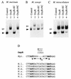
Binding of M. leprae OxyR to the oxyR-ahpC intergenic region of M. marinum, M. xenopi, and M. intracellulare and the consensus mycobacterial OxyR binding sequence. Purified M. leprae OxyR (see Materials and Methods) was incubated with 32P-labeled DNA fragments containing the oxyR-ahpC intergenic region from M. marinum (−120 to +50 relative to the oxyR start codon) (A), M. xenopi (−193 to +96) (B), and M. intracellulare (−108 to +193) (C). Protein-DNA complexes (open triangles) were separated from unbound probes (filled triangles) by electrophoresis on a 4% native polyacrylamide gel and analyzed by autoradiography. The specificity of the binding was tested by competition assays using specific and nonspecific competitor DNAs in the reactions. Lanes: 1, radiolabeled probe alone; 2, probe incubated with His10-OxyR; 3, same as lane 2 plus 0.5 μg of cold specific competitor (unlabeled DNA identical to the radiolabeled probe); 4, same as lane 2 plus 0.5 μg of cold nonspecific competitor DNA (a 324-bp fragment from the M. bovis BCG ahpC structural gene). (D) Consensus sequence of the mycobacterial OxyR binding site within the oxyR-ahpC region (Myc.) and OxyR sequences from M. leprae (M.l.), M. tuberculosis (M.t.), M. marinum (M.m.), M. xenopi (M.x.), M. avium (M.a.), and M. intracellulare (M.i.). The sequence exhibits twofold dyad symmetry (ATC-N9-GAT; bold letters) and contains the T-N11-A core motif typical of recognition sequences of the LysR-type transcriptional regulators (34). Init., initiation.
FIG. 6
FIG. 6
DNase I footprinting analysis of OxyR contacts with the recognition sequence. OxyR was bound to a probe containing the OxyR binding site within the M. marinum ahpC-oxyR intergenic region and subjected to DNase I footprinting as described in Materials and Methods. Lanes: OxyR +, OxyR-bound probe; OxyR −, free probe; G, A, T, and C, sequencing ladder generated with the primer used to produce the probe (see Materials and Methods). Circles, protected bases; asterisks, hypersensitive sites. The nucleotide positions of the protected and hypersensitive sites are indicated on the right; bold letters highlight the core of the proposed OxyR binding recognition sequence.
FIG. 7
FIG. 7
Mutational analysis of the OxyR recognition sequence within the oxyR-ahpC region. DNA fragments containing mutations (listed at the bottom; introduced by site-specific mutagenesis as described in Materials and Methods) were subjected to DNA binding mobility shift assays. Open triangles, DNA-OxyR complex; closed arrows, unbound probe. Lanes: 1 and 4, radiolabeled probe alone; 2, 3, 5, and 6, probe incubated with His10-OxyR. Lanes 2 and 3 and lanes 5 and 6 represent duplicate samples. The M.t. sequence is proposed to contain a natural mutation of the left half site. Quantitation of DNA-OxyR complexes by densitometric analysis: (A) lanes 2 and 3, 2% probe bound; lanes 5 and 6, 60% probe bound; (B) lanes 2 and 3, 80% probe bound; lanes 5 and 6, binding below detection limit; (C) lanes 2 and 3, 1% probe bound; lanes 5 and 6, 1% DNA-OxyR probe bound.
FIG. 8
FIG. 8
M. marinum FurA and multiple sequence alignment of mycobacterial FurA homologs. Mm-FurA, M. marinum FurA (accession no. AF038027); Mt-FurA, M. tuberculosis FurA (accession no. AF002194); Ml-FurA, M. leprae FurA (accession no. AF013983 for annotation); Mt-Fur, M. tuberculosis Fur (accession no. Z95208); Ec-Fur, E. coli Fur (33).
FIG. 9
FIG. 9
Southern blot analysis of PstI- or SacII-digested M. marinum genomic DNA probed with M. tuberculosis katG (22) (A), furA (accession no. AF002194) (B), fur (accession no. Z95208) (C), and ideR (35) (D).

Similar articles

Cited by

References

    1. Altuvia S, Almirom M, Huisman G, Kolter R, Storz G. The dps promoter is activated by OxyR during growth and by IHF and ςS in stationary phase. Mol Microbiol. 1994;13:265–272. - PubMed
    1. Ausubel F M, Brent R, Kingston R E, Moore D D, Seidman J G, Smith J A, Struhl K, editors. Current protocols in molecular biology. New York, N.Y: John Wiley & Sons; 1989.
    1. Barker L P, George K M, Falkow S, Small P L C. Differential trafficking of live and dead Mycobacterium marinum organisms in macrophages. Infect Immun. 1997;65:1497–1504. - PMC - PubMed
    1. Chae H Z, Robinson K, Poole L B, Church G, Storz G, Rhee S G. Cloning and sequencing of thiol-specific antioxidant from mammalian brain: alkyl hydroperoxide reductase and thiol-specific antioxidant define a large family of antioxidant enzymes. Proc Natl Acad Sci USA. 1994;91:7017–7021. - PMC - PubMed
    1. Chan J, Kaufmann H E. Immune mechanisms of protection. In: Bloom B R, editor. Tuberculosis: pathogenesis, protection, and control. Washington, D.C: ASM Press; 1994. pp. 389–415.

Publication types

Associated data

LinkOut - more resources